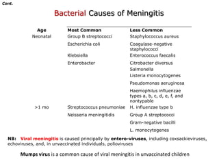
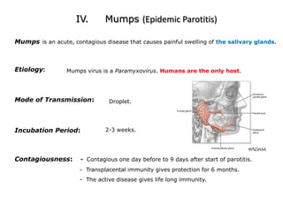
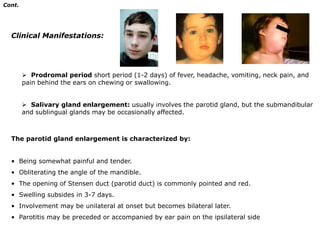
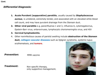
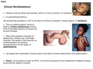
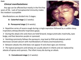
Infectious DiseasesPrepared byDR.SHERIF REDAProf. of PediatricsAL- AZHAR UNIVERSITYCAIRO  -  EGYPT
Introduction
I.        MeningitisII.       EncephalitisIII.      PoliomyelitisIV.      MumpsV.       Tetanus NeonatorumVI.      Pertussis
I.      MeningitisThe meninges are membranes that cover and protect the brain and spinal cord. There are 3 layers known as the:Dura mater is the tough outer membraneArachnoid mater is the thin, delicate, web-like membranePia mater is the innermost membraneThe arachnoid & pia mater are together known the as leptomeninges      Inflammation of the leptomeninges, caused byBacteriaViruses                          Or rarely,   FungiThe term ASEPTIC MENINGITIS refers principally to viral meningitis, but a similar picture may be seen with:Other infectious organisms (Lyme disease, syphilis, tuberculosis) Parameningeal infections (brain abscess, epidural abscess, venous sinus empyema) Chemical exposure (nonsteroidal anti-inflammatory drug, IV immunoglobulin) Autoimmune disordersAlterations of host defense:  (anatomic defects or immune deficits) increase the risk of meningitis from less common pathogens e.g. Pseudomonasaeruginosa, Staphylococcus aureus, Salmonella spp., and L. monocytogenes.
Cont.Bacterial Causes of MeningitisNB:   Viral meningitis is caused principally by entero-viruses, including coxsackieviruses, echoviruses, and, in unvaccinated individuals, poliovirusesMumps virus is a common cause of viral meningitis in unvaccinated children
Cont.Mode of infection:Bacterial meningitis is most commonly results from hematogenous dissemination of microorganisms from a distant site of infectionClinical Manifestations:The onset of acute bacterial meningitis has two predominant patterns: The more dramatic and fortunately less common presentation is sudden onset with rapidly progressive manifestations of shock, purpura, DIC, unconsciousness, and frequently resulting in death within 24 hours. More often, meningitis: is preceded by several days of fever with upper respiratory or gastrointestinal symptoms followed by nonspecific signs of CNS infection such as lethargy or irritability.
Cont.1)  Non specific findings:Fever, anorexia, poor feeding, myalgia, arthralgia, tachycardia, hypotension and various cutaneous signs such as petechiae, purpura or an erythematous macular rush. 2)  Signs of meningeal irritation:Nuchal rigidity and back pain
Kernig’s sign: flexion of the hip 90 degrees with subsequent pain with extension of the leg.
Brudzinski sign: involuntary flexion of the knees and hips after passive flexion of the neck while supine.3)  Symptoms and Signs of increased ICP: 1- Headache and vomiting 		2- Bulging fontanel or widening of the sutures3- Cranial neuropathies.		4- Hypertension with bradycardia5- Apnea or hyperventilation, stupor and coma.4)  Seizures: (focal or generalized) due to, cerebritis, infarction or electrolyte disturbances. Seizures that occur on presentation or within the first 4 days of onset are usually of no prognostic significance
Cont.Diagnosis:Lumbar puncture for CSF analysis should be performed when bacterial meningitis is suspected:1- Microorganisms on gram stain and culture. 	2- Neutrophilpleocytosis (300 - 2000/mm3). 3- Elevated protein (100 - 500mg/dL)			4- Reduced glucose concentration (< 50% of serum glucose)5- Physical manifestations (Turbid with elevated pressure,100 - 300 mm H2O).Normal CSF shows:Normal pressure (50 - 80 mm H2O), leucocytes (< 5/mm3), proteins (20 -45 mg/dl) and glucose (75 % of serum glucose)
Cont.Treatment:     A)Initial Antibiotic Therapy:Vancomycin 60 mg/kg/24hr given every 6 hr in combinationWith either cefotaxime (200 mg/kg/24hr given every 6 hours) or ceftriaxone (100 mg/kg/24hr single dose or given every 12 hour). Patients allergic to β- Lactam antibiotics can be treated with chloramphenicol, 100 mg/kg/24 hr given every 6 hr. Duration of antibiotic therapy:  At least for 7-14 days I.V.     B)  Corticosteroids:  I.V dexamethasone 0.15 mg/kg/dose given every 6hr for 2 days in the treatment of children older than 6wk with acute bacterial meningitis caused by H. influenzae type b to decrease the permanent auditory nerve damage.   C)  Supportive and symptomatic therapy:    - Good evaluation and monitoring are essential.    -Correction of dehydration and electrolyte disturbances and proper nutrition.    -Control of seizures  - Management of neurological complications
Cont.Prevention: Vaccination and antibiotic prophylaxis of susceptible at – risk contacts:Close contact:Should be treated with RIFAMPIN 10mg/kg/dose every 12hr, for 2 days (in N. meningitides) and 20mg/kg/24  for 4 days (in H. influenzae type b).Meningococcal quadrivalent Vaccine: Avaccine is recommended for high-risk children older than 2yr. Also Vaccines for H. influenzae type b should be given to all children beginning at 2 mo of age 3 doses (2, 4, 6 months).
II.    Encephalitis  Encephalitis is an inflammatory process of the brain parenchyma that usually is an acute infectious process, but may be a postinfectious encephalomyelitis, a chronic degenerative disease, or a slow viral infection.
  Organisms cause encephalitis by one of two mechanisms: direct infection of the brain parenchyma  an apparent immune-mediated response in the CNS that usually begins several days after the appearance of extraneural manifestations of the infection  Encephalitis may be diffuse or localizedEtiology:  Viruses are the principal causes of acute infectious encephalitis.
  The most common viral causes of encephalitis are the arboviruses (St. Louis, LaCrosse, California, West Nile encephalitis viruses), enteroviruses, and herpesviruses.
  Encephalitis also may result from other types of infection, metabolic, toxic, and neoplastic disorders. Cont.Clinical Manifestations:  Acute infectious encephalitis usually is preceded by a prodrome of several days of nonspecific symptoms, such as cough, sore throat, fever, headache, and abdominal complaints.
  The characteristic symptoms of progressive lethargy, behavioral changes, and neurologic deficits, followed.
Seizures are common at presentation.
  Children with encephalitis also may have a maculopapular rash and severe complications, such as fulminant coma, transverse myelitis, anterior horn cell disease (polio-like illness), or peripheral neuropathy.
  Acute DisseminatedEncephaloMyelitis (ADEM):    - Is the abrupt development of multiple neurologic signs related to an inflammatory, demyelinating disorder of the brain and spinal cord.     - Acute disseminated encephalomyelitis follows childhood viral infections, such as measles and chickenpox or vaccinations.
Cont.Laboratory And Imaging Studies: The diagnosis of viral encephalitis is supported by examination of the CSF, which typically shows a lymphocytic pleocytosis, slight elevation in protein content, and normal glucose level. Increased erythrocytes and CSF protein may occur with HSV.
 The CSF occasionally may be normal.
Neuroimaging studies (CT, MRI) may be normal or may show diffuse cerebral swelling of the parenchyma or focal abnormalities.
 The EEG is the definitive test and shows diffuse, slow wave activity, although focal changes may be present.
Brain biopsy may be appropriate for patients with severe encephalopathy who show no clinical improvement if the diagnosis remains obscure.Cont.Treatment: With the exception of HSV and HIV, there is no specific therapy for viral encephalitis.
 Management is supportive and frequently requires ICU admission, which allows aggressive therapy for seizures, timely detection of electrolyte abnormalities, and, when necessary, airway monitoring and protection and reduction of increased intracranial pressure.
 IV acyclovir is the treatment of choice for HSV infections.
 ADEM has been treated with high-dose IV corticosteroids.III.       PoliomyelitisEtiology: The polioviruses are non-enveloped, RNA viruses belonging to the Picornaviridae family,   the genus Enterovirus. It  includes three antigenically distinct serotypes (types 1, 2, and 3).
 There is no cross immunity between the three types of the virus. Most extensive epidemics of the disease are due to type 1.
 The polioviruses can retain activity for several days at room temperature, and can be stored indefinitely frozen at -20°C.
 They are rapidly inactivated by heat (>56°C), formaldehyde, chlorination, and ultraviolet light.
  Humans are the only known reservoir for the polioviruses.Cont.Incubation Period:7-14 daysMode of Infection:Feco-oral mainlyPathogenesis:-  Poliovirus gain entry through the gastrointestinal tract, multiplies in the alimentary tract (in Peyer's patches) and in lymph nodes of the intestine.-  Virus will invade the CNS along the peripheral nerves.
Cont.Clinical Manifestations:1-  Unapparent infection, which occurs in 90-95% of cases and causes no disease and no sequelae.2-  Abortive poliomyelitis occursin about 5% of patients (nonspecific influenza-like syndrome). The illness is short-lived (up to 2-3 days). 3-  Nonparalytic poliomyelitis:In about 1% of infected patients, the signs of abortive poliomyelitis are present but headache, nausea and vomiting are more intense. In addition there is the following: Signs of meningeal irritation.
 Generalized muscle pain and tenderness.
Tripod sign. The patient can sit only with hands thrown far behind for support.

Infectious diseases

  • 2.
    Infectious DiseasesPrepared byDR.SHERIFREDAProf. of PediatricsAL- AZHAR UNIVERSITYCAIRO - EGYPT
  • 3.
  • 4.
    I. MeningitisII. EncephalitisIII. PoliomyelitisIV. MumpsV. Tetanus NeonatorumVI. Pertussis
  • 5.
    I. MeningitisThe meninges are membranes that cover and protect the brain and spinal cord. There are 3 layers known as the:Dura mater is the tough outer membraneArachnoid mater is the thin, delicate, web-like membranePia mater is the innermost membraneThe arachnoid & pia mater are together known the as leptomeninges Inflammation of the leptomeninges, caused byBacteriaViruses Or rarely, FungiThe term ASEPTIC MENINGITIS refers principally to viral meningitis, but a similar picture may be seen with:Other infectious organisms (Lyme disease, syphilis, tuberculosis) Parameningeal infections (brain abscess, epidural abscess, venous sinus empyema) Chemical exposure (nonsteroidal anti-inflammatory drug, IV immunoglobulin) Autoimmune disordersAlterations of host defense: (anatomic defects or immune deficits) increase the risk of meningitis from less common pathogens e.g. Pseudomonasaeruginosa, Staphylococcus aureus, Salmonella spp., and L. monocytogenes.
  • 6.
    Cont.Bacterial Causes ofMeningitisNB: Viral meningitis is caused principally by entero-viruses, including coxsackieviruses, echoviruses, and, in unvaccinated individuals, poliovirusesMumps virus is a common cause of viral meningitis in unvaccinated children
  • 7.
    Cont.Mode of infection:Bacterialmeningitis is most commonly results from hematogenous dissemination of microorganisms from a distant site of infectionClinical Manifestations:The onset of acute bacterial meningitis has two predominant patterns: The more dramatic and fortunately less common presentation is sudden onset with rapidly progressive manifestations of shock, purpura, DIC, unconsciousness, and frequently resulting in death within 24 hours. More often, meningitis: is preceded by several days of fever with upper respiratory or gastrointestinal symptoms followed by nonspecific signs of CNS infection such as lethargy or irritability.
  • 8.
    Cont.1) Nonspecific findings:Fever, anorexia, poor feeding, myalgia, arthralgia, tachycardia, hypotension and various cutaneous signs such as petechiae, purpura or an erythematous macular rush. 2) Signs of meningeal irritation:Nuchal rigidity and back pain
  • 9.
    Kernig’s sign: flexionof the hip 90 degrees with subsequent pain with extension of the leg.
  • 10.
    Brudzinski sign: involuntaryflexion of the knees and hips after passive flexion of the neck while supine.3) Symptoms and Signs of increased ICP: 1- Headache and vomiting 2- Bulging fontanel or widening of the sutures3- Cranial neuropathies. 4- Hypertension with bradycardia5- Apnea or hyperventilation, stupor and coma.4) Seizures: (focal or generalized) due to, cerebritis, infarction or electrolyte disturbances. Seizures that occur on presentation or within the first 4 days of onset are usually of no prognostic significance
  • 11.
    Cont.Diagnosis:Lumbar puncture forCSF analysis should be performed when bacterial meningitis is suspected:1- Microorganisms on gram stain and culture. 2- Neutrophilpleocytosis (300 - 2000/mm3). 3- Elevated protein (100 - 500mg/dL) 4- Reduced glucose concentration (< 50% of serum glucose)5- Physical manifestations (Turbid with elevated pressure,100 - 300 mm H2O).Normal CSF shows:Normal pressure (50 - 80 mm H2O), leucocytes (< 5/mm3), proteins (20 -45 mg/dl) and glucose (75 % of serum glucose)
  • 12.
    Cont.Treatment: A)Initial Antibiotic Therapy:Vancomycin 60 mg/kg/24hr given every 6 hr in combinationWith either cefotaxime (200 mg/kg/24hr given every 6 hours) or ceftriaxone (100 mg/kg/24hr single dose or given every 12 hour). Patients allergic to β- Lactam antibiotics can be treated with chloramphenicol, 100 mg/kg/24 hr given every 6 hr. Duration of antibiotic therapy: At least for 7-14 days I.V. B) Corticosteroids: I.V dexamethasone 0.15 mg/kg/dose given every 6hr for 2 days in the treatment of children older than 6wk with acute bacterial meningitis caused by H. influenzae type b to decrease the permanent auditory nerve damage. C) Supportive and symptomatic therapy: - Good evaluation and monitoring are essential. -Correction of dehydration and electrolyte disturbances and proper nutrition. -Control of seizures - Management of neurological complications
  • 13.
    Cont.Prevention: Vaccination andantibiotic prophylaxis of susceptible at – risk contacts:Close contact:Should be treated with RIFAMPIN 10mg/kg/dose every 12hr, for 2 days (in N. meningitides) and 20mg/kg/24 for 4 days (in H. influenzae type b).Meningococcal quadrivalent Vaccine: Avaccine is recommended for high-risk children older than 2yr. Also Vaccines for H. influenzae type b should be given to all children beginning at 2 mo of age 3 doses (2, 4, 6 months).
  • 14.
    II. Encephalitis Encephalitis is an inflammatory process of the brain parenchyma that usually is an acute infectious process, but may be a postinfectious encephalomyelitis, a chronic degenerative disease, or a slow viral infection.
  • 15.
    Organismscause encephalitis by one of two mechanisms: direct infection of the brain parenchyma an apparent immune-mediated response in the CNS that usually begins several days after the appearance of extraneural manifestations of the infection Encephalitis may be diffuse or localizedEtiology: Viruses are the principal causes of acute infectious encephalitis.
  • 16.
    Themost common viral causes of encephalitis are the arboviruses (St. Louis, LaCrosse, California, West Nile encephalitis viruses), enteroviruses, and herpesviruses.
  • 17.
    Encephalitisalso may result from other types of infection, metabolic, toxic, and neoplastic disorders. Cont.Clinical Manifestations: Acute infectious encephalitis usually is preceded by a prodrome of several days of nonspecific symptoms, such as cough, sore throat, fever, headache, and abdominal complaints.
  • 18.
    Thecharacteristic symptoms of progressive lethargy, behavioral changes, and neurologic deficits, followed.
  • 19.
    Seizures are commonat presentation.
  • 20.
    Childrenwith encephalitis also may have a maculopapular rash and severe complications, such as fulminant coma, transverse myelitis, anterior horn cell disease (polio-like illness), or peripheral neuropathy.
  • 21.
    AcuteDisseminatedEncephaloMyelitis (ADEM): - Is the abrupt development of multiple neurologic signs related to an inflammatory, demyelinating disorder of the brain and spinal cord. - Acute disseminated encephalomyelitis follows childhood viral infections, such as measles and chickenpox or vaccinations.
  • 22.
    Cont.Laboratory And ImagingStudies: The diagnosis of viral encephalitis is supported by examination of the CSF, which typically shows a lymphocytic pleocytosis, slight elevation in protein content, and normal glucose level. Increased erythrocytes and CSF protein may occur with HSV.
  • 23.
    The CSFoccasionally may be normal.
  • 24.
    Neuroimaging studies (CT,MRI) may be normal or may show diffuse cerebral swelling of the parenchyma or focal abnormalities.
  • 25.
    The EEGis the definitive test and shows diffuse, slow wave activity, although focal changes may be present.
  • 26.
    Brain biopsy maybe appropriate for patients with severe encephalopathy who show no clinical improvement if the diagnosis remains obscure.Cont.Treatment: With the exception of HSV and HIV, there is no specific therapy for viral encephalitis.
  • 27.
    Management issupportive and frequently requires ICU admission, which allows aggressive therapy for seizures, timely detection of electrolyte abnormalities, and, when necessary, airway monitoring and protection and reduction of increased intracranial pressure.
  • 28.
    IV acycloviris the treatment of choice for HSV infections.
  • 29.
    ADEM hasbeen treated with high-dose IV corticosteroids.III. PoliomyelitisEtiology: The polioviruses are non-enveloped, RNA viruses belonging to the Picornaviridae family, the genus Enterovirus. It includes three antigenically distinct serotypes (types 1, 2, and 3).
  • 30.
    There isno cross immunity between the three types of the virus. Most extensive epidemics of the disease are due to type 1.
  • 31.
    The poliovirusescan retain activity for several days at room temperature, and can be stored indefinitely frozen at -20°C.
  • 32.
    They arerapidly inactivated by heat (>56°C), formaldehyde, chlorination, and ultraviolet light.
  • 33.
    Humansare the only known reservoir for the polioviruses.Cont.Incubation Period:7-14 daysMode of Infection:Feco-oral mainlyPathogenesis:- Poliovirus gain entry through the gastrointestinal tract, multiplies in the alimentary tract (in Peyer's patches) and in lymph nodes of the intestine.- Virus will invade the CNS along the peripheral nerves.
  • 34.
    Cont.Clinical Manifestations:1- Unapparent infection, which occurs in 90-95% of cases and causes no disease and no sequelae.2- Abortive poliomyelitis occursin about 5% of patients (nonspecific influenza-like syndrome). The illness is short-lived (up to 2-3 days). 3- Nonparalytic poliomyelitis:In about 1% of infected patients, the signs of abortive poliomyelitis are present but headache, nausea and vomiting are more intense. In addition there is the following: Signs of meningeal irritation.
  • 35.
    Generalized musclepain and tenderness.
  • 36.
    Tripod sign. Thepatient can sit only with hands thrown far behind for support.

Editor's Notes

  • #19 Recovery is complete, and no neurologic signs or sequelae develop. stiffness of the neck and back with positive Kernig&apos;s and Brudzinski&apos;s signs. Nuchal rigidity in polio is characterized by being present in the supine position and disappears in the prone position. This differentiates it from pyogenic meningitis where it is present in prone position. Paralysis of motor cranial nerve nuclei. Involvement of 9th, 10th, and 12th cranial nerves is the most important. Hence, there is paralysis of tongue, pharynx, and larynx which results in airway obstruction.Involvement of vital centers leads to:Respiratory centers: irregularity of rate, depth and rhythm of respiration.Circulatory center: hypertension and cardiac arrhythmias.Heat regulating center: rapid changes is body temperature.
  • #22 T