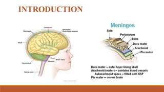
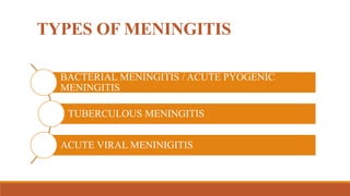
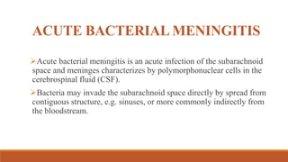
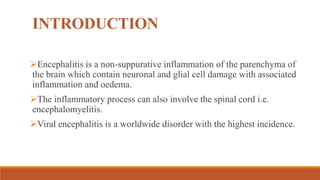
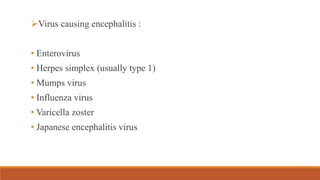
MENINGITIS
BY – AYUSHI KOYANI (FY MPT)
GUIDED BY – DR. VAISHALI SUTHAR MA’AM
CONTENTS
Introduction
Pathology
Clinical features
Investigations
Complications
Differential diagnosis
Prognosis
Treatment
INTRODUCTION
TYPES OF MENINGITIS
BACTERIAL MENINGITIS / ACUTE PYOGENIC
MENINGITIS
TUBERCULOUS MENINGITIS
ACUTE VIRAL MENINIGITIS
ACUTE BACTERIAL MENINGITIS
Acute bacterial meningitis is an acute infection of the subarachnoid
space and meninges characterizes by polymorphonuclear cells in the
cerebrospinal fluid (CSF).
Bacteria may invade the subarachnoid space directly by spread from
contiguous structure, e.g. sinuses, or more commonly indirectly from
the bloodstream.
Causative organisms
In neonates – gram -ve bacilli, e.g. E. coli, Klebsiella.
In children – Haemophiles influenzae. Pneumococcus,
meningococcus
In adults – Pneumococcus, Meningococcus
Host factors – congenital or acquired immune deficiency, predispose
to infection, overcrowding and poverty.
May occur following head injury, mastoiditis or after lumbar
puncture.
Nasopharyngeal colonisation
Replication & growth
Blood
Subarachnoid Space invasion
Replication of subarachnoid space
Infection & inflammation of sub arachnoid space
Pathology
Clinical features
c/f are present on the basis of 3 main causes:
• Infection
• Increased ICP
• Meningeal inflammation & irritation
Meningitis symptoms
• Severe frontal/occipital headache
• Stiff neck
• Fever, vomiting
• Photophobia
Meningitis sign :
•Neck stiffness
•Kernig's sign +ve
•Brudzinski's sign +ve
C/FACCORDING TO STAGE OF
MENINGITIS :
STAGE-1 STAGE OF INVASION:
Sudden onset
Severe head ache
Vomiting
Fever
Neck and back pain
Convulsion
Insomnia
Severe confusion
STAGE-2 MENINGEAL STAGE :
More severe head ache
Lumbar pain
Muscular stiffness & neck stiffness
Kernig's & Brudzinski's signs +ve
Muscular twitching & tenderness
Temperature variable
Exaggerated DTR
Sensory loss
Sensory neural deafness
Pulse : slow & irregular
Continue…
Pupils
Unequal & sluggish reaction
Dilated & fixed
Ptosis
Diplopia- double vision
Seizures
Other features
Facial paralysis
Dysphagia
Incoordination
Urine retention
constipation
INVESTIGATION
1) CT SCAN :
 Excludes focal neurological deficit, increased ICP or other
complications
2) CSF EXAMINATION :
 Increased pressure (>300 mm of H20)
 Purulent appearance
 Thick & greenish- pneumococcal
 Thin & yellow- strepto/ staphylococcal
 Increased total cell count >1000/ml
 Increased protein 1.0-5.0gm/lit
 Sugar decreased or absent
 Chloride & lactate decreased
3) Blood :
 Increased total blood count
 Increased ESR
 Culture isolate organism
4) Serum electrolytes :
Increased frequency
5) X-ray :
 chest- pneumonia
 Skull- #
COMPLICATIONS
1) Septicaemia
2) Arthritis
3) neurological complications
Cerebral oedema
Focal neurological damage
Psychiatric problems
Hydrocephalus
Cranial nerve palsies
4) Cardio-vascular complications
Myocarditis
Bacterial endocarditis
5) Electrolyte imbalance
6) Shock
DIFFERENTIAL DIAGNOSIS
Poliomyelitis
Acute disseminated encephalomyelitis
Acute encephalitis
Cerebral abscess
Sub arachnoid haemorrhage
PROGNOSIS
Over all prognosis- good
Depends upon different factors
E.g. Infective organism
Response to antibiotics
Stages of illness when pt comes
Presence of complications
Over all mortality- 15-25%
TREATMENT
PT EVALUATION OF THE
PATIENT WITH MENINGITIS
1. Observation of current functional status of the patient
2. Assessment of standard vital signs
3. Assessment of cognitive status
4. Assessment of sensory integrity
5. Assessment of movement abilities
6. Assessment of functional abilities
AIMS OF THE PT INTERVENTIONS
Optimization of postural control
Optimization of functional activities with selective voluntary
movement pattern control
Improvement of performance of functional activities
Enhancement of integration of sensory information
Optimization of cognitive status and psychosocial responses
ENCEPHALITIS
INTRODUCTION
Encephalitis is a non-suppurative inflammation of the parenchyma of
the brain which contain neuronal and glial cell damage with associated
inflammation and oedema.
The inflammatory process can also involve the spinal cord i.e.
encephalomyelitis.
Viral encephalitis is a worldwide disorder with the highest incidence.
Virus causing encephalitis :
• Enterovirus
• Herpes simplex (usually type 1)
• Mumps virus
• Influenza virus
• Varicella zoster
• Japanese encephalitis virus
The most common virus responsible for large-scale epidemic in
India is Japanese B encephalitis virus.
It is spread by mosquito that extensively in rice ecosystem.
In adults, >90% of cases of herpes simplex encephalitis result from
infection with HSV-1.
In children, influenza virus is an important cause of encephalitis.
CLINICAL FEATURES
GENERAL : pyrexia, myalgia
Specific to causative virus,
Meningeal involvement (slight) – neck stiffness, cellular response in
CSF.
Sign and symptoms of parenchymal involvement – focal / diffuse.
INVESTIGATION
CT SCAN : In temporal lobes with surrounding oedema.
CSF : Increased pressure
•Protein levels are mildly elevated
•Cell count is increased
EEG : periodic high voltage slow wave complexes over the
involved temporal lobe.
DIFFERNTIAL DIAGNOSIS
Post infectious encephalomyelitis
Bacterial or tubercular infections of the brain
PROGNOSIS
It is highly variable, in chronic cases high mortality is seen.
Many may be left with residual deficit like dementia, focal deficits
and epilepsy.
Complications remain such as flexion deformities of arms,
hyperextension of the legs, language impairment, learning difficulties
and behavioral problems.
Many recover completely if the illness is mild.
TREATMENT
General measures to care for the unconscious patient should be started.
Anticonvulsants are often necessary.
Dexamethasone 4 mg 6 hourly for brain oedema.
HSE responds well to acyclovir (10 mg/kg IV 8 hourly for 14-21 days)
The goals of therapeutic intervention program
for patients with inflammatory CNS disorders :
REFERENCE
Lindsay – Neurology & Neurosurgery Illustrated, 4th edition
Mathew – Medicine, 5th edition
Darcy – Umphred’s neurological rehabilitation, 6th edition
MENINGITIS & ENCEPHALITIS - Ayushi.pptx pdf

MENINGITIS & ENCEPHALITIS - Ayushi.pptx pdf

  • 1.
    MENINGITIS BY – AYUSHIKOYANI (FY MPT) GUIDED BY – DR. VAISHALI SUTHAR MA’AM
  • 2.
  • 3.
  • 4.
    TYPES OF MENINGITIS BACTERIALMENINGITIS / ACUTE PYOGENIC MENINGITIS TUBERCULOUS MENINGITIS ACUTE VIRAL MENINIGITIS
  • 5.
    ACUTE BACTERIAL MENINGITIS Acutebacterial meningitis is an acute infection of the subarachnoid space and meninges characterizes by polymorphonuclear cells in the cerebrospinal fluid (CSF). Bacteria may invade the subarachnoid space directly by spread from contiguous structure, e.g. sinuses, or more commonly indirectly from the bloodstream.
  • 6.
    Causative organisms In neonates– gram -ve bacilli, e.g. E. coli, Klebsiella. In children – Haemophiles influenzae. Pneumococcus, meningococcus In adults – Pneumococcus, Meningococcus Host factors – congenital or acquired immune deficiency, predispose to infection, overcrowding and poverty. May occur following head injury, mastoiditis or after lumbar puncture.
  • 7.
    Nasopharyngeal colonisation Replication &growth Blood Subarachnoid Space invasion Replication of subarachnoid space Infection & inflammation of sub arachnoid space Pathology
  • 8.
    Clinical features c/f arepresent on the basis of 3 main causes: • Infection • Increased ICP • Meningeal inflammation & irritation Meningitis symptoms • Severe frontal/occipital headache • Stiff neck • Fever, vomiting • Photophobia
  • 9.
    Meningitis sign : •Neckstiffness •Kernig's sign +ve •Brudzinski's sign +ve
  • 11.
    C/FACCORDING TO STAGEOF MENINGITIS : STAGE-1 STAGE OF INVASION: Sudden onset Severe head ache Vomiting Fever Neck and back pain Convulsion Insomnia Severe confusion
  • 12.
    STAGE-2 MENINGEAL STAGE: More severe head ache Lumbar pain Muscular stiffness & neck stiffness Kernig's & Brudzinski's signs +ve Muscular twitching & tenderness Temperature variable Exaggerated DTR Sensory loss Sensory neural deafness Pulse : slow & irregular
  • 13.
    Continue… Pupils Unequal & sluggishreaction Dilated & fixed Ptosis Diplopia- double vision Seizures Other features Facial paralysis Dysphagia Incoordination Urine retention constipation
  • 14.
    INVESTIGATION 1) CT SCAN:  Excludes focal neurological deficit, increased ICP or other complications
  • 15.
    2) CSF EXAMINATION:  Increased pressure (>300 mm of H20)  Purulent appearance  Thick & greenish- pneumococcal  Thin & yellow- strepto/ staphylococcal  Increased total cell count >1000/ml  Increased protein 1.0-5.0gm/lit  Sugar decreased or absent  Chloride & lactate decreased
  • 16.
    3) Blood : Increased total blood count  Increased ESR  Culture isolate organism 4) Serum electrolytes : Increased frequency 5) X-ray :  chest- pneumonia  Skull- #
  • 17.
    COMPLICATIONS 1) Septicaemia 2) Arthritis 3)neurological complications Cerebral oedema Focal neurological damage Psychiatric problems Hydrocephalus Cranial nerve palsies
  • 18.
    4) Cardio-vascular complications Myocarditis Bacterialendocarditis 5) Electrolyte imbalance 6) Shock
  • 19.
    DIFFERENTIAL DIAGNOSIS Poliomyelitis Acute disseminatedencephalomyelitis Acute encephalitis Cerebral abscess Sub arachnoid haemorrhage
  • 20.
    PROGNOSIS Over all prognosis-good Depends upon different factors E.g. Infective organism Response to antibiotics Stages of illness when pt comes Presence of complications Over all mortality- 15-25%
  • 21.
  • 22.
    PT EVALUATION OFTHE PATIENT WITH MENINGITIS 1. Observation of current functional status of the patient 2. Assessment of standard vital signs 3. Assessment of cognitive status 4. Assessment of sensory integrity 5. Assessment of movement abilities 6. Assessment of functional abilities
  • 23.
    AIMS OF THEPT INTERVENTIONS Optimization of postural control Optimization of functional activities with selective voluntary movement pattern control Improvement of performance of functional activities Enhancement of integration of sensory information Optimization of cognitive status and psychosocial responses
  • 24.
  • 25.
    INTRODUCTION Encephalitis is anon-suppurative inflammation of the parenchyma of the brain which contain neuronal and glial cell damage with associated inflammation and oedema. The inflammatory process can also involve the spinal cord i.e. encephalomyelitis. Viral encephalitis is a worldwide disorder with the highest incidence.
  • 26.
    Virus causing encephalitis: • Enterovirus • Herpes simplex (usually type 1) • Mumps virus • Influenza virus • Varicella zoster • Japanese encephalitis virus
  • 27.
    The most commonvirus responsible for large-scale epidemic in India is Japanese B encephalitis virus. It is spread by mosquito that extensively in rice ecosystem. In adults, >90% of cases of herpes simplex encephalitis result from infection with HSV-1. In children, influenza virus is an important cause of encephalitis.
  • 28.
    CLINICAL FEATURES GENERAL :pyrexia, myalgia Specific to causative virus, Meningeal involvement (slight) – neck stiffness, cellular response in CSF. Sign and symptoms of parenchymal involvement – focal / diffuse.
  • 30.
    INVESTIGATION CT SCAN :In temporal lobes with surrounding oedema. CSF : Increased pressure •Protein levels are mildly elevated •Cell count is increased EEG : periodic high voltage slow wave complexes over the involved temporal lobe.
  • 31.
    DIFFERNTIAL DIAGNOSIS Post infectiousencephalomyelitis Bacterial or tubercular infections of the brain
  • 32.
    PROGNOSIS It is highlyvariable, in chronic cases high mortality is seen. Many may be left with residual deficit like dementia, focal deficits and epilepsy. Complications remain such as flexion deformities of arms, hyperextension of the legs, language impairment, learning difficulties and behavioral problems. Many recover completely if the illness is mild.
  • 33.
    TREATMENT General measures tocare for the unconscious patient should be started. Anticonvulsants are often necessary. Dexamethasone 4 mg 6 hourly for brain oedema. HSE responds well to acyclovir (10 mg/kg IV 8 hourly for 14-21 days)
  • 34.
    The goals oftherapeutic intervention program for patients with inflammatory CNS disorders :
  • 35.
    REFERENCE Lindsay – Neurology& Neurosurgery Illustrated, 4th edition Mathew – Medicine, 5th edition Darcy – Umphred’s neurological rehabilitation, 6th edition