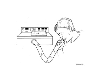
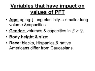
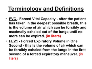
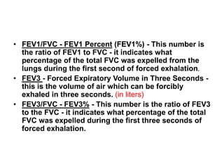
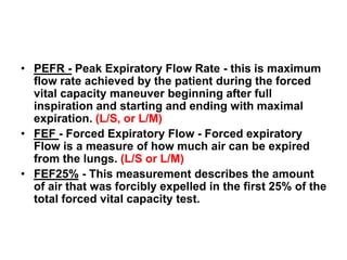
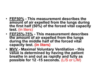
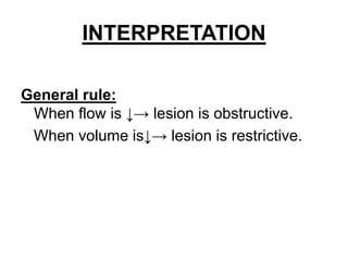
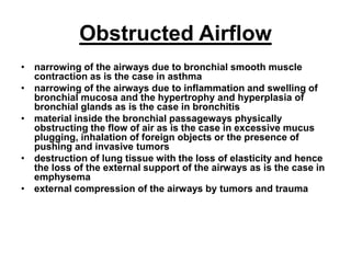
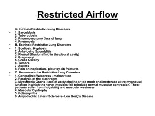
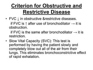
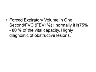
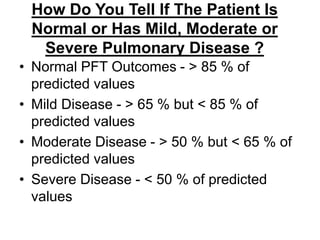
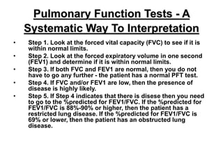
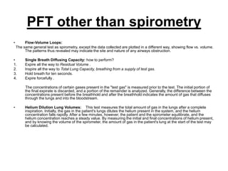
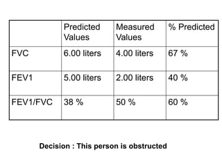
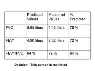
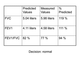
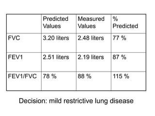
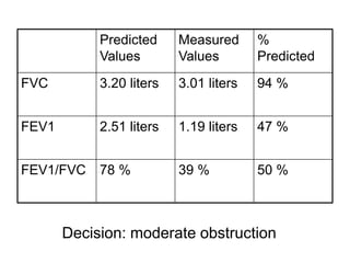
Clinical application of
pulmonary function tests
By
Prof. Dr.
Nermin Sadek
Information to gain from PFT
• How much air volume can be moved in and
out of the lungs?
• How fast the air in the lungs can be moved
in and out ?
• How stiff are the lungs and chest wall - a
question about compliance?
• The diffusion characteristics of the
membrane through which the gas moves
(determined by special tests) .
• How the lungs respond to chest physical
therapy procedures?
Reasons to use PFT
• Screening for the presence of obstructive and
restrictive diseases
• Evaluating the patient prior to surgery
• Evaluating the patient's condition for weaning
from a ventilator. If the patient on a ventilator can
demonstrate a vital capacity (VC) of 10 - 15 ml/Kg
of body weight, it is generally thought that there is
enough ventilatory reserve to permit (try) weaning
and extubation.
• Documenting the progression of pulmonary
disease - restrictive or obstructive
• Documenting the effectiveness of therapeutic
intervention
.
PFT are specially helpful when
patients :
• a. are older than 60-65 years of age
b. are known to have pulmonary disease
c. are obese (as in pathologically obese)
d. have a history of smoking, cough or
wheezing
e. will be under anesthesia for a lengthy
period of time
f. are undergoing an abdominal or a thoracic
operation
Equipment
• The primary instrument used in pulmonary
function testing is the spirometer. It is
designed to measure changes in volume and
can only measure lung volume
compartments that exchange gas with the
atmosphere . Spirometers with electronic
signal outputs (pneumotachs) also measure
flow (volume per unit of time). A device is
usually always attached to the spirometer
which measures the movement of gas in and
out of the chest and is referred to as a
spirograph.
Variables that have impact on
values of PFT
• Age: aging ↓ lung elasticity→ smaller lung
volume &capacities.
• Gender: volumes & capacities in ♂ > ♀.
• Body height & size:
• Race: blacks, Hispanics,& native
Americans differ from Caucasians.
Terminology and Definitions
• FVC - Forced Vital Capacity - after the patient
has taken in the deepest possible breath, this
is the volume of air which can be forcibly and
maximally exhaled out of the lungs until no
more can be expired. (in liters)
• FEV1 - Forced Expiratory Volume in One
Second - this is the volume of air which can
be forcibly exhaled from the lungs in the first
second of a forced expiratory maneuver. (in
liters)
• FEV1/FVC - FEV1 Percent (FEV1%) - This number is
the ratio of FEV1 to FVC - it indicates what
percentage of the total FVC was expelled from the
lungs during the first second of forced exhalation.
• FEV3 - Forced Expiratory Volume in Three Seconds -
this is the volume of air which can be forcibly
exhaled in three seconds. (in liters)
• FEV3/FVC - FEV3% - This number is the ratio of FEV3
to the FVC - it indicates what percentage of the total
FVC was expelled during the first three seconds of
forced exhalation.
• PEFR - Peak Expiratory Flow Rate - this is maximum
flow rate achieved by the patient during the forced
vital capacity maneuver beginning after full
inspiration and starting and ending with maximal
expiration. (L/S, or L/M)
• FEF - Forced Expiratory Flow - Forced expiratory
Flow is a measure of how much air can be expired
from the lungs. (L/S or L/M)
• FEF25% - This measurement describes the amount
of air that was forcibly expelled in the first 25% of the
total forced vital capacity test.
• FEF50% - This measurement describes the
amount of air expelled from the lungs during
the first half (50%) of the forced vital capacity
test. (in liters)
• FEF25%-75% - This measurement describes
the amount of air expelled from the lungs
during the middle half of the forced vital
capacity test. (in liters)
• MVV - Maximal Voluntary Ventilation - this
value is determined by having the patient
breathe in and out as rapidly and fully as
possible for 12 -15 seconds. (L/S or L/M)
INTERPRETATION
General rule:
When flow is ↓→ lesion is obstructive.
When volume is↓→ lesion is restrictive.
Obstructed Airflow
• narrowing of the airways due to bronchial smooth muscle
contraction as is the case in asthma
• narrowing of the airways due to inflammation and swelling of
bronchial mucosa and the hypertrophy and hyperplasia of
bronchial glands as is the case in bronchitis
• material inside the bronchial passageways physically
obstructing the flow of air as is the case in excessive mucus
plugging, inhalation of foreign objects or the presence of
pushing and invasive tumors
• destruction of lung tissue with the loss of elasticity and hence
the loss of the external support of the airways as is the case in
emphysema
• external compression of the airways by tumors and trauma
Restricted Airflow
• A. Intrinsic Restrictive Lung Disorders
• 1. Sarcoidosis
2. Tuberculosis
3. Pnuemonectomy (loss of lung)
4. Pneumonia
• B. Extrinsic Restrictive Lung Disorders
• 1. Scoliosis, Kyphosis
2. Ankylosing Spondylitis
3. Pleural Effusion (fluid in the pleural cavity)
4. Pregnancy
5. Gross Obesity
6. Tumors
7. Ascites
8. Pain on inspiration - pleurisy, rib fractures
• C. Neuromuscular Restrictive Lung Disorders
• 1. Generalized Weakness - malnutrition
2. Paralysis of the diaphragm
3. Myasthenia Gravis - lack of acetylcholine or too much cholinesterase at the myoneural
junction in which the nerve impulses fail to induce normal muscular contraction. These
patients suffer from fatigability and muscular weakness.
4. Muscular Dystrophy
5. Poliomyelitis
6. Amyotrophic Lateral Sclerosis - Lou Gerig's Disease
Criterion for Obstructive and
Restrictive Disease
• FVC ↓ in obstructive &restrictive diseases.
if FVC is ↑ after use of bronchodilator → it is
obstruction.
if FVC is the same after bronchodilator → it is
restriction.
• Slow Vital Capacity (SVC): This test is
performed by having the patient slowly and
completely blow out all of the air from their
lungs. This eliminates bronchoconstrictive effect
of rapid exhalation.
• Forced Expiratory Volume in One
Second/FVC (FEV1%) : normally it is75%
- 80 % of the vital capacity. Highly
diagnostic of obstructive lesions.
How Do You Tell If The Patient Is
Normal or Has Mild, Moderate or
Severe Pulmonary Disease ?
• Normal PFT Outcomes - > 85 % of
predicted values
• Mild Disease - > 65 % but < 85 % of
predicted values
• Moderate Disease - > 50 % but < 65 % of
predicted values
• Severe Disease - < 50 % of predicted
values
Pulmonary Function Tests - A
Systematic Way To Interpretation
• Step 1. Look at the forced vital capacity (FVC) to see if it is
within normal limits.
• Step 2. Look at the forced expiratory volume in one second
(FEV1) and determine if it is within normal limits.
• Step 3. If both FVC and FEV1 are normal, then you do not
have to go any further - the patient has a normal PFT test.
• Step 4. If FVC and/or FEV1 are low, then the presence of
disease is highly likely.
• Step 5. If Step 4 indicates that there is disese then you need
to go to the %predicted for FEV1/FVC. If the %predicted for
FEV1/FVC is 88%-90% or higher, then the patient has a
restricted lung disease. If the %predicted for FEV1/FVC is
69% or lower, then the patient has an obstructed lung
disease.
PFT other than spirometry
• Flow-Volume Loops:
The same general test as spirometry, except the data collected are plotted in a different way, showing flow vs. volume.
The patterns thus revealed may indicate the site and nature of any airways obstruction.
• Single Breath Diffusing Capacity: how to perform?
1. Expire all the way to Residual Volume .
2. Inspire all the way to Total Lung Capacity, breathing from a supply of test gas.
3. Hold breath for ten seconds.
4. Expire forcefully .
The concentrations of certain gases present in the "test gas" is measured prior to the test. The initial portion of
the final expirate is discarded, and a portion of the remainder is analyzed. Generally, the difference between the
concentrations present before the breathhold and after the breathhold indicates the amount of gas that diffuses
through the lungs and into the bloodstream.
• Helium Dilution Lung Volumes: This test measures the total amount of gas in the lungs after a complete
inspiration. Initially, the gas in the patient's lungs dilutes the helium present in the system, and the helium
concentration falls rapidly. After a few minutes, however, the patient and the spirometer equilibrate, and the
helium concentration reaches a steady value. By measuring the initial and final concentrations of helium present,
and by knowing the volume of the spirometer, the amount of gas in the patient's lung at the start of the test may
be calculated.
Predicted
Values
Measured
Values
% Predicted
FVC 6.00 liters 4.00 liters 67 %
FEV1 5.00 liters 2.00 liters 40 %
FEV1/FVC 38 % 50 % 60 %
Decision : This person is obstructed
Predicted
Values
Measured
Values
%
Predicted
FVC 5.68 liters 4.43 liters 78 %
FEV1 4.90 liters 3.52 liters 72 %
FEV1/FVC 84 % 79 % 94 %
Decision : This person is restricted
Decision: normal
Predicted
Values
Measured
Values
%
Predicted
FVC 5.04 liters 5.98 liters 119 %
FEV1 4.11 liters 4.58 liters 111 %
FEV1/FVC 82 % 77 % 94 %
Decision: mild restrictive lung disease
Predicted
Values
Measured
Values
%
Predicted
FVC 3.20 liters 2.48 liters 77 %
FEV1 2.51 liters 2.19 liters 87 %
FEV1/FVC 78 % 88 % 115 %
Decision: moderate obstruction
Predicted
Values
Measured
Values
%
Predicted
FVC 3.20 liters 3.01 liters 94 %
FEV1 2.51 liters 1.19 liters 47 %
FEV1/FVC 78 % 39 % 50 %
THANK YOU

clinical approach Pulmonary function test .ppt

  • 1.
    Clinical application of pulmonaryfunction tests By Prof. Dr. Nermin Sadek
  • 2.
    Information to gainfrom PFT • How much air volume can be moved in and out of the lungs? • How fast the air in the lungs can be moved in and out ? • How stiff are the lungs and chest wall - a question about compliance? • The diffusion characteristics of the membrane through which the gas moves (determined by special tests) . • How the lungs respond to chest physical therapy procedures?
  • 3.
    Reasons to usePFT • Screening for the presence of obstructive and restrictive diseases • Evaluating the patient prior to surgery • Evaluating the patient's condition for weaning from a ventilator. If the patient on a ventilator can demonstrate a vital capacity (VC) of 10 - 15 ml/Kg of body weight, it is generally thought that there is enough ventilatory reserve to permit (try) weaning and extubation. • Documenting the progression of pulmonary disease - restrictive or obstructive • Documenting the effectiveness of therapeutic intervention .
  • 4.
    PFT are speciallyhelpful when patients : • a. are older than 60-65 years of age b. are known to have pulmonary disease c. are obese (as in pathologically obese) d. have a history of smoking, cough or wheezing e. will be under anesthesia for a lengthy period of time f. are undergoing an abdominal or a thoracic operation
  • 5.
    Equipment • The primaryinstrument used in pulmonary function testing is the spirometer. It is designed to measure changes in volume and can only measure lung volume compartments that exchange gas with the atmosphere . Spirometers with electronic signal outputs (pneumotachs) also measure flow (volume per unit of time). A device is usually always attached to the spirometer which measures the movement of gas in and out of the chest and is referred to as a spirograph.
  • 7.
    Variables that haveimpact on values of PFT • Age: aging ↓ lung elasticity→ smaller lung volume &capacities. • Gender: volumes & capacities in ♂ > ♀. • Body height & size: • Race: blacks, Hispanics,& native Americans differ from Caucasians.
  • 8.
    Terminology and Definitions •FVC - Forced Vital Capacity - after the patient has taken in the deepest possible breath, this is the volume of air which can be forcibly and maximally exhaled out of the lungs until no more can be expired. (in liters) • FEV1 - Forced Expiratory Volume in One Second - this is the volume of air which can be forcibly exhaled from the lungs in the first second of a forced expiratory maneuver. (in liters)
  • 9.
    • FEV1/FVC -FEV1 Percent (FEV1%) - This number is the ratio of FEV1 to FVC - it indicates what percentage of the total FVC was expelled from the lungs during the first second of forced exhalation. • FEV3 - Forced Expiratory Volume in Three Seconds - this is the volume of air which can be forcibly exhaled in three seconds. (in liters) • FEV3/FVC - FEV3% - This number is the ratio of FEV3 to the FVC - it indicates what percentage of the total FVC was expelled during the first three seconds of forced exhalation.
  • 10.
    • PEFR -Peak Expiratory Flow Rate - this is maximum flow rate achieved by the patient during the forced vital capacity maneuver beginning after full inspiration and starting and ending with maximal expiration. (L/S, or L/M) • FEF - Forced Expiratory Flow - Forced expiratory Flow is a measure of how much air can be expired from the lungs. (L/S or L/M) • FEF25% - This measurement describes the amount of air that was forcibly expelled in the first 25% of the total forced vital capacity test.
  • 11.
    • FEF50% -This measurement describes the amount of air expelled from the lungs during the first half (50%) of the forced vital capacity test. (in liters) • FEF25%-75% - This measurement describes the amount of air expelled from the lungs during the middle half of the forced vital capacity test. (in liters) • MVV - Maximal Voluntary Ventilation - this value is determined by having the patient breathe in and out as rapidly and fully as possible for 12 -15 seconds. (L/S or L/M)
  • 12.
    INTERPRETATION General rule: When flowis ↓→ lesion is obstructive. When volume is↓→ lesion is restrictive.
  • 13.
    Obstructed Airflow • narrowingof the airways due to bronchial smooth muscle contraction as is the case in asthma • narrowing of the airways due to inflammation and swelling of bronchial mucosa and the hypertrophy and hyperplasia of bronchial glands as is the case in bronchitis • material inside the bronchial passageways physically obstructing the flow of air as is the case in excessive mucus plugging, inhalation of foreign objects or the presence of pushing and invasive tumors • destruction of lung tissue with the loss of elasticity and hence the loss of the external support of the airways as is the case in emphysema • external compression of the airways by tumors and trauma
  • 14.
    Restricted Airflow • A.Intrinsic Restrictive Lung Disorders • 1. Sarcoidosis 2. Tuberculosis 3. Pnuemonectomy (loss of lung) 4. Pneumonia • B. Extrinsic Restrictive Lung Disorders • 1. Scoliosis, Kyphosis 2. Ankylosing Spondylitis 3. Pleural Effusion (fluid in the pleural cavity) 4. Pregnancy 5. Gross Obesity 6. Tumors 7. Ascites 8. Pain on inspiration - pleurisy, rib fractures • C. Neuromuscular Restrictive Lung Disorders • 1. Generalized Weakness - malnutrition 2. Paralysis of the diaphragm 3. Myasthenia Gravis - lack of acetylcholine or too much cholinesterase at the myoneural junction in which the nerve impulses fail to induce normal muscular contraction. These patients suffer from fatigability and muscular weakness. 4. Muscular Dystrophy 5. Poliomyelitis 6. Amyotrophic Lateral Sclerosis - Lou Gerig's Disease
  • 15.
    Criterion for Obstructiveand Restrictive Disease • FVC ↓ in obstructive &restrictive diseases. if FVC is ↑ after use of bronchodilator → it is obstruction. if FVC is the same after bronchodilator → it is restriction. • Slow Vital Capacity (SVC): This test is performed by having the patient slowly and completely blow out all of the air from their lungs. This eliminates bronchoconstrictive effect of rapid exhalation.
  • 16.
    • Forced ExpiratoryVolume in One Second/FVC (FEV1%) : normally it is75% - 80 % of the vital capacity. Highly diagnostic of obstructive lesions.
  • 17.
    How Do YouTell If The Patient Is Normal or Has Mild, Moderate or Severe Pulmonary Disease ? • Normal PFT Outcomes - > 85 % of predicted values • Mild Disease - > 65 % but < 85 % of predicted values • Moderate Disease - > 50 % but < 65 % of predicted values • Severe Disease - < 50 % of predicted values
  • 18.
    Pulmonary Function Tests- A Systematic Way To Interpretation • Step 1. Look at the forced vital capacity (FVC) to see if it is within normal limits. • Step 2. Look at the forced expiratory volume in one second (FEV1) and determine if it is within normal limits. • Step 3. If both FVC and FEV1 are normal, then you do not have to go any further - the patient has a normal PFT test. • Step 4. If FVC and/or FEV1 are low, then the presence of disease is highly likely. • Step 5. If Step 4 indicates that there is disese then you need to go to the %predicted for FEV1/FVC. If the %predicted for FEV1/FVC is 88%-90% or higher, then the patient has a restricted lung disease. If the %predicted for FEV1/FVC is 69% or lower, then the patient has an obstructed lung disease.
  • 19.
    PFT other thanspirometry • Flow-Volume Loops: The same general test as spirometry, except the data collected are plotted in a different way, showing flow vs. volume. The patterns thus revealed may indicate the site and nature of any airways obstruction. • Single Breath Diffusing Capacity: how to perform? 1. Expire all the way to Residual Volume . 2. Inspire all the way to Total Lung Capacity, breathing from a supply of test gas. 3. Hold breath for ten seconds. 4. Expire forcefully . The concentrations of certain gases present in the "test gas" is measured prior to the test. The initial portion of the final expirate is discarded, and a portion of the remainder is analyzed. Generally, the difference between the concentrations present before the breathhold and after the breathhold indicates the amount of gas that diffuses through the lungs and into the bloodstream. • Helium Dilution Lung Volumes: This test measures the total amount of gas in the lungs after a complete inspiration. Initially, the gas in the patient's lungs dilutes the helium present in the system, and the helium concentration falls rapidly. After a few minutes, however, the patient and the spirometer equilibrate, and the helium concentration reaches a steady value. By measuring the initial and final concentrations of helium present, and by knowing the volume of the spirometer, the amount of gas in the patient's lung at the start of the test may be calculated.
  • 20.
    Predicted Values Measured Values % Predicted FVC 6.00liters 4.00 liters 67 % FEV1 5.00 liters 2.00 liters 40 % FEV1/FVC 38 % 50 % 60 % Decision : This person is obstructed
  • 21.
    Predicted Values Measured Values % Predicted FVC 5.68 liters4.43 liters 78 % FEV1 4.90 liters 3.52 liters 72 % FEV1/FVC 84 % 79 % 94 % Decision : This person is restricted
  • 22.
    Decision: normal Predicted Values Measured Values % Predicted FVC 5.04liters 5.98 liters 119 % FEV1 4.11 liters 4.58 liters 111 % FEV1/FVC 82 % 77 % 94 %
  • 23.
    Decision: mild restrictivelung disease Predicted Values Measured Values % Predicted FVC 3.20 liters 2.48 liters 77 % FEV1 2.51 liters 2.19 liters 87 % FEV1/FVC 78 % 88 % 115 %
  • 24.
    Decision: moderate obstruction Predicted Values Measured Values % Predicted FVC3.20 liters 3.01 liters 94 % FEV1 2.51 liters 1.19 liters 47 % FEV1/FVC 78 % 39 % 50 %
  • 25.