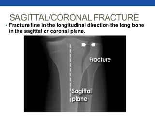
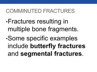
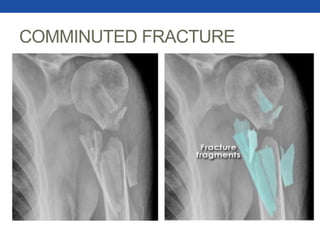
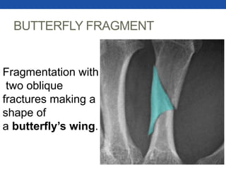
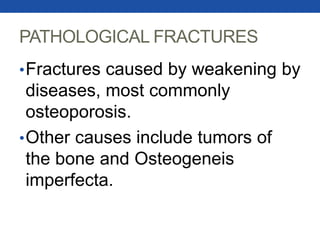
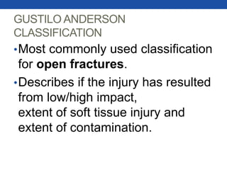
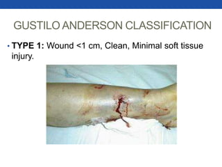
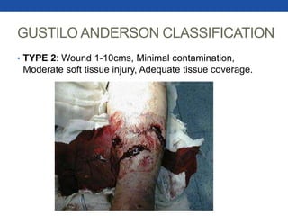
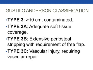
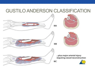
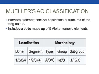
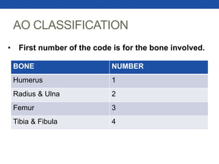
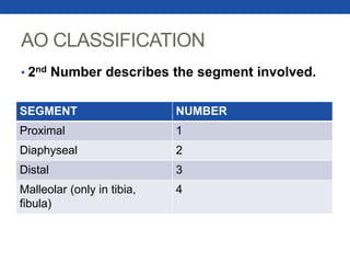
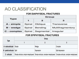
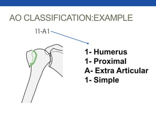
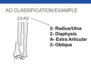
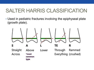
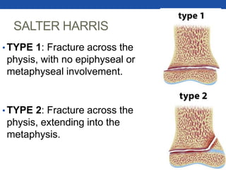
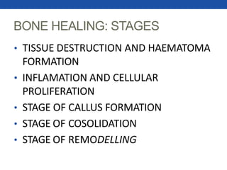
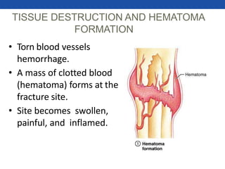
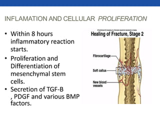
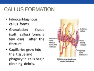
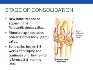
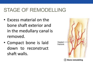
FRACTURESJamal Alvi (5-1/2013/116)
Hamza Rana (5-1/2013/115)
OBJECTIVES
•Defining a fracture.
•Describing fractures and their
classifications.
•Fracture healing and its
staging.
FRACTURE
•A break/damage in the structural
continuity of the bone resulting
from trauma/stress OR an
underlying medical condition which
weakens the bone (Osteoporosis,
Bone cancer, Osteogenesis
Imperfecta,etc) in which case they
are called Pathological fractures.
FRACTURES
•Closed (Simple) fracture: With
the overlying skin and soft tissue
intact.
•Open (Compound) fracture: A
break in the overlying skin/ soft
tissue allowing contact between
external and internal environment.
OPEN AND CLOSED FRACTURES
LOCATION - ANATOMY
•The location of the fracture on
its anatomical location is
described, with the direction of
the fracture line.
TYPES
• Transverse: Right angle, to the shaft of the long bone.
OBLIQUE FRACTURE
• Fracture line running oblique to the shaft of the long bone.
SPIRAL FRACTURE
• Fracture line going spirally around long bone, Caused
by twisting/rotational injury.
SAGITTAL/CORONAL FRACTURE
• Fracture line in the longitudinal direction the long bone
in the sagittal or coronal plane.
COMMINUTED FRACTURES
•Fractures resulting in
multiple bone fragments.
•Some specific examples
include butterfly fractures
and segmental fractures.
COMMINUTED FRACTURE
BUTTERFLY FRAGMENT
Fragmentation with
two oblique
fractures making a
shape of
a butterfly’s wing.
SEGMENTAL FRACTURE
• Segments formed by proximal and distal separation of the shaft.
FRACTURE DISPLACEMENT
•Abnormal position of the distal
fragment in relation to the proximal
fragment.
•Types include Angulation,
Rotation, Change in bone length
and Loss of Alignment.
DISPLACEMENTAND SHORTENING
• Displacement is loss of alignment along the long
axis, while shortening of the overall bone length
occurs after proximal migration after fracture.
ANGULATION AND ROTATION
•Angulation is the displaced angle in
relation to long axis of the proximal
bone, Medial angulation is called
varus while lateral angulation is called
valgus.
•Rotation in relation to the proximal
bone, internally (inward) or
externally (outward).
ANGULATION AND ROTATIONAL
PATHOLOGICAL FRACTURES
•Fractures caused by weakening by
diseases, most commonly
osteoporosis.
•Other causes include tumors of
the bone and Osteogeneis
imperfecta.
OSTEOPOROTIC FRACTURE
•Decreased bone
density, cortical
thickness reduced.
•Subtle impact
fracture of
Ulna.
GUSTILO ANDERSON
CLASSIFICATION
•Most commonly used classification
for open fractures.
•Describes if the injury has resulted
from low/high impact,
extent of soft tissue injury and
extent of contamination.
GUSTILO ANDERSON CLASSIFICATION
• TYPE 1: Wound <1 cm, Clean, Minimal soft tissue
injury.
GUSTILO ANDERSON CLASSIFICATION
• TYPE 2: Wound 1-10cms, Minimal contamination,
Moderate soft tissue injury, Adequate tissue coverage.
GUSTILO ANDERSON CLASSIFICATION
•TYPE 3: >10 cm, contaminated..
•TYPE 3A: Adequate soft tissue
coverage.
•TYPE 3B: Extensive periosteal
stripping with requirement of free flap.
•TYPE 3C: Vascular injury, requiring
vascular repair.
GUSTILO ANDERSON CLASSIFICATION
MUELLER’S AO CLASSIFICATION
• Provides a comprehensive description of fractures of the
long bones.
• Includes a code made up of 5 Alpha-numeric elements.
AO CLASSIFICATION
BONE NUMBER
Humerus 1
Radius & Ulna 2
Femur 3
Tibia & Fibula 4
• First number of the code is for the bone involved.
AO CLASSIFICATION
• 2nd Number describes the segment involved.
SEGMENT NUMBER
Proximal 1
Diaphyseal 2
Distal 3
Malleolar (only in tibia,
fibula)
4
AO CLASSIFICATION
• 3rd Number describes the type involved.
SEGMENT A B C
1 Extra-
articular
Partial
Articular
Complete
Articular
2 Simple Wedge Complex
3 Extra-
articular
Partial
Articular
Complete
Articular
AO CLASSIFICATION
FOR DIAPHYSEAL FRACTURES
FOR EPIPHYSEAL FRACTURES
AO CLASSIFICATION:EXAMPLE
1- Humerus
1- Proximal
A- Extra Articular
1- Simple
AO CLASSIFICATION:EXAMPLE
2- Radius/Ulna
2- Diaphysis
A- Extra Articular
2- Oblique
SALTER HARRIS CLASSIFICATION
• Used in pediatric fractures involving the epiphyseal plate
(growth plate).
SALTER HARRIS
• TYPE 1: Fracture across the
physis, with no epiphyseal or
metaphyseal involvement.
• TYPE 2: Fracture across the
physis, extending into the
metaphysis.
SALTER HARRIS
• TYPE 3: Fracture through the physis and
extends into the epiphysis.
• TYPE 4: Fracture across the metaphysis,
physis and epiphysis.
SALTER HARRIS
• TYPE 5: Crush injury to the physis.
BONE HEALING: STAGES
• TISSUE DESTRUCTION AND HAEMATOMA
FORMATION
• INFLAMATION AND CELLULAR
PROLIFERATION
• STAGE OF CALLUS FORMATION
• STAGE OF COSOLIDATION
• STAGE OF REMODELLING
TISSUE DESTRUCTION AND HEMATOMA
FORMATION
• Torn blood vessels
hemorrhage.
• A mass of clotted blood
(hematoma) forms at the
fracture site.
• Site becomes swollen,
painful, and inflamed.
INFLAMATION AND CELLULAR PROLIFERATION
• Within 8 hours
inflammatory reaction
starts.
• Proliferation and
Differentiation of
mesenchymal stem
cells.
• Secretion of TGF-B
, PDGF and various BMP
factors.
CALLUS FORMATION
• Fibrocartilaginous
callus forms.
• Granulation tissue
(soft callus) forms a
few days after the
fracture.
• Capillaries grow into
the tissue and
phagocytic cells begin
cleaning debris.
STAGE OF CONSOLIDATION
• New bone trabeculae
appear in the
fibrocartilaginous callus.
• Fibrocartilaginous callus
converts into a bony (hard)
callus.
• Bone callus begins 3-4
weeks after injury, and
continues until firm union
is formed 2-3 months
later.
STAGE OF REMODELLING
• Excess material on the
bone shaft exterior and
in the medullary canal is
removed.
• Compact bone is laid
down to reconstruct
shaft walls.
•THANK
YOU
REFERENCES
• www.RadiologyMasterclass.co.uk
• www.Wikipedia.org.
• www.Orthobullets.org.
• Muellers AO classification manual.

Fractures By Dr Jamal Alvi

Editor's Notes

  • #4 Trauma: External force or physical violence. Stress fracture: Resulting from less severe accumulating minor stress overtime. Trauma and pathological are also classifications in etiology.
  • #5 Basic classification of fractures. First thing mentioned while describing fractures.
  • #7 After describing whether its simple or compound, we describe the location anatomically. Lcoation – Distal humerus, mid-shaft of the femur.
  • #8 Simple, Mid-tibeal transverse fracture
  • #9 Simple, distal metatarsal, oblique fracture.
  • #10 Simple, mid- tibeal , oblique fracture. Example of twisty injury is skiing or hand twisting in cases of abuse.
  • #11 Simple, proximal tibeal , sagittal fracture.
  • #13 Simple, proximal humerus , comminuted fracture.
  • #15 Simple, segmental fracture of the tibia and fibula.
  • #17 Translation is dplacement of the bones where the whole end is not approximated at all and is completely moved to the side.
  • #20 OI= genetic disease with abnormality of production of collagen type 1.
  • #21 Cortical bone on the boundaries is less white= less denser.
  • #24 Tissue coverage means flaps don’t need to be
  • #27 AO- association of osteogenesis in germany. This was established in 1974 and is still in use. Gives us an idea of the anatomic and functional outcomes of the injury.
  • #30 Extra-articular: doesn’t involved the joints. Complex; comminuted fracture with soft tissue injury.
  • #40 TGF-B – Transforming growth factor. BMP – Bone morphogenic protein- basically like cytokine , their function is to induce bone growth.
  • #41 Granulation tissue- connective tissue and microscopic blood vessels growing in order for healing to occur.
  • #42 Trabeculae, is cancellous bone with bands and spaces for bone marrow.