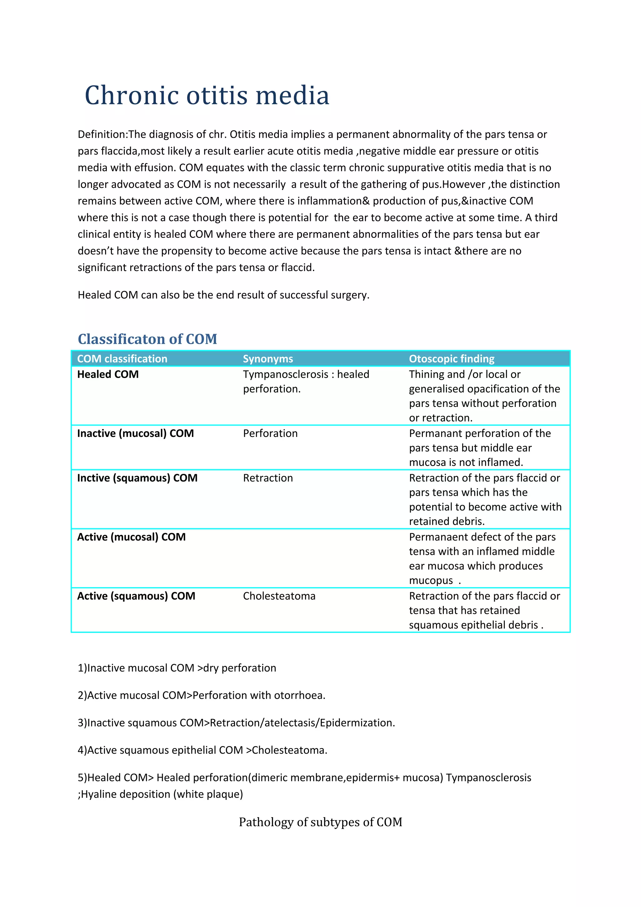
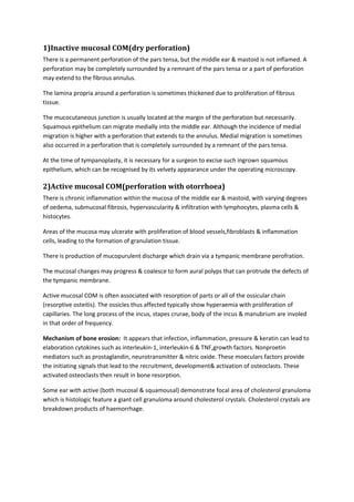
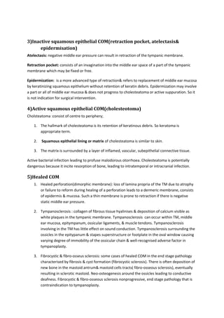
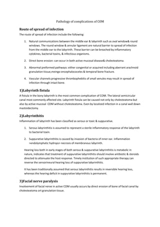
Chronic otitis media 
Definition:The diagnosis of chr. Otitis media implies a permanent abnormality of the pars tensa or 
pars flaccida,most likely a result earlier acute otitis media ,negative middle ear pressure or otitis 
media with effusion. COM equates with the classic term chronic suppurative otitis media that is no 
longer advocated as COM is not necessarily a result of the gathering of pus.However ,the distinction 
remains between active COM, where there is inflammation& production of pus,&inactive COM 
where this is not a case though there is potential for the ear to become active at some time. A third 
clinical entity is healed COM where there are permanent abnormalities of the pars tensa but ear 
doesn’t have the propensity to become active because the pars tensa is intact &there are no 
significant retractions of the pars tensa or flaccid. 
Healed COM can also be the end result of successful surgery. 
Classificaton of COM 
COM classification Synonyms Otoscopic finding 
Healed COM Tympanosclerosis : healed 
perforation. 
Thining and /or local or 
generalised opacification of the 
pars tensa without perforation 
or retraction. 
Inactive (mucosal) COM Perforation Permanant perforation of the 
pars tensa but middle ear 
mucosa is not inflamed. 
Inctive (squamous) COM Retraction Retraction of the pars flaccid or 
pars tensa which has the 
potential to become active with 
retained debris. 
Active (mucosal) COM Permanaent defect of the pars 
tensa with an inflamed middle 
ear mucosa which produces 
mucopus . 
Active (squamous) COM Cholesteatoma Retraction of the pars flaccid or 
tensa that has retained 
squamous epithelial debris . 
1)Inactive mucosal COM >dry perforation 
2)Active mucosal COM>Perforation with otorrhoea. 
3)Inactive squamous COM>Retraction/atelectasis/Epidermization. 
4)Active squamous epithelial COM >Cholesteatoma. 
5)Healed COM> Healed perforation(dimeric membrane,epidermis+ mucosa) Tympanosclerosis 
;Hyaline deposition (white plaque) 
Pathology of subtypes of COM
1)Inactive mucosal COM(dry perforation) 
There is a permanent perforation of the pars tensa, but the middle ear & mastoid is not inflamed. A 
perforation may be completely surrounded by a remnant of the pars tensa or a part of perforation 
may extend to the fibrous annulus. 
The lamina propria around a perforation is sometimes thickened due to proliferation of fibrous 
tissue. 
The mucocutaneous junction is usually located at the margin of the perforation but necessarily. 
Squamous epithelium can migrate medially into the middle ear. Although the incidence of medial 
migration is higher with a perforation that extends to the annulus. Medial migration is sometimes 
also occurred in a perforation that is completely surrounded by a remnant of the pars tensa. 
At the time of tympanoplasty, it is necessary for a surgeon to excise such ingrown squamous 
epithelium, which can be recognised by its velvety appearance under the operating microscopy. 
2)Active mucosal COM(perforation with otorrhoea) 
There is chronic inflammation within the mucosa of the middle ear & mastoid, with varying degrees 
of oedema, submucosal fibrosis, hypervascularity & infiltration with lymphocytes, plasma cells & 
histocytes. 
Areas of the mucosa may ulcerate with proliferation of blood vessels,fibroblasts & inflammation 
cells, leading to the formation of granulation tissue. 
There is production of mucopurulent discharge which drain via a tympanic membrane perofration. 
The mucosal changes may progress & coalesce to form aural polyps that can protrude the defects of 
the tympanic membrane. 
Active mucosal COM is often associated with resorption of parts or all of the ossicular chain 
(resorptive osteitis). The ossicles thus affected typically show hyperaemia with proliferation of 
capillaries. The long process of the incus, stapes crurae, body of the incus & manubrium are involed 
in that order of frequency. 
Mechanism of bone erosion: It appears that infection, inflammation, pressure & keratin can lead to 
elaboration cytokines such as interleukin-1, interleukin-6 & TNF,growth factors. Nonproetin 
mediators such as prostaglandin, neurotransmitter & nitric oxide. These moeculars factors provide 
the initiating signals that lead to the recruitment, development& activation of osteoclasts. These 
activated osteoclasts then result in bone resorption. 
Some ear with active (both mucosal & squamousal) demonstrate focal area of cholesterol granuloma 
which is histologic feature a giant cell granuloma around cholesterol crystals. Cholesterol crystals are 
breakdown products of haemorrhage.
3)Inactive squamous epithelial COM(retraction pocket, atelectasis& 
epidermisation) 
Atelectasis: negative middle ear pressure can result in retraction of the tympanic membrane. 
Retraction pocket: consists of an invagination into the middle ear space of a part of the tympanic 
membrane which may be fixed or free. 
Epidermization: is a more advanced type of retraction& refers to replacement of middle ear mucosa 
by keratinizing squamous epithelium without retention of keratin debris. Epidermization may involve 
a part or all of middle ear mucosa & does not progress to cholesteatoma or active suppuration. So it 
is not indication for surgical intervention. 
4)Active squamous epithelial COM(cholesteotoma) 
Cholsteatoma :consist of centre to periphery; 
1. The hallmark of cholesteatoma is its retention of keratinous debris. So keratoma is 
appropriate term. 
2. Squamous epithelial lining or matrix of cholesteatoma is similar to skin. 
3. The matrix is surrounded by a layer of inflamed, vascular, subepithelial connective tissue. 
Active bacterial infection leading to profuse malodorous otorrhoea. Cholesteatoma is potentially 
dangerous because it incite resorption of bone, leading to intratemporal or intracranial infection. 
5)Healed COM 
1. Healed perforation(dimorphic membrane): loss of lamina propria of the TM due to atrophy 
or failure to reform during healing of a perforation leads to a dermeric membrane, consists 
of epidermis & mucosa. Such a thin membrane is prone to retraction if there is negative 
static middle ear pressure. 
2. Tympanosclerosis : collagen of fibrous tissue hyalinises & deposition of calcium visible as 
white plaques in the tympanic membrane. Tympanosclerosis can occur within TM, middle 
ear mucosa, epitympanum, ossicular ligaments, & muscle tendons. Tympanosclerosis 
involving in the TM has little effect on sound conduction. Tympanosclerosis surrounding the 
ossicles in the epitypanum & stapes superstructure or footplate in the oval window causing 
varying degree of immobility of the ossicular chain & well-recognised adverse factor in 
tympanoplasty. 
3. Fibrocystic & fibro-osseus sclerosis: some cases of healed COM in the end stage pathology 
characterised by fibrosis & cyst formation (fibrocystic sclerosis). There is often deposition of 
new bone in the mastoid antrum& mastoid cells tracts( fibro-osseous sclerosis), eventually 
resulting in sclerotic mastoid. Neo-osteogenesis around the ossicles leading to conductive 
deafness. Fibrocystic & fibro-osseous sclerosis nonprogressive, end stage pathology that is 
contraindication to tympanoplasty.
Pathology of complications of COM 
Route of spread of infection 
The route of spread of infection include the following: 
1. Natural communications between the middle ear & labyrinth such as oval window& round 
windows. The round window & annular ligament are natural barrier to spread of infection 
from the middle ear to the labyrinth. These barrier can be breached by inflammatory 
cytokines, bacterial toxins, & infectious organisms. 
2. Direct bone erosion: can occur in both active mucosal disease& cholesteatoma. 
3. Abnormal preformed pathways: either congenital or acquired including aberrant arachnoid 
granulation tissue,meingo-encephalococeles & temporal bone fracture. 
4. Vascular channels:progressive thrombophebitis of small venules may result in spread of 
infection through intact bone. 
1)Labyrinth fistula 
A fistula in the bony labyrinth is the most common complication of COM. The lateral semicircular 
canal most commonly affected site. Labyrinth fistula can be caused not only by cholesteatoma but 
also by active mucosal COM without cholesteatoma. Even by localized infection in a canal wall down 
mastoidectomy. 
2)Labyrinthitis 
Inflammation of labyrinth has been classified as serous or toxic & suppurative. 
1. Serous labyrinthitis is assumed to represent a sterile inflammatory response of the labyrinth 
to bacterial toxin. 
2. Suppurative labyrinthitis is caused by invasion of bacteria of inner ear. Inflammation 
>endolymphatic hydrops> necrosis of membranous labyrinth. 
Hearing loss both in early stages of both serous & suppurative labyrinthitis is metabolic in 
nature, indicates that treatment of suppurative labyrinthitis should involve antibiotic & steroids 
directed to attenuate the host response. Timely institution of such appropriate therapy can 
reverse the sensorineural hearing loss of suppurative labyrinthitis. 
It has been traditionally assumed that serous labyrinthitis results in reversible hearing loss, 
whereas the hearing deficit in suppurative labyrinthitis is permanent. 
3)Facial nerve paralysis 
Involvement of facial nerve in active COM usually occurs by direct erosion of bone of facial canal by 
cholesteatoma ort granulation tissue.
4)Petrositis 
In 1904, Gradenigo recognized that the triad of persistent otorrhoea,severe periorbital pain, & 
abducent nerve paralysis is characteristic of infection of the petrous apex. 
1. Acute petrositis: acute inflammation in the mucous membrane of petrous apex cells, it may 
progress to an abscess . 
2. Chronic petrositis: chronic inflammatory changes & there is resorption of bone as well as 
new bone formation. Chronic petrositis may be an indolent infection that can persist for 
month ort year. Chronic petrositis causes serous complications because of anatomical 
proximity of the petrous apex to the meninges, trigeminal nerve, & carotid artery. Serous 
complication including meningitis, extradural abscess, multiple cranial nerve paralysis & 
carotid artery thrombosis. 
5) Otogenic intracranial complications 
The dura covering is a strong barrier that protect the intracranial complications. Intracranial 
complications depends on including the type & nature of the COM, offending organism& anatomical 
location. These complication are meningitis, extradural abscess,subdural abscess, cerebellar & 
temporal lobe abscess, sigmoid sinus thrombophlebitis. 
Surgical pathology 
A)Tympanic membrane grafts 
Tympanic membrane perforations can be successful repair using a variety of graft materials. 
Commonly used grafts include autologous temporalis fascia, perichondrium, cartilage, adipose 
tissue. 
Histologically , tympanic membrane grafts become lined by squamous epithilum on the ear canal 
side& middle ear mucosa on the tympanic cavity side. The graft becomes middle or connective 
tissue portion of reconstructed drum. 
B)Ossicular grafts & implants 
Autologous ossicle grafts (incus or head of malleus) maintain morphologic contour, size, shape& 
physical integrity for long periods of time over 25 years. They do not incite formation of new bone 
nor do they undergo resorption(in absence of infection). They undergo slow replacement of non-viable 
bone by new bone formation through a process of creeping substitution. 
Cartilage grafts are not optimal for ossicular reconstruction. Cartilage often develop chondromalacia 
with loss of stiffness & tendency to resorbed over time. 
Porous high density polyethylene( plastipore) elicit an intense foreign body giant cell reaction that 
becomes well established within a few weeks. Microdegradation of plastic material also occurs. 
More recently many others synthetic material are used bioactive glass(bioglass), aluminium oxide 
ceramic, carbon, hydroxylapatite, gold, titanium.
C)Mastoidectomy cavities 
A canal wall-down mastoidectomy cavity that is dry & well healed characterized histologically by a 
lining of keratinized epithelium on a layer of subepithelial fibrous tissue & underlying sclerotic 
mastoid bone. 
Discharging mastoid due to 
1. Recurrent or residual cholesteatoma; 
2. A high facial ridge, meatal or canal stenosis & 
3. Suppuration in unexenterated cells of the mastoid & middle ear especially the tegmental 
cells, cells of sinodural angle, mastoid tip & facial recess. 
If part of cholesteatoma matrix is left in the middle ear cleft: 
1. It may undergo metaplasia to middle ear mucosa type. 
2. Squamous epithelial retention cyst or cholesteatoma pearl. 
Biological & pathological factors 
determining success of 
tympanomastoid surgery for COM 
1. The primary objective of surgery for COM is to eradicate infection, disease & make the ear 
safe & dry. 
2. A second objective of surgery for COM is to restore hearing to serviceable levels by mean of 
tympanoplasty. 
Success after tympanoplasty depend on 
1. Operation after controlling infection 
2. Surgeon’s technical skill. 
3. As a sequel to surgical trauma or healing response to COM, deposition of fibrous 
tissue, formation of adhesions, neo-osteogenesis. These tissues cause fixation of 
stapes footplate, ankylosis or displacement of ossicle strut. Gelfom prevent the 
formation of adhesion or fibrous tissue. 
4. Total or partial non-aeration of the middle ear & development of negative 
middle ear pressure due to Eustachian tube dysfunction> OME> severe 
atelectasis. It causes displacement or exclusion of ossicular grafts. The negative 
pressure can also lead to recurrent cholesteatoma which is disadvantage for 
canal wall-up procedure.
In some patients the problem is selective non-aeration of the posterior 
mesotympanonum while anterior mesotympanonum remain aerated.

1)chronic otitis media

  • 1.
    Chronic otitis media Definition:The diagnosis of chr. Otitis media implies a permanent abnormality of the pars tensa or pars flaccida,most likely a result earlier acute otitis media ,negative middle ear pressure or otitis media with effusion. COM equates with the classic term chronic suppurative otitis media that is no longer advocated as COM is not necessarily a result of the gathering of pus.However ,the distinction remains between active COM, where there is inflammation& production of pus,&inactive COM where this is not a case though there is potential for the ear to become active at some time. A third clinical entity is healed COM where there are permanent abnormalities of the pars tensa but ear doesn’t have the propensity to become active because the pars tensa is intact &there are no significant retractions of the pars tensa or flaccid. Healed COM can also be the end result of successful surgery. Classificaton of COM COM classification Synonyms Otoscopic finding Healed COM Tympanosclerosis : healed perforation. Thining and /or local or generalised opacification of the pars tensa without perforation or retraction. Inactive (mucosal) COM Perforation Permanant perforation of the pars tensa but middle ear mucosa is not inflamed. Inctive (squamous) COM Retraction Retraction of the pars flaccid or pars tensa which has the potential to become active with retained debris. Active (mucosal) COM Permanaent defect of the pars tensa with an inflamed middle ear mucosa which produces mucopus . Active (squamous) COM Cholesteatoma Retraction of the pars flaccid or tensa that has retained squamous epithelial debris . 1)Inactive mucosal COM >dry perforation 2)Active mucosal COM>Perforation with otorrhoea. 3)Inactive squamous COM>Retraction/atelectasis/Epidermization. 4)Active squamous epithelial COM >Cholesteatoma. 5)Healed COM> Healed perforation(dimeric membrane,epidermis+ mucosa) Tympanosclerosis ;Hyaline deposition (white plaque) Pathology of subtypes of COM
  • 2.
    1)Inactive mucosal COM(dryperforation) There is a permanent perforation of the pars tensa, but the middle ear & mastoid is not inflamed. A perforation may be completely surrounded by a remnant of the pars tensa or a part of perforation may extend to the fibrous annulus. The lamina propria around a perforation is sometimes thickened due to proliferation of fibrous tissue. The mucocutaneous junction is usually located at the margin of the perforation but necessarily. Squamous epithelium can migrate medially into the middle ear. Although the incidence of medial migration is higher with a perforation that extends to the annulus. Medial migration is sometimes also occurred in a perforation that is completely surrounded by a remnant of the pars tensa. At the time of tympanoplasty, it is necessary for a surgeon to excise such ingrown squamous epithelium, which can be recognised by its velvety appearance under the operating microscopy. 2)Active mucosal COM(perforation with otorrhoea) There is chronic inflammation within the mucosa of the middle ear & mastoid, with varying degrees of oedema, submucosal fibrosis, hypervascularity & infiltration with lymphocytes, plasma cells & histocytes. Areas of the mucosa may ulcerate with proliferation of blood vessels,fibroblasts & inflammation cells, leading to the formation of granulation tissue. There is production of mucopurulent discharge which drain via a tympanic membrane perofration. The mucosal changes may progress & coalesce to form aural polyps that can protrude the defects of the tympanic membrane. Active mucosal COM is often associated with resorption of parts or all of the ossicular chain (resorptive osteitis). The ossicles thus affected typically show hyperaemia with proliferation of capillaries. The long process of the incus, stapes crurae, body of the incus & manubrium are involed in that order of frequency. Mechanism of bone erosion: It appears that infection, inflammation, pressure & keratin can lead to elaboration cytokines such as interleukin-1, interleukin-6 & TNF,growth factors. Nonproetin mediators such as prostaglandin, neurotransmitter & nitric oxide. These moeculars factors provide the initiating signals that lead to the recruitment, development& activation of osteoclasts. These activated osteoclasts then result in bone resorption. Some ear with active (both mucosal & squamousal) demonstrate focal area of cholesterol granuloma which is histologic feature a giant cell granuloma around cholesterol crystals. Cholesterol crystals are breakdown products of haemorrhage.
  • 3.
    3)Inactive squamous epithelialCOM(retraction pocket, atelectasis& epidermisation) Atelectasis: negative middle ear pressure can result in retraction of the tympanic membrane. Retraction pocket: consists of an invagination into the middle ear space of a part of the tympanic membrane which may be fixed or free. Epidermization: is a more advanced type of retraction& refers to replacement of middle ear mucosa by keratinizing squamous epithelium without retention of keratin debris. Epidermization may involve a part or all of middle ear mucosa & does not progress to cholesteatoma or active suppuration. So it is not indication for surgical intervention. 4)Active squamous epithelial COM(cholesteotoma) Cholsteatoma :consist of centre to periphery; 1. The hallmark of cholesteatoma is its retention of keratinous debris. So keratoma is appropriate term. 2. Squamous epithelial lining or matrix of cholesteatoma is similar to skin. 3. The matrix is surrounded by a layer of inflamed, vascular, subepithelial connective tissue. Active bacterial infection leading to profuse malodorous otorrhoea. Cholesteatoma is potentially dangerous because it incite resorption of bone, leading to intratemporal or intracranial infection. 5)Healed COM 1. Healed perforation(dimorphic membrane): loss of lamina propria of the TM due to atrophy or failure to reform during healing of a perforation leads to a dermeric membrane, consists of epidermis & mucosa. Such a thin membrane is prone to retraction if there is negative static middle ear pressure. 2. Tympanosclerosis : collagen of fibrous tissue hyalinises & deposition of calcium visible as white plaques in the tympanic membrane. Tympanosclerosis can occur within TM, middle ear mucosa, epitympanum, ossicular ligaments, & muscle tendons. Tympanosclerosis involving in the TM has little effect on sound conduction. Tympanosclerosis surrounding the ossicles in the epitypanum & stapes superstructure or footplate in the oval window causing varying degree of immobility of the ossicular chain & well-recognised adverse factor in tympanoplasty. 3. Fibrocystic & fibro-osseus sclerosis: some cases of healed COM in the end stage pathology characterised by fibrosis & cyst formation (fibrocystic sclerosis). There is often deposition of new bone in the mastoid antrum& mastoid cells tracts( fibro-osseous sclerosis), eventually resulting in sclerotic mastoid. Neo-osteogenesis around the ossicles leading to conductive deafness. Fibrocystic & fibro-osseous sclerosis nonprogressive, end stage pathology that is contraindication to tympanoplasty.
  • 4.
    Pathology of complicationsof COM Route of spread of infection The route of spread of infection include the following: 1. Natural communications between the middle ear & labyrinth such as oval window& round windows. The round window & annular ligament are natural barrier to spread of infection from the middle ear to the labyrinth. These barrier can be breached by inflammatory cytokines, bacterial toxins, & infectious organisms. 2. Direct bone erosion: can occur in both active mucosal disease& cholesteatoma. 3. Abnormal preformed pathways: either congenital or acquired including aberrant arachnoid granulation tissue,meingo-encephalococeles & temporal bone fracture. 4. Vascular channels:progressive thrombophebitis of small venules may result in spread of infection through intact bone. 1)Labyrinth fistula A fistula in the bony labyrinth is the most common complication of COM. The lateral semicircular canal most commonly affected site. Labyrinth fistula can be caused not only by cholesteatoma but also by active mucosal COM without cholesteatoma. Even by localized infection in a canal wall down mastoidectomy. 2)Labyrinthitis Inflammation of labyrinth has been classified as serous or toxic & suppurative. 1. Serous labyrinthitis is assumed to represent a sterile inflammatory response of the labyrinth to bacterial toxin. 2. Suppurative labyrinthitis is caused by invasion of bacteria of inner ear. Inflammation >endolymphatic hydrops> necrosis of membranous labyrinth. Hearing loss both in early stages of both serous & suppurative labyrinthitis is metabolic in nature, indicates that treatment of suppurative labyrinthitis should involve antibiotic & steroids directed to attenuate the host response. Timely institution of such appropriate therapy can reverse the sensorineural hearing loss of suppurative labyrinthitis. It has been traditionally assumed that serous labyrinthitis results in reversible hearing loss, whereas the hearing deficit in suppurative labyrinthitis is permanent. 3)Facial nerve paralysis Involvement of facial nerve in active COM usually occurs by direct erosion of bone of facial canal by cholesteatoma ort granulation tissue.
  • 5.
    4)Petrositis In 1904,Gradenigo recognized that the triad of persistent otorrhoea,severe periorbital pain, & abducent nerve paralysis is characteristic of infection of the petrous apex. 1. Acute petrositis: acute inflammation in the mucous membrane of petrous apex cells, it may progress to an abscess . 2. Chronic petrositis: chronic inflammatory changes & there is resorption of bone as well as new bone formation. Chronic petrositis may be an indolent infection that can persist for month ort year. Chronic petrositis causes serous complications because of anatomical proximity of the petrous apex to the meninges, trigeminal nerve, & carotid artery. Serous complication including meningitis, extradural abscess, multiple cranial nerve paralysis & carotid artery thrombosis. 5) Otogenic intracranial complications The dura covering is a strong barrier that protect the intracranial complications. Intracranial complications depends on including the type & nature of the COM, offending organism& anatomical location. These complication are meningitis, extradural abscess,subdural abscess, cerebellar & temporal lobe abscess, sigmoid sinus thrombophlebitis. Surgical pathology A)Tympanic membrane grafts Tympanic membrane perforations can be successful repair using a variety of graft materials. Commonly used grafts include autologous temporalis fascia, perichondrium, cartilage, adipose tissue. Histologically , tympanic membrane grafts become lined by squamous epithilum on the ear canal side& middle ear mucosa on the tympanic cavity side. The graft becomes middle or connective tissue portion of reconstructed drum. B)Ossicular grafts & implants Autologous ossicle grafts (incus or head of malleus) maintain morphologic contour, size, shape& physical integrity for long periods of time over 25 years. They do not incite formation of new bone nor do they undergo resorption(in absence of infection). They undergo slow replacement of non-viable bone by new bone formation through a process of creeping substitution. Cartilage grafts are not optimal for ossicular reconstruction. Cartilage often develop chondromalacia with loss of stiffness & tendency to resorbed over time. Porous high density polyethylene( plastipore) elicit an intense foreign body giant cell reaction that becomes well established within a few weeks. Microdegradation of plastic material also occurs. More recently many others synthetic material are used bioactive glass(bioglass), aluminium oxide ceramic, carbon, hydroxylapatite, gold, titanium.
  • 6.
    C)Mastoidectomy cavities Acanal wall-down mastoidectomy cavity that is dry & well healed characterized histologically by a lining of keratinized epithelium on a layer of subepithelial fibrous tissue & underlying sclerotic mastoid bone. Discharging mastoid due to 1. Recurrent or residual cholesteatoma; 2. A high facial ridge, meatal or canal stenosis & 3. Suppuration in unexenterated cells of the mastoid & middle ear especially the tegmental cells, cells of sinodural angle, mastoid tip & facial recess. If part of cholesteatoma matrix is left in the middle ear cleft: 1. It may undergo metaplasia to middle ear mucosa type. 2. Squamous epithelial retention cyst or cholesteatoma pearl. Biological & pathological factors determining success of tympanomastoid surgery for COM 1. The primary objective of surgery for COM is to eradicate infection, disease & make the ear safe & dry. 2. A second objective of surgery for COM is to restore hearing to serviceable levels by mean of tympanoplasty. Success after tympanoplasty depend on 1. Operation after controlling infection 2. Surgeon’s technical skill. 3. As a sequel to surgical trauma or healing response to COM, deposition of fibrous tissue, formation of adhesions, neo-osteogenesis. These tissues cause fixation of stapes footplate, ankylosis or displacement of ossicle strut. Gelfom prevent the formation of adhesion or fibrous tissue. 4. Total or partial non-aeration of the middle ear & development of negative middle ear pressure due to Eustachian tube dysfunction> OME> severe atelectasis. It causes displacement or exclusion of ossicular grafts. The negative pressure can also lead to recurrent cholesteatoma which is disadvantage for canal wall-up procedure.
  • 7.
    In some patientsthe problem is selective non-aeration of the posterior mesotympanonum while anterior mesotympanonum remain aerated.