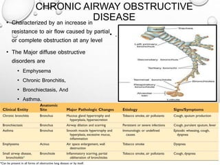
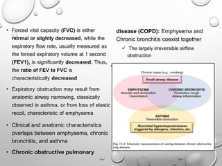
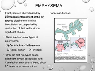
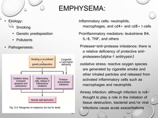
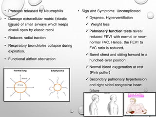
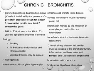
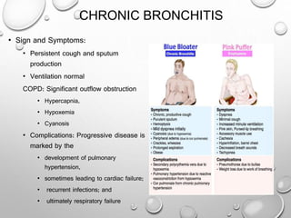
2. CHRONIC AIRWAY OBSTRUCTIVE
DISEASE• Characterized by an increase in
resistance to air flow caused by partial
or complete obstruction at any level
• The Major diffuse obstructive
disorders are
• Emphysema
• Chronic Bronchitis,
• Bronchiectasis, And
• Asthma.
• .
3. • Forced vital capacity (FVC) is either
normal or slightly decreased, while the
expiratory flow rate, usually measured as
the forced expiratory volume at 1 second
(FEV1), is significantly decreased. Thus,
the ratio of FEV to FVC is
characteristically decreased
• Expiratory obstruction may result from
anatomic airway narrowing, classically
observed in asthma, or from loss of elastic
recoil, characteristic of emphysema
• Clinical and anatomic characteristics
overlaps between emphysema, chronic
bronchitis, and asthma
• Chronic obstructive pulmonary
disease (COPD): Emphysema and
Chronic bronchitis coexist together
The largely irreversible airflow
obstruction
• Asthma: reversible airflow obstruction
4. EMPHYSEMA:
• Emphysema is characterized by
permanent enlargement of the air
spaces distal to the terminal
bronchioles, accompanied by
destruction of their walls without
significant fibrosis.
• There are four major types of
emphysema:
(1) Centriacinar (2) Panacinar
(3) distal acinar (4) irregular
• Only the first two types cause
significant airway obstruction, with
Centriacinar emphysema being about
20 times more common than
Panacinar disease.
5. EMPHYSEMA:
• Etiology:
• Smoking
• Genetic predisposition
• Pollutants
• Pathogensesis:
Inflammatory cells: neutrophils,
macrophages, and cd4+ and cd8+ t cells
Proinflammatory mediators: leukotriene B4,
IL-8, TNF, and others
Protease–anti-protease imbalance: there is
a relative deficiency of protective anti-
proteasesv(alpha-1 antitrypsin)
oxidative stress: reactive oxygen species
are generated by cigarette smoke and
other inhaled particles and released from
activated inflammatory cells such as
macrophages and neutrophils
Airway infection: although infection is not
thought to play a role in the initiation of
tissue destruction, bacterial and/or viral
infections cause acute exacerbations
6. • Protease released by Neutrophills
• Damage extracellular matrix (elastic
tissue) of small airways which keeps
alveoli open by elastic recoil
• Reduces radial traction
• Respiratory bronchioles collapse during
expiration.
• Functional airflow obstruction
• Sign and Symptoms: Uncomplicated
Dyspnea, Hyperventillation
Weight loss
Pulmonary function tests reveal
reduced FEV1 with normal or near-
normal FVC. Hence, the FEV1 to
FVC ratio is reduced.
Barrel chest and sitting forward in a
hunched-over position
Normal blood oxygenation at rest
(Pink puffer)
Secondary pulmonary hypertension
and right sided congestive heart
failure
7. CHRONIC BRONCHITIS
• Chronic bronchitis is diagnosed on clinical
grounds: it is defined by the presence of a
persistent productive cough for at least
3 consecutive months in at least 2
consecutive years.
• 20% to 25% of men in the 40- to 65-
year-old age group are prone to develop
Etiology:
• Smoking
• Air Pollutants (sulfur dioxide and
nitrogen dioxide)
• Microbial Infection may be present
• Pathogenesis:
Irritant induced Mucus gland hypertrophy
In trachea and bronchi (large bronchi)
Increase in number of mucin secreating
goblet cell
inflammation marked by the infiltration of
macrophages, neutrophils, and
lymphocytes
the airflow obstruction in chronic bronchitis
results from
(1) small airway disease, induced by
mucous plugging of the bronchiolar lumen,
inflammation, and bronchiolar wall
fibrosis, and (2) coexistent emphysema.
Bronchiolitis: mild obstruction
Emphysema: Significant obstruction
8. CHRONIC BRONCHITIS
• Sign and Symptoms:
• Persistent cough and sputum
production
• Ventilation normal
COPD: Significant outflow obstruction
• Hypercapnia,
• Hypoxemia
• Cyanosis
• Complications: Progressive disease is
marked by the
• development of pulmonary
hypertension,
• sometimes leading to cardiac failure;
• recurrent infections; and
• ultimately respiratory failure