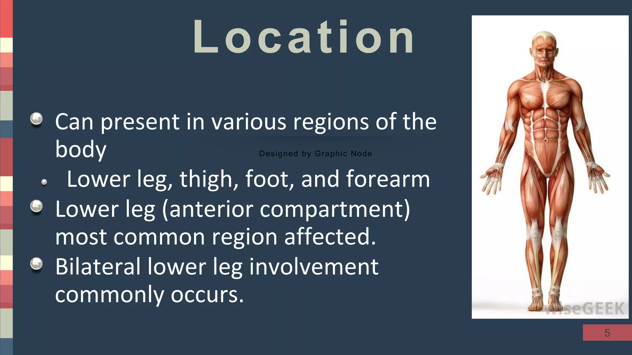
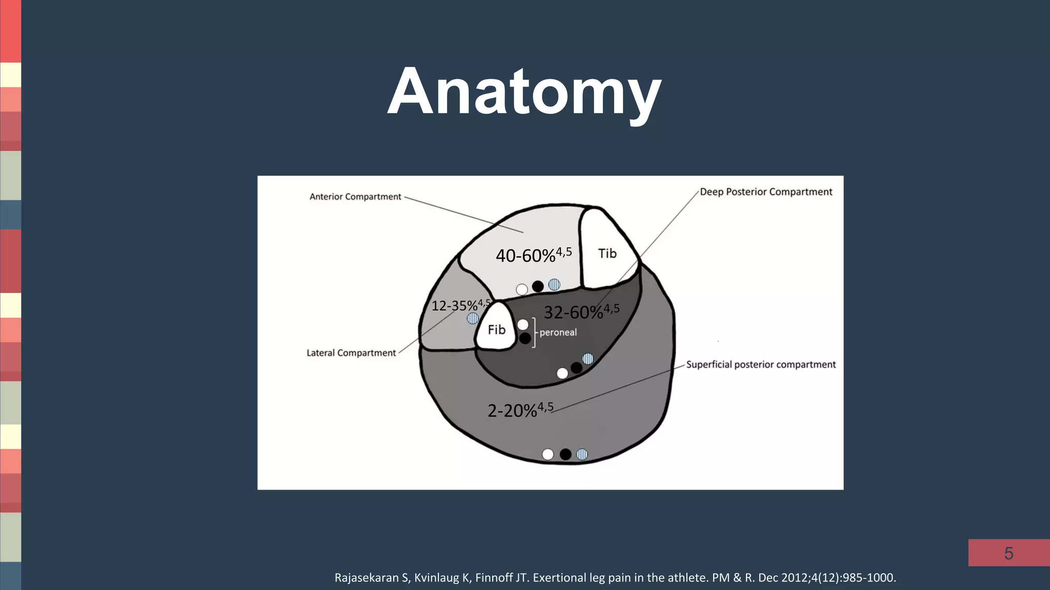
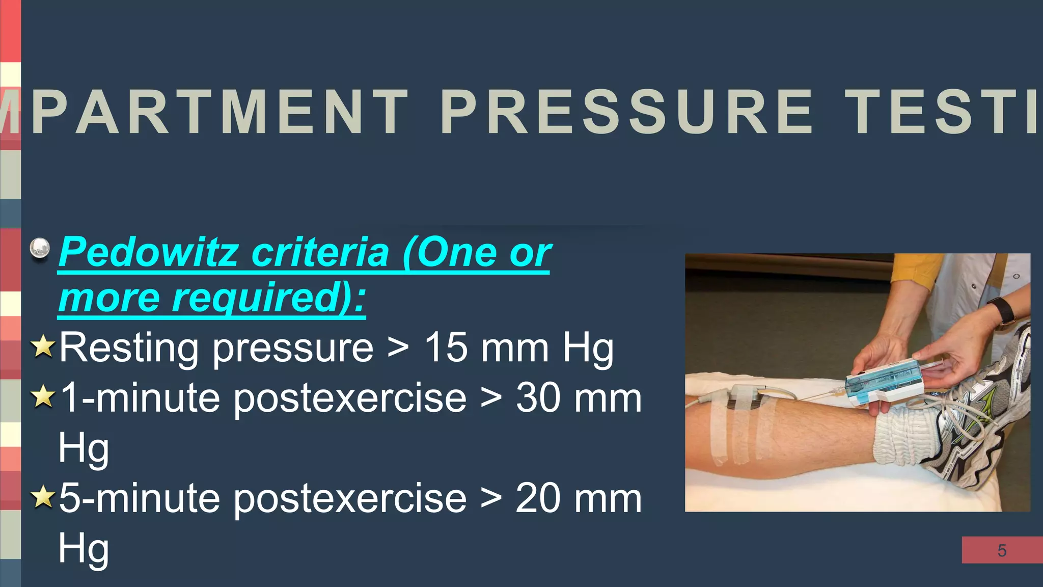
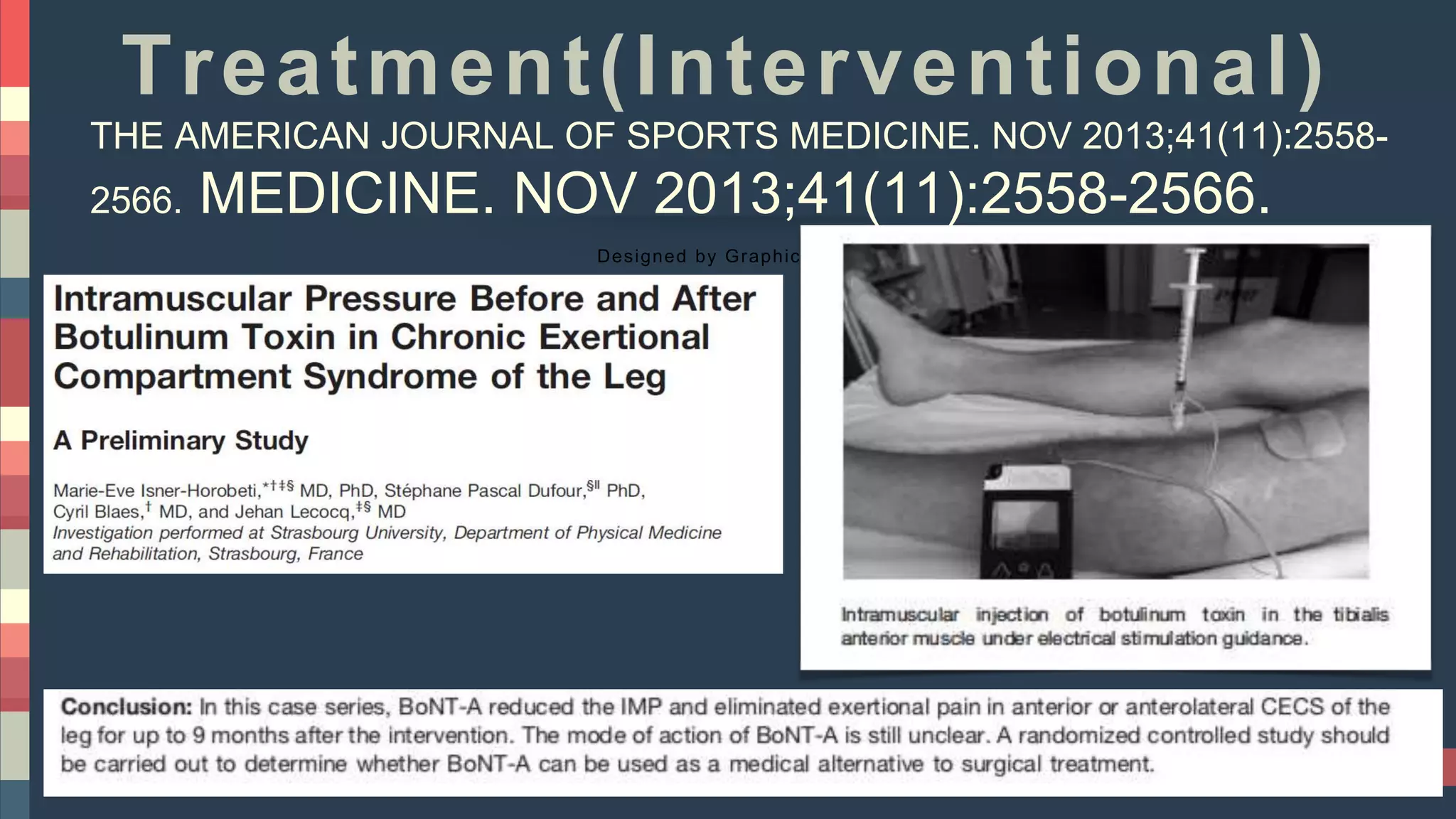
1
Chronic Exertional Compartment
Syndrome
Ahmed Youssef
Mubarak Alkabeer Hospital
5
Outline
Designed by Graphic Node
Definition
Anatomy
Pathophysiology
History and Physical Exam
Diagnostic Evaluation
DD
Treatment
5
Definition
Designed by Graphic Node
•Reversible ischemia secondary to a
noncompliant osteofascial compartment
that is unresponsive to the expansion of
muscle volume that occurs with exercise
5
Epidemiology
Designed by Graphic Node
CECS is most common in young adult
recreational runners, elite athletes, and military
recruits
Bimodal distribution (20 vs. 48 years).
Male>Female
Associated with sports (Running)
Diabetic patients with exertional leg pain and
normal vascular studies may have CECS
5
Location
Designed by Graphic Node
Can present in various regions of the
body
Lower leg, thigh, foot, and forearm
Lower leg (anterior compartment)
most common region affected.
Bilateral lower leg involvement
commonly occurs.
5
40-60%4,5
32-60%4,512-35%4,5
2-20%4,5
Rajasekaran S, Kvinlaug K, Finnoff JT. Exertional leg pain in the athlete. PM & R. Dec 2012;4(12):985-1000.
Anatomy
5
Pathophysiology
Designed by Graphic Node
↓↓Compliance of facial structures → ↑ ↑
Compartment pressure
Reduced microcirculatory capacity
Vascular congestion as a result of
decreased venous return
↑↑ muscle volume (Anabolic steroid and
creatine use)
5
CLINICAL EVALUATION
Designed by Graphic Node
Typical patient with CECS is
Competitive runner, 20 to 30 years old,
Exercise-induced burning pain ↑↑ after 20 to 30
minutes of running.
The pain usually resolves within 15 to 30 minutes of
cessation of exercise.
Paresthesias of the nerves running through the
involved compartment often are reported.
5
Signs of CECS
Designed by Graphic Node
Patients should be examined after completing the
exercise
Tenderness over the musculature of the involved
compartment
Muscle herniation through defects in fascia may be
palpated
Diminished sensation along the affected nerve.
Weakness is often reported
5
MPARTMENT PRESSURE TESTI
Pedowitz criteria (One or
more required):
Resting pressure > 15 mm Hg
1-minute postexercise > 30 mm
Hg
5-minute postexercise > 20 mm
Hg
5
Other investigations
Designed by Graphic Node
Useful to eliminate other
pathology
Xrays,MRI,Bone scan
MRI in-scanner exercise protocol
—>>(smoothing and
segmentation of bone and blood
vessels useful for screening)
5
Differential Diagnosis
Designed by Graphic Node
Medial tibial stress syndrome (shin splints) —vague
diffuse pain reduced with training
Stress fracture—Xrays & Bone scan(localized, intense
uptake)
Periostitis—Bone scan, with diffuse uptake often
covering outer third of the bone.
Superficial peroneal nerve syndrome pain during
active, resisted dorsiflexion and eversion of the ankle,
Tinel sign also may be positive
5
Differential Diagnosis
Designed by Graphic Node
Tenosynovitis (Achilles tendon, peroneal tendon, or
tibialis posterior)
Lumbosacral radiculopathy — Lumbar tension signs
Popliteal artery entrapment syndrome —(vascular
studies)
Deep venous thrombosis
Neurogenic & Vascular claudication
Others (Infection, Myopathy, Tumors)
5
Treatment
Designed by Graphic Node
Conservative
Interventional
Surgery
5
Conservative Treatment
Designed by Graphic Node
Limiting activity to a level that avoids all
but minimal symptoms.
Antiinflammatory medications
Stretching and strengthening of the
involved muscles
Orthotics
5
Treatment(Interventional)
Designed by Graphic Node
THE AMERICAN JOURNAL OF SPORTS MEDICINE. NOV 2013;41(11):2558-
2566. MEDICINE. NOV 2013;41(11):2558-2566.
5
OPERATIVE TREATMENT
Designed by Graphic Node
Single incision (open) technique
One or two incision (subcutaneous) technique
Endoscopic Compartment release (Removal of a
strip of fascia)
5
SINGLE-INCISION FASCIOTOMY for Ant.&Lat. CECS
Designed by Graphic Node
A. Incision between tibial crest
and fibular shaft, over
anterolateral intermuscular
septum, when no fascial
hernia exists.
B. In presence of fascial hernia,
incision is directly over
fascial defect.
C. Defect is enlarged across
intermuscular septum (1).
D. and E, Complete longitudinal
release of anterior
compartment (2 and 3) and
lateral compartment (4 and
5
DOUBLE MINI-INCISION FASCIOTOMY for anterior CECS
Designed by Graphic Node
A. Two vertical 2-cm skin incisions (15cm apart).
B. Development of subcutaneous flap with blunt dissection.
C. Skin retraction to allow fasciotomy under direct vision.
5
Designed by Graphic Node
A transverse incision at the
anterolateral aspect of the
knee between the fibular
head and Gerdy’s tubercle is
used to access the anterior
and lateral compartments.
The deep fascia encasing the
compartment is exposed.
The balloon dissector is
inserted down to the level of
the ankle under direct
palpation
The balloon is inflated.
Endoscopic
Compartment
release for CECS
5
Endoscopic Compartment
release for CECS
Designed by Graphic Node
Anterior compartment release in a left
leg. The black arrow points to the
intermuscular septum between the
anterior and lateral compartments.
The white arrow denotes the
superficial peroneal nerve exiting the
fascia of the lateral compartment
distally.
Endoscopic visualization of the
posterior fascia of a left leg. The black
arrow denotes the deep posterior
release directly off the tibia. The
white arrow denotes the superficial
posterior compartment release.
5
Designed by Graphic Node
POD 1-2
The limb is elevated for 24 to 48 hours and ice is applied
Gentle active and passive ROM, weight bearing as tolerated
Basic activities of daily living
POD 3-4
Achieve independence with activities of daily living, and begin unassisted
ambulation
Weeks 1-4 Add stair climbing and increase walking distance
Weeks 4-6 Begin non-impact lower extremity aerobic exercise
Weeks 6+ Initiate unrestricted impact lower extremity activities
Rajasekaran S, Kvinlaug K, Finnoff JT. Exertional leg pain in the athlete. PM & R. Dec 2012;4(12):985-1000.
CARE
5
Complications of Surgery
Designed by Graphic Node
Infection
Nerve (SPN) most common or vascular injury
Deep vein thrombosis
Wound dehiscence
Complex regional pain syndrome
Scar hypersensitivity
Seroma/hematoma formation
5
Tibial Stress Syndrome (Shin Splints)
Overuse injury or
repetitive-load injury of
the shin area that
includes:
medial (posteromedial)
tibial stress syndrome
(most common)
anterior (anterolateral)
tibial stress syndrome
5
60% of leg pain syndromes
Vague, diffuse pain along
middle-distal tibia that
decreases with running
Radiographs to exclude stress
fracture
Differentiate from stress
fracture, which shows
"dreaded black line"
Tibial Stress Syndrome (Shin Splints)
5
Triphasic bone scan: to
exclude stress fracture
Diffuse, longitudinal
increased uptake along
posteromedial border of
tibia in delayed phase
MRI:periosteal edema,
progressive marrow
involvement
Tibial Stress Syndrome (Shin Splints)
5
Treatment
Nonoperative treatment:
First line of treatment and successful in
vast majority
Activity modification with shoe modification
Operative:
Failed non operative treatment
Deep posterior compartment fasciotomy +
release of painful portion of periosteum
5
Take Home Messages
Designed by Graphic Node
CECS is most common in young adult
recreational runners, elite athletes, and military
recruits.
Diabetic patients with exertional leg pain and
normal vascular studies may have CECS.
Anterior compartment of lower leg most common
region affected. Bilateral affection is common.
5
Take Home Messages
Designed by Graphic Node
Exercise-induced burning pain↑↑after 20 to 30
minutes of running.
The pain usually resolves within 15 to 30 minutes
of cessation of exercise.
D.D.: Tibial stress syndrome—-> pain ↓↓ with
activity.
Fasciotomy (SC,open,endoscopic ) will resolve
the problem.
29

Chronic Exertional Compartment Syndrome

  • 1.
    1 Chronic Exertional Compartment Syndrome AhmedYoussef Mubarak Alkabeer Hospital
  • 2.
    5 Outline Designed by GraphicNode Definition Anatomy Pathophysiology History and Physical Exam Diagnostic Evaluation DD Treatment
  • 3.
    5 Definition Designed by GraphicNode •Reversible ischemia secondary to a noncompliant osteofascial compartment that is unresponsive to the expansion of muscle volume that occurs with exercise
  • 4.
    5 Epidemiology Designed by GraphicNode CECS is most common in young adult recreational runners, elite athletes, and military recruits Bimodal distribution (20 vs. 48 years). Male>Female Associated with sports (Running) Diabetic patients with exertional leg pain and normal vascular studies may have CECS
  • 5.
    5 Location Designed by GraphicNode Can present in various regions of the body Lower leg, thigh, foot, and forearm Lower leg (anterior compartment) most common region affected. Bilateral lower leg involvement commonly occurs.
  • 6.
    5 40-60%4,5 32-60%4,512-35%4,5 2-20%4,5 Rajasekaran S, KvinlaugK, Finnoff JT. Exertional leg pain in the athlete. PM & R. Dec 2012;4(12):985-1000. Anatomy
  • 7.
    5 Pathophysiology Designed by GraphicNode ↓↓Compliance of facial structures → ↑ ↑ Compartment pressure Reduced microcirculatory capacity Vascular congestion as a result of decreased venous return ↑↑ muscle volume (Anabolic steroid and creatine use)
  • 8.
    5 CLINICAL EVALUATION Designed byGraphic Node Typical patient with CECS is Competitive runner, 20 to 30 years old, Exercise-induced burning pain ↑↑ after 20 to 30 minutes of running. The pain usually resolves within 15 to 30 minutes of cessation of exercise. Paresthesias of the nerves running through the involved compartment often are reported.
  • 9.
    5 Signs of CECS Designedby Graphic Node Patients should be examined after completing the exercise Tenderness over the musculature of the involved compartment Muscle herniation through defects in fascia may be palpated Diminished sensation along the affected nerve. Weakness is often reported
  • 10.
    5 MPARTMENT PRESSURE TESTI Pedowitzcriteria (One or more required): Resting pressure > 15 mm Hg 1-minute postexercise > 30 mm Hg 5-minute postexercise > 20 mm Hg
  • 11.
    5 Other investigations Designed byGraphic Node Useful to eliminate other pathology Xrays,MRI,Bone scan MRI in-scanner exercise protocol —>>(smoothing and segmentation of bone and blood vessels useful for screening)
  • 12.
    5 Differential Diagnosis Designed byGraphic Node Medial tibial stress syndrome (shin splints) —vague diffuse pain reduced with training Stress fracture—Xrays & Bone scan(localized, intense uptake) Periostitis—Bone scan, with diffuse uptake often covering outer third of the bone. Superficial peroneal nerve syndrome pain during active, resisted dorsiflexion and eversion of the ankle, Tinel sign also may be positive
  • 13.
    5 Differential Diagnosis Designed byGraphic Node Tenosynovitis (Achilles tendon, peroneal tendon, or tibialis posterior) Lumbosacral radiculopathy — Lumbar tension signs Popliteal artery entrapment syndrome —(vascular studies) Deep venous thrombosis Neurogenic & Vascular claudication Others (Infection, Myopathy, Tumors)
  • 14.
    5 Treatment Designed by GraphicNode Conservative Interventional Surgery
  • 15.
    5 Conservative Treatment Designed byGraphic Node Limiting activity to a level that avoids all but minimal symptoms. Antiinflammatory medications Stretching and strengthening of the involved muscles Orthotics
  • 16.
    5 Treatment(Interventional) Designed by GraphicNode THE AMERICAN JOURNAL OF SPORTS MEDICINE. NOV 2013;41(11):2558- 2566. MEDICINE. NOV 2013;41(11):2558-2566.
  • 17.
    5 OPERATIVE TREATMENT Designed byGraphic Node Single incision (open) technique One or two incision (subcutaneous) technique Endoscopic Compartment release (Removal of a strip of fascia)
  • 18.
    5 SINGLE-INCISION FASCIOTOMY forAnt.&Lat. CECS Designed by Graphic Node A. Incision between tibial crest and fibular shaft, over anterolateral intermuscular septum, when no fascial hernia exists. B. In presence of fascial hernia, incision is directly over fascial defect. C. Defect is enlarged across intermuscular septum (1). D. and E, Complete longitudinal release of anterior compartment (2 and 3) and lateral compartment (4 and
  • 19.
    5 DOUBLE MINI-INCISION FASCIOTOMYfor anterior CECS Designed by Graphic Node A. Two vertical 2-cm skin incisions (15cm apart). B. Development of subcutaneous flap with blunt dissection. C. Skin retraction to allow fasciotomy under direct vision.
  • 20.
    5 Designed by GraphicNode A transverse incision at the anterolateral aspect of the knee between the fibular head and Gerdy’s tubercle is used to access the anterior and lateral compartments. The deep fascia encasing the compartment is exposed. The balloon dissector is inserted down to the level of the ankle under direct palpation The balloon is inflated. Endoscopic Compartment release for CECS
  • 21.
    5 Endoscopic Compartment release forCECS Designed by Graphic Node Anterior compartment release in a left leg. The black arrow points to the intermuscular septum between the anterior and lateral compartments. The white arrow denotes the superficial peroneal nerve exiting the fascia of the lateral compartment distally. Endoscopic visualization of the posterior fascia of a left leg. The black arrow denotes the deep posterior release directly off the tibia. The white arrow denotes the superficial posterior compartment release.
  • 22.
    5 Designed by GraphicNode POD 1-2 The limb is elevated for 24 to 48 hours and ice is applied Gentle active and passive ROM, weight bearing as tolerated Basic activities of daily living POD 3-4 Achieve independence with activities of daily living, and begin unassisted ambulation Weeks 1-4 Add stair climbing and increase walking distance Weeks 4-6 Begin non-impact lower extremity aerobic exercise Weeks 6+ Initiate unrestricted impact lower extremity activities Rajasekaran S, Kvinlaug K, Finnoff JT. Exertional leg pain in the athlete. PM & R. Dec 2012;4(12):985-1000. CARE
  • 23.
    5 Complications of Surgery Designedby Graphic Node Infection Nerve (SPN) most common or vascular injury Deep vein thrombosis Wound dehiscence Complex regional pain syndrome Scar hypersensitivity Seroma/hematoma formation
  • 24.
    5 Tibial Stress Syndrome(Shin Splints) Overuse injury or repetitive-load injury of the shin area that includes: medial (posteromedial) tibial stress syndrome (most common) anterior (anterolateral) tibial stress syndrome
  • 25.
    5 60% of legpain syndromes Vague, diffuse pain along middle-distal tibia that decreases with running Radiographs to exclude stress fracture Differentiate from stress fracture, which shows "dreaded black line" Tibial Stress Syndrome (Shin Splints)
  • 26.
    5 Triphasic bone scan:to exclude stress fracture Diffuse, longitudinal increased uptake along posteromedial border of tibia in delayed phase MRI:periosteal edema, progressive marrow involvement Tibial Stress Syndrome (Shin Splints)
  • 27.
    5 Treatment Nonoperative treatment: First lineof treatment and successful in vast majority Activity modification with shoe modification Operative: Failed non operative treatment Deep posterior compartment fasciotomy + release of painful portion of periosteum
  • 28.
    5 Take Home Messages Designedby Graphic Node CECS is most common in young adult recreational runners, elite athletes, and military recruits. Diabetic patients with exertional leg pain and normal vascular studies may have CECS. Anterior compartment of lower leg most common region affected. Bilateral affection is common.
  • 29.
    5 Take Home Messages Designedby Graphic Node Exercise-induced burning pain↑↑after 20 to 30 minutes of running. The pain usually resolves within 15 to 30 minutes of cessation of exercise. D.D.: Tibial stress syndrome—-> pain ↓↓ with activity. Fasciotomy (SC,open,endoscopic ) will resolve the problem.
  • 30.

Editor's Notes

  • #7 CECS involves the anterior compartment in 40%-60% of patients deep posterior compartment in 32%-60% lateral compartment in 12%-35% Superficial posterior compartment in 2%-20%
  • #17 Abstract published in 16th European congress of PM&R (2008)