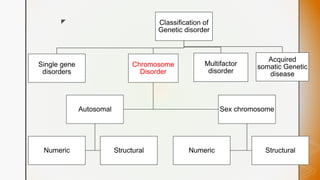
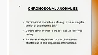
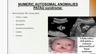
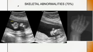
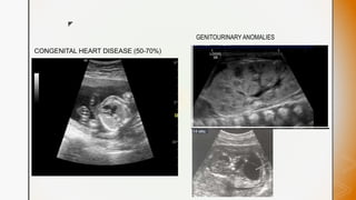
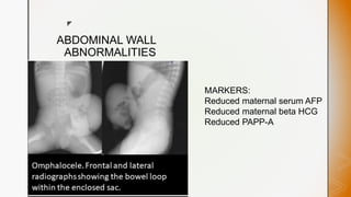
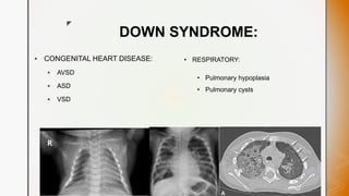
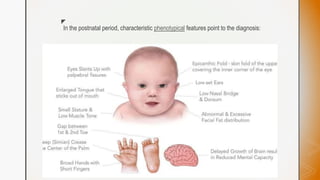
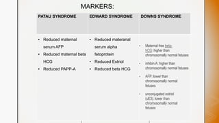
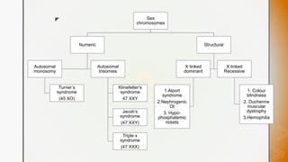
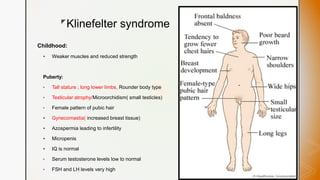
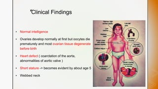
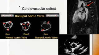
z
CHROMOSOMAL
ANOMALIES
Dr.Niranjan patil
Department of Radiodiagnosis
DYP Kolhapur
z Classification of
Genetic disorder
Single gene
disorders
Chromosome
Disorder
Autosomal
Numeric Structural
Sex chromosome
Numeric Structural
Multifactor
disorder
Acquired
somatic Genetic
disease
z
CHROMOSOMAL ANOMALIES
 Chromosomal anomalies = Missing , extra or irregular
portion of chromosomal DNA
 Chromosomal anomalies are detected via karyotype
testing
 Abnormalities depends on type of chromosome
affected due to non- disjunction chromosomes.
z
Chromosome
affected
Autosomes
Numeric
Autosomal
Trisomies
Autosomal
Monosomy
Structural
Deletions
Sex
chromosomes
Numeric Structural
1.Cri du chat
syndrome
2. Prader Willi
syndrome
3. Angelman
syndrome
z
Autosomal
anomalies (
NUMERIC)
Monosomy
Single copy of
an autosome,
Lethal inearly
pregnancy
Trisomy
DOWN syndrome
(47+ 21)
EDWARD’s syndrome
(47 + 18)
PATAU syndrome
(47 + 13)
z
NUMERIC AUTOSOMAL ANOMALIES
PATAU syndrome:
• Abnormal facies: 90%, strong marker
• Cleft lip +/- palate
• Micropthalmia
• Micrognathia
• Hypotelorism/hypertelorism
• Cyclopia
• proboscis
z
SKELETAL ABNORMALITIES (70%)
z
CNS ABNORMALITIES (70%)
z
CONGENITAL HEART DISEASE (50-70%)
GENITOURINARY ANOMALIES
z
ABDOMINAL WALL
ABNORMALITIES
MARKERS:
Reduced maternal serum AFP
Reduced maternal beta HCG
Reduced PAPP-A
z
Antenatal ultrasound: general features
 Intrauterine growth restriction (IUGR) tends to be early
 Abnormal liquor volume : mostly polyhydramnios or oligo
 Increased nuchal thickness
 Evidence of hydrops fetalis
z
EDWARD SYNDROME:
CONGENITAL HEART DISEASE (90-95%)
z
Single umbilical
artery (80%)
z
CNS anomalies: (70%)
DOLICHOCEPHALY
z
z
Skeletal abnormalities:
z
Markers :
 Reduced materanal serum alpha fetoprotein
 Reduced Estriol
 Reduced beta HCG
z
DOWN SYNDROME:
 CONGENITAL HEART DISEASE:
 AVSD
 ASD
 VSD
• RESPIRATORY:
• Pulmonary hypoplasia
• Pulmonary cysts
z
CNS MANIFESTATIONS:
 Intellectual disability : avg IQ 50-70%
 Hearing loss from inner ear anomalies ( eg. Semicircular canal
dysplasia and dehiscence, narrowed internal acoustic canal ,
cochlear nerve canal stensosis)
 Cerebellar and vermian hypoplasia
 Alzheimer disease developing in all patients older than 40 yrs
z
z MUSCULOSKELETAL
z
In the postnatal period, characteristic phenotypical features point to the diagnosis:
z
MARKERS:
 Reduced
materanal serum
alpha fetoprotein
 Reduced Estriol
 Reduced beta
HCG
PATAU SYNDROME EDWARD SYNDROME DOWNS SYNDROME
• Reduced maternal
serum AFP
• Reduced maternal beta
HCG
• Reduced PAPP-A
• Reduced materanal
serum alpha
fetoprotein
• Reduced Estriol
• Reduced beta HCG
• Maternal free beta-
hCG: higher than
chromosomally normal fetuses
• inhibin A: higher than
chromosomally normal fetuses
• AFP: lower than
chromosomally normal
fetuses
• unconjugated estriol
(uE3): lower than
chromosomally normal
fetuses
z
STRUCTURAL AUTOSOMAL ANOMALIES :
CRI DU CHAT SYNDROME
 Cri du chat syndrome is a rare congenital disorder caused by the deletion of
the short arm of chromosome 5.
 A high-pitched monotonous cry is the characteristic finding.
 The patient also usually has craniofacial malformations such as microcephaly,
bridge
 Newborns also have microcephaly and have an increased risk of asphyxia
z
Prader-Willi and Angelman Syndrome
Prader-Willi Syndrome
 Microdeletion of chromosome15
 Lack of muscle tone in newborn
 As adult – gross obesity
 Radiographic features:
 Abnormalities of calvarium and
sella turcica
 Coxa valga
 Scoliosis
Angelman Syndrome
 Microdeletion of chromosome 15.
 Development delay
 Jerky movements
 Stiff, fixed smile
 Epilepsy
z Sex
chromosomes
Numeric
Autosomal
monosomy
Turner’s
syndrome
(45 XO)
Autosomal
trisomies
Klinefelter’s
syndrome
47 XXY
Jacob’s
syndrome
(47 XXY)
Triple x
syndrome
(47 XXX)
Structural
X linked
dominant
1.Alport
syndrome
2.Nephrogenic
DI
3. Hypo-
phosphatemic
rickets
X linked
Recessive
1. Colour
blindness
2. Duchenne
muscular
dystrophy
3.Hemophilia
z
Klinefelter syndrome
Childhood:
 Weaker muscles and reduced strength
Puberty:
• Tall stature ; long lower limbs, Rounder body type
• Testicular atrophy/Microorchidism( small testicles)
• Female pattern of pubic hair
 Gynecomastia( increased breast tissue)
 Azospermia leading to infertility
 Micropenis
 IQ is normal
• Serum testosterone levels low to normal
• FSH and LH levels very high
z
Radiographic features:
 USG: Klinefelter syndrome should be suspected in postpubertal males with bilateral
symmetrical small testicular volume (usually 3~4 mL in volume) usually presented by
subfertility/primary infertility or assessment of small testicular size
z
TURNER SYNDROME
 Absence of one chromosome x (45 XO )
 Most common sex abnormality in females
 GENE SHOX -> important for bone development and growth
 Loss of one copy of this gene -> short stature
z
Clinical Findings
 Normal intelligence
 Ovaries develop normally at first but oocytes die
prematurely and most ovarian tissue degenerate
before birth
 Heart defect ( coarctation of the aorta,
abnormalities of aortic valve )
 Short stature -> becomes evident by about age 5
 Webbed neck
z Radiological features:
 Skeletal features -> inconstant and non specific
 Decreased density of the skeleton ( esp . Hands and feet )
ANTENATAL ULTRASOUND
1. Cystic hygroma
2. Increased nuchal thickness
3. Increased nuchal translucency
4. Hydrops fetalis
5. Mild IUGR
6. Short fetal limbs
z
Nuchal translucency
INCREASED nuchal translucency
 A finding during first trimester
 Measured on saggital image through the fetal neck
 Rate of aneuploidy when the nuchal thickness is < 2mm
--> less than 1 %
Back of neck
z
Nuchal thickness:
INCREASED nuchal thickness
 Measured in a second trimester
 Abnormal value > 6mm
 Should not be measured after 20.6 weeks
Back of head
z Cystic hygroma
 A cystic lymphatic lesion
 Septation indicate poorer outcome
 Volumes > 75mm3 correlate with increased
abnormality and poorer fetal outcome
z Hydrops fetalis
 Excessive accumulation of fluid in fetal subcutaneous tissue or
atleast 2 fetal serous cavities
 Fetal pleural effusion
 Fetal pericardial effusion
 Fetal ascites
 Placental enlargement
 Polyhydromnios
 Fetal anasarca
z
Hand and feet :
Post partum and Adulthood
 HAND:
1. Positive metacarpal sign
2. Increased carrying angle of elbow / cubitus
valgus.
FEET:
1. Abnormal medial tibial condyle -> depressed
and beak like
2. Abnormal medial femoral condyle -> project
downward
z
Positive metacarpal sign
 A line drawn along the heads of 4 th and 5 th metacarpals will intersect the
head of the 3 rd metacarpal if shortening is present
z
Cubitus valgus
 Increased carrying angle of the elbow
 Carrying angle ( Cubital angle ) -> formed
between the humerus and the forearm axis
 NORMAL – 5-15 degree away from the body
 <5 degree : cubitus varus
 >15 degree: cubitus valgus
z
Cardiovascular defect
z
Pelvic ultrasound
z
Dd
TURNER’S SYNDROME DOWN SYNDROME
Normal intelligence Mental retardation
Cardiac defect : coartation of aorta, bicuspid
aortic valve
Cardiac defect: intracardial septal defect
Metacarpal sign
Cubitus valgus
Clinodactily
Mickey mouse pelvis ( large ilium)
Absence of last rib
Webbed neck Orbital hypo/ hypertelorism
Cystic hygroma
Increased Nuchal thickness
Increased Nuchal translucency
Cystic hygroma
Increased Nuchal thickness
Increased Nuchal translucency
z
GENOTYPE GENDER SYNDROME PHYSICAL TRAITS
XY MALE -
XXY MALE Klinefelter’s
syndrome
Sterility, small testicles, breast
enlargement
XYY MALE Jacob’s syndrome Normal male traits
XX FEMALE -
XO FEMALE Turner syndrome Sex organs don’t mature at
adolescence , sterility, short
stature
XXX FEMALE Triple X Tall stature , normal intelligence
with learning disabilities, limited
fertility
z
Chromosome
affected
Autosomes
Numeric
Autosomal
monosomy
Autosomal
Trisomies
Patau syndrome
Edward syndrome
Down syndrome
Structural
Deletions
1.Cri du chat
syndrome
2. Prader Willi
syndrome
3. Angelman
syndrome
Sex
chromosomes
Numeric
Autosomal
monosomy
Autosomal
trisomies
1. Klinefelter’s
syndrome
2. Jacob’s
syndrome
3. Triple x
syndrome
Structural
X linked
dominant
X linked
recessive
1.Alport
syndrome
2.Nephrogenic DI
3. Hypo-
phosphatemic
rickets
Turner’s
syndrome
(45 XO)
1. Colour
blindness
2. Duchenne
muscular
dystrophy
3.Hemophilia
SUMMARY
z
 THANK YOU

chromosomal anomalies.pptx

  • 1.
  • 2.
    z Classification of Geneticdisorder Single gene disorders Chromosome Disorder Autosomal Numeric Structural Sex chromosome Numeric Structural Multifactor disorder Acquired somatic Genetic disease
  • 3.
    z CHROMOSOMAL ANOMALIES  Chromosomalanomalies = Missing , extra or irregular portion of chromosomal DNA  Chromosomal anomalies are detected via karyotype testing  Abnormalities depends on type of chromosome affected due to non- disjunction chromosomes.
  • 4.
  • 5.
    z Autosomal anomalies ( NUMERIC) Monosomy Single copyof an autosome, Lethal inearly pregnancy Trisomy DOWN syndrome (47+ 21) EDWARD’s syndrome (47 + 18) PATAU syndrome (47 + 13)
  • 6.
    z NUMERIC AUTOSOMAL ANOMALIES PATAUsyndrome: • Abnormal facies: 90%, strong marker • Cleft lip +/- palate • Micropthalmia • Micrognathia • Hypotelorism/hypertelorism • Cyclopia • proboscis
  • 7.
  • 8.
  • 9.
    z CONGENITAL HEART DISEASE(50-70%) GENITOURINARY ANOMALIES
  • 10.
    z ABDOMINAL WALL ABNORMALITIES MARKERS: Reduced maternalserum AFP Reduced maternal beta HCG Reduced PAPP-A
  • 11.
    z Antenatal ultrasound: generalfeatures  Intrauterine growth restriction (IUGR) tends to be early  Abnormal liquor volume : mostly polyhydramnios or oligo  Increased nuchal thickness  Evidence of hydrops fetalis
  • 12.
  • 13.
  • 14.
  • 15.
  • 16.
  • 17.
    z Markers :  Reducedmateranal serum alpha fetoprotein  Reduced Estriol  Reduced beta HCG
  • 18.
    z DOWN SYNDROME:  CONGENITALHEART DISEASE:  AVSD  ASD  VSD • RESPIRATORY: • Pulmonary hypoplasia • Pulmonary cysts
  • 19.
    z CNS MANIFESTATIONS:  Intellectualdisability : avg IQ 50-70%  Hearing loss from inner ear anomalies ( eg. Semicircular canal dysplasia and dehiscence, narrowed internal acoustic canal , cochlear nerve canal stensosis)  Cerebellar and vermian hypoplasia  Alzheimer disease developing in all patients older than 40 yrs
  • 20.
  • 21.
  • 22.
    z In the postnatalperiod, characteristic phenotypical features point to the diagnosis:
  • 23.
    z MARKERS:  Reduced materanal serum alphafetoprotein  Reduced Estriol  Reduced beta HCG PATAU SYNDROME EDWARD SYNDROME DOWNS SYNDROME • Reduced maternal serum AFP • Reduced maternal beta HCG • Reduced PAPP-A • Reduced materanal serum alpha fetoprotein • Reduced Estriol • Reduced beta HCG • Maternal free beta- hCG: higher than chromosomally normal fetuses • inhibin A: higher than chromosomally normal fetuses • AFP: lower than chromosomally normal fetuses • unconjugated estriol (uE3): lower than chromosomally normal fetuses
  • 24.
    z STRUCTURAL AUTOSOMAL ANOMALIES: CRI DU CHAT SYNDROME  Cri du chat syndrome is a rare congenital disorder caused by the deletion of the short arm of chromosome 5.  A high-pitched monotonous cry is the characteristic finding.  The patient also usually has craniofacial malformations such as microcephaly, bridge  Newborns also have microcephaly and have an increased risk of asphyxia
  • 25.
    z Prader-Willi and AngelmanSyndrome Prader-Willi Syndrome  Microdeletion of chromosome15  Lack of muscle tone in newborn  As adult – gross obesity  Radiographic features:  Abnormalities of calvarium and sella turcica  Coxa valga  Scoliosis Angelman Syndrome  Microdeletion of chromosome 15.  Development delay  Jerky movements  Stiff, fixed smile  Epilepsy
  • 26.
    z Sex chromosomes Numeric Autosomal monosomy Turner’s syndrome (45 XO) Autosomal trisomies Klinefelter’s syndrome 47XXY Jacob’s syndrome (47 XXY) Triple x syndrome (47 XXX) Structural X linked dominant 1.Alport syndrome 2.Nephrogenic DI 3. Hypo- phosphatemic rickets X linked Recessive 1. Colour blindness 2. Duchenne muscular dystrophy 3.Hemophilia
  • 27.
    z Klinefelter syndrome Childhood:  Weakermuscles and reduced strength Puberty: • Tall stature ; long lower limbs, Rounder body type • Testicular atrophy/Microorchidism( small testicles) • Female pattern of pubic hair  Gynecomastia( increased breast tissue)  Azospermia leading to infertility  Micropenis  IQ is normal • Serum testosterone levels low to normal • FSH and LH levels very high
  • 28.
    z Radiographic features:  USG:Klinefelter syndrome should be suspected in postpubertal males with bilateral symmetrical small testicular volume (usually 3~4 mL in volume) usually presented by subfertility/primary infertility or assessment of small testicular size
  • 29.
    z TURNER SYNDROME  Absenceof one chromosome x (45 XO )  Most common sex abnormality in females  GENE SHOX -> important for bone development and growth  Loss of one copy of this gene -> short stature
  • 30.
    z Clinical Findings  Normalintelligence  Ovaries develop normally at first but oocytes die prematurely and most ovarian tissue degenerate before birth  Heart defect ( coarctation of the aorta, abnormalities of aortic valve )  Short stature -> becomes evident by about age 5  Webbed neck
  • 31.
    z Radiological features: Skeletal features -> inconstant and non specific  Decreased density of the skeleton ( esp . Hands and feet ) ANTENATAL ULTRASOUND 1. Cystic hygroma 2. Increased nuchal thickness 3. Increased nuchal translucency 4. Hydrops fetalis 5. Mild IUGR 6. Short fetal limbs
  • 32.
    z Nuchal translucency INCREASED nuchaltranslucency  A finding during first trimester  Measured on saggital image through the fetal neck  Rate of aneuploidy when the nuchal thickness is < 2mm --> less than 1 % Back of neck
  • 33.
    z Nuchal thickness: INCREASED nuchalthickness  Measured in a second trimester  Abnormal value > 6mm  Should not be measured after 20.6 weeks Back of head
  • 34.
    z Cystic hygroma A cystic lymphatic lesion  Septation indicate poorer outcome  Volumes > 75mm3 correlate with increased abnormality and poorer fetal outcome
  • 35.
    z Hydrops fetalis Excessive accumulation of fluid in fetal subcutaneous tissue or atleast 2 fetal serous cavities  Fetal pleural effusion  Fetal pericardial effusion  Fetal ascites  Placental enlargement  Polyhydromnios  Fetal anasarca
  • 36.
    z Hand and feet: Post partum and Adulthood  HAND: 1. Positive metacarpal sign 2. Increased carrying angle of elbow / cubitus valgus. FEET: 1. Abnormal medial tibial condyle -> depressed and beak like 2. Abnormal medial femoral condyle -> project downward
  • 37.
    z Positive metacarpal sign A line drawn along the heads of 4 th and 5 th metacarpals will intersect the head of the 3 rd metacarpal if shortening is present
  • 38.
    z Cubitus valgus  Increasedcarrying angle of the elbow  Carrying angle ( Cubital angle ) -> formed between the humerus and the forearm axis  NORMAL – 5-15 degree away from the body  <5 degree : cubitus varus  >15 degree: cubitus valgus
  • 39.
  • 40.
  • 41.
    z Dd TURNER’S SYNDROME DOWNSYNDROME Normal intelligence Mental retardation Cardiac defect : coartation of aorta, bicuspid aortic valve Cardiac defect: intracardial septal defect Metacarpal sign Cubitus valgus Clinodactily Mickey mouse pelvis ( large ilium) Absence of last rib Webbed neck Orbital hypo/ hypertelorism Cystic hygroma Increased Nuchal thickness Increased Nuchal translucency Cystic hygroma Increased Nuchal thickness Increased Nuchal translucency
  • 42.
    z GENOTYPE GENDER SYNDROMEPHYSICAL TRAITS XY MALE - XXY MALE Klinefelter’s syndrome Sterility, small testicles, breast enlargement XYY MALE Jacob’s syndrome Normal male traits XX FEMALE - XO FEMALE Turner syndrome Sex organs don’t mature at adolescence , sterility, short stature XXX FEMALE Triple X Tall stature , normal intelligence with learning disabilities, limited fertility
  • 43.
    z Chromosome affected Autosomes Numeric Autosomal monosomy Autosomal Trisomies Patau syndrome Edward syndrome Downsyndrome Structural Deletions 1.Cri du chat syndrome 2. Prader Willi syndrome 3. Angelman syndrome Sex chromosomes Numeric Autosomal monosomy Autosomal trisomies 1. Klinefelter’s syndrome 2. Jacob’s syndrome 3. Triple x syndrome Structural X linked dominant X linked recessive 1.Alport syndrome 2.Nephrogenic DI 3. Hypo- phosphatemic rickets Turner’s syndrome (45 XO) 1. Colour blindness 2. Duchenne muscular dystrophy 3.Hemophilia SUMMARY
  • 44.

Editor's Notes

  • #3 Numeric autosomal anomalies Structural autosomal anomalies
  • #4 Karyotype = full set of chromosomes from an individual. Non dysjunction can occur either during meiosis 1 or meiosis 2
  • #5 numeric autosomal anomalies
  • #6 Down ,Edward and down syndrome are the only three trisomies compatible with extrauterine life.
  • #7 Normal upper lip Cleft lip with cleft palate Micropthalmia- small eyes micrognathia- small mandible’ Hypotelorism- abnormal decrease in distance between 2 eyes Cyclopia- single palpebral fissure and single midline orbit which may contain either a single globe or two separate globe. Proboscis- anterior appendage like structure is seen projecting from the midline forehead
  • #8 Polydactily in hand and feet. Frontal radiograph of the hand showing polydactyly of the fifth finger ( post axial polydactyly – where additional digit is on the ulnar margin of hand or lateral to 5 th little toe ) SKELETAL ABNORMALITIES: Polydactyly Rock bottom feet
  • #9 CNS ABNORMALITIES : Holoprosencephaly is a disorder caused by failure 1.Holoprosencephaly – most commonly assosciated cns anomaly In alobar holoprosencephaly, the thalami are fused and there is a single large posteriorly located ventricle. Most commonly associated with facial abnormalities  Semilobar holoprosencephaly The basic structure of the cerebral lobes are present but are fused most commonly anteriorly and at the thalami Monoventricle Fused central thalami Fused choroid plexus 4th ventricle Nuchal edema- subcutaneous accumulation of extracellular fluid in fetal neck is detected as increased nuchal translucency (NT) by ultrasonography in 1 st trimester of pregnancy LOBAR TYPE- RARE 1.holoprosencephaly 2. Microcephaly 3. Fetal hydrocephaly 4. Enlarged cisterna magna 4. Agenesis of corpus callosum
  • #10 Congenital heart disease : 1.Hypoplastic left heart syndrome 2. Ventricular septal defect (VSD) GENITOURINARY ANOMALIES; Cystic renal dysplasia cryptorchidism
  • #11 ABDOMINAL WALL ABNORMALITIES: Omphalocele Bladder exstrophy Alfa feto protein Pregnancy assosciated plasma protein A
  • #12 Hydrops is defined as the accumulation of fluid +/- edema involving at least two fetal components, fetal pleural effusion fetal pericardial effusion fetal ascites generalized body edema: fetal anasarca/nuchal edema/cystic hygroma placental enlargement polyhydramnios
  • #13 Atrial septal defect VSD PDA dextrocardia
  • #14 Other umbilical cord anomalies: Include1, umbilical cord cysts 2. Umbilical cord pseudocysts
  • #15 Flatennning of occipital bones With relative hypoplasia of the frontal bones causing pointing of frontal lobes Strawberry skull is seen associated with DOLICHOCEPHALY ( most common form of craniosynostosis ) where premature closure of sagittal suture results in impediment of lateral growth of the skull while anteroposterior growth continues , producing a classic elongated yet narrow skull
  • #17 Hand abnormalities: Clenched hands : with an overlap of 2 nd and 3 rd digits in 80% Rocker bottom feet
  • #19 Atrioventricular septal defect Atrial septal defect Ventricular septal defect Hypoplastic appearing thorax with reduced lung volume - pulmonary hypoplasia Extensive lung disease with interstitial thickening and cystic change peripherally. Perihilar collapse. subpleural cysts bilaterally (small arrows).  variable-sized cysts in the middle
  • #21  Axial US image of a hypoplastic cerebellum with a transcerebellar diameter of 19 mm (at the 22 weeks of gestation, the 5th and 95th percentile the transcerebellar diameter is between 21.6 mm and 25 mm)
  • #22 DEVELOPMENT DYSPLASIA OF HIP : Hilgenreiner's line is drawn horizontally, connecting the inferior aspect of the tri-radiate cartilages bilaterally. The femoral head should be below this line Perkin's line is drawn vertically though the lateral most aspect of the acetabular roof, perpendicular to Hilgenreiner's line. The ossified femoral head should be located in the inferomedial quadrant created. Shenton line along the inferior aspect of the superior pubic ramus and the inferiomedial femoral neck.  The acetabular angle is measured between Hilgenreiner's line and a line parallel to the acetabular roof. At birth it should be less than 28 degrees, and should progressively reduce with maturation of the hip. Classic "Mickey Mouse" appearance of the pelvis in Down syndrome with flared iliac wings.
  • #23 Depressed nasal bridge and low set ears Epicanthal folds Abundant neck skin Macroglossia Simian crease( single palmar crease)
  • #24 Pregnancy assosciated plasma protein A
  • #25 Cri-du-chat syndrome Micro deletion of chromosome 5 Di-George syndrome Micro deletion of chromosome 22 Schizophrenia & Obsessive Compulsive Disorder Micro deletion of chromosome 22 associated Angelman syndrome Micro deletion of chromosome 15 Prader-Willi syndrome Micro deletion of chromosome 15
  • #26 Both occur due to microdeletion of chromosome 15
  • #27 Numeric sex chromosomal anomalies Structural sex chromosomal anomalies.
  • #33 It should be measured between 11- 13.6weeks
  • #34 It should be measured between 14-21weeks Cavum septum pellucidum Thalami Cerebellum Cisterna magna Nuchal fold thickness
  • #40 Mc cardiovascular defect : Bicuspid aortic valve Coartation of aorta
  • #41 Ultrasound showing streak ovaries with no follicles Horse shoe kidney
  • #43 Jacobs – normal physically and mentally , increase in testosterone and more aggressive