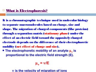
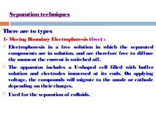
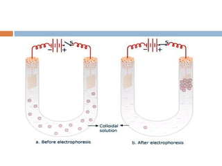
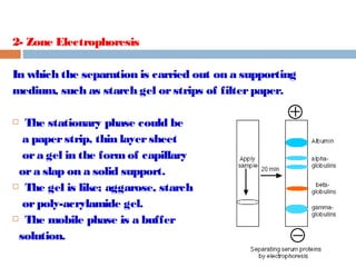
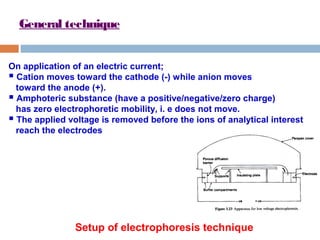
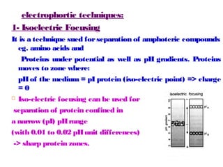
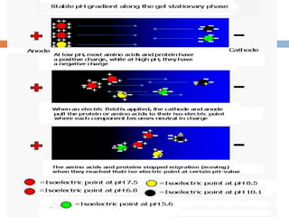
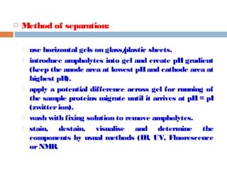
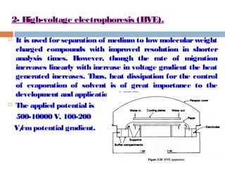
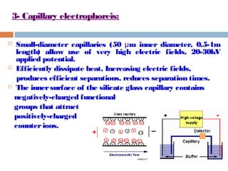
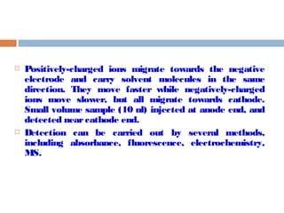
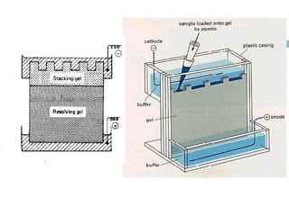
This document provides an overview of electrophoresis techniques. It defines electrophoresis as a method used to separate macromolecules like proteins based on their charge, size, and shape under the influence of an electric field. There are two main types - moving boundary electrophoresis where components separate in solution, and zone electrophoresis where separation occurs on a supporting medium like a gel or paper. Key factors that affect electrophoretic mobility and separation include the electric field strength, characteristics of the sample, properties of the supporting medium, and buffer composition and pH. Common electrophoresis methods include isoelectric focusing, high-voltage electrophoresis, capillary electrophoresis, and continuous versus discontinuous gel systems.
1. CHROMATOGRAPHIC
METHODS OF
ANALYSIS
ELECTROPHORESIS
By: Prof / Dr. Tarek Fayed
Represented by : Mahmoud Galal Zidan
E-Mail : M7moud.zidan@yahoo.com
E-Mail : m7moud.zidan@aol.com
E-Mail : M7moud.zidan@Gmail.com
Facebook : www.fb.com/m7moud.zidan
Twitter : www.twitter.com/m7moudzidane
2. What is Electrophoresis?
It is a chromatographic technique used in molecular biology
to separate macromolecules based on charge, size and
shape. The migration of charged components (like proteins)
through a separation matrix (stationary phase) under the
effect of an electric field toward the oppositely charged
electrode depends on the difference of their electrophoretic
mobility (net effect of charge and size).
The electrophoretic mobility of an analyte me is
proportional to the electric field strength (E).
me = n/E
n is the velocity of migration of ions
3. Separation techniques
There are to types
1- Moving Boundary Electrophoresis (free) :
Electrophoresis in a free solution in which the separated
components are in solution, and are therefore free to diffuse
the moment the current is switched off.
The apparatus includes a U-shaped cell filled with buffer
solution and electrodes immersed at its ends. On applying
voltage, the compounds will migrate to the anode or cathode
depending on their charges.
Used for the separation of colloids.
4.
5. 2- Zone Electrophoresis
In which the separation is carried out on a supporting
medium, such as starch gel or strips of filter paper.
The stationary phase could be
a paper strip, thin layer sheet
or a gel in the form of capillary
or a slap on a solid support.
The gel is like; aggarose, starch
or poly-acrylamide gel.
The mobile phase is a buffer
solution.
6. General technique
On application of an electric current;
Cation moves toward the cathode (-) while anion moves
toward the anode (+).
Amphoteric substance (have a positive/negative/zero charge)
has zero electrophoretic mobility, i. e does not move.
The applied voltage is removed before the ions of analytical interest
reach the electrodes
Setup of electrophoresis technique
7. During the course of electrophoresis current flows, and as in
electrolysis, the products are oxygen and hydrogen;
H2O + 2e → 2OH- + H2↑ at cathode, and pH increases
2H+ + 0.5O2 + 2e → H2O at anode, and pH decreases
To keep the pH constant during separation a porous
diffusion barrier or diaphragm is used near the electrode.
8. Factors affecting mobility (sensitivity of technique)
1. Electric Field:
Voltage:
The velocity of migration of a molecule is directly proportional to the
potential gradient across the stationary phase;
n (V/d) V= volt, d= distance α from the electrode
For low voltage electrophoresis (LVE); V = 200-500 V and for high
voltage electrophoresis (HVE); V = 500-1000 V
Current:
The current is carried mainly by the buffer but small amount is carried by
the sample components. Due to resistance of the supporting medium
heat is evolved. So the current intensity has to be controlled.
9. 2. The sample
Size and molecular weight:
smaller molecules migrate faster than larger molecules carrying the same
charge.
Shape:
molecules with lots of side-chains experiences more frictional resistance than
a linear molecule of the same mass and charge, and will therefore move more
slowly.
Charge:
The type and number of net charge determine the direction and mobility of
components.
3- solid support (stationary phase):
It could affect the mobility via adsorption (as in paper or thin layer),
molecular sieving (as in gel) or electro-osmosis (due to adsorption of
ions on the surface of stationary phase). Both adsorption and
molecular sieving causes decrease of mobility, while electro-osmosis
Increase mobility.
10. 4- Buffer:
pH (protonation - deprotonation process):
proteins and amino acids exist as zwitter ions like amino
acids, and can be either positively or negatively charged because
they contain both acidic and basic groups. The extent, and
direction, of ionization depends on the pH of the buffer.
+ cation moves → toward cathode
at low pH; R-CH(COOH)NH3
at high pH; R-CH(COO-)NH2 → anion moves toward anode
at neutral pH; R-CH(COO-)NH3
+ → zwitter ions do not move (iso-electric
point).
ionic strength (m):
low m
few counter-ions
low charge shielding for current carriers.
high m
many counter-ions
high charge shielding for current carriers.
11. Procedures of separation by electrophoresis
1- Preparation and saturation of the supporting medium:
If the supporting medium is not a gel, it should be
saturated with buffer before separation is started to
conduct the electric current.
Saturation is best done before the sample is applied.
2- sample application:
The sample is applied as a solution with a micro-pipette,
as a small spot or narrow band on th stationary phase.
Sample containing positively charged ion, is applied near
the anode, but negatively charge ions are applied near
the cathode.
Mixed ions sample (containing positive and negative
ions) is applied to the middle.
12. 3- Running of sample
After sample application, the power is switched on at the required and
adjusted potential for a period of time (usually 2 hours). The current has to be
constant.
4- Removal of supporting medium
After running of the ample and elution process, the supporting medium is
removed and dried in an oven at 110 °C to remove the buffer. Gels are
squeezed by hypodermic syringe.
5- Staining of components:
Most biological compounds are colorless, and therefore is necessary
to detect them and determine their position on the supporting medium
by using coloring reagents. Organic dyes are used for this purpose.
6- Identification and determination of components:
Using of specific coloring reagents helps in identification of components
(RNA gives blue color with pyronine, while DNA gives red color).
For estimation of components, complete elution and estimation using
spectral methods is used.
13. electrophortic techniques:
1- Isoelectric Focusing
It is a technique sued for separation of amphoteric compounds
eg. amino acids and
Proteins under potential as well as pH gradients. Proteins
moves to zone where:
pH of the medium = pI protein (iso-electric point) => charge
= 0
Iso-electric focusing can be used for
separation of protein confined in
a narrow (pI) pH range
(with 0.01 to 0.02 pH unit differences)
-> sharp protein zones.
14.
15. Method of separation:
use horizontal gels on glass/plastic sheets.
introduce ampholytes into gel and create pH gradient
(keep the anode area at lowest pH and cathode area at
highest pH).
apply a potential difference across gel for running of
the sample proteins migrate until it arrives at pH = pI
(zwitter ion).
wash with fixing solution to remove ampholytes.
stain, destain, visualise and determine the
components by usual methods (IR, UV, Fluorescence
or NMR.
16. 2- High-voltage electrophoresis (HVE).
It is used for separation of medium to low molecular weight
charged compounds with improved resolution in shorter
analysis times. However, though the rate of migration
increases linearly with increase in voltage gradient the heat
generated increases. Thus, heat dissipation for the control
of evaporation of solvent is of great importance to the
development and application of HVE.
The applied potential is
500-10000 V, 100-200
V/cm potential gradient.
17. 3- Capillary electrophoreis:
Small-diameter capillaries (50 m inner μ diameter, 0.5-1m
length) allow use of very high electric fields, 20-30kV
applied potential.
Efficiently dissipate heat, Increasing electric fields,
produces efficient separations, reduces separation times.
The inner surface of the silicate glass capillary contains
negatively-charged functional
groups that attract
positively-charged
counter ions.
18. Positively-charged ions migrate towards the negative
electrode and carry solvent molecules in the same
direction. They move faster while negatively-charged
ions move slower, but all migrate towards cathode.
Small volume sample (10 nl) injected at anode end, and
detected near cathode end.
Detection can be carried out by several methods,
including absorbance, fluorescence, electrochemistry,
MS.
19. 4- Continuous and Discontinuous electrophoresis
In a continuous system, only a single separating gel is used
with the same buffer in the tanks.
In a discontinuous system a nonrestrictive large pore gel
(as agarose gel), called a stacking gel, is layered on top of a
separating small pore gel (as polyacrylamide gel).
The stacking gel don’t restrict the migration of the
proteins, and deposits proteins in a stack at top of
separating gel
The resolution obtainable in a discontinuous system is
much greater than that obtainable in a continuous one.
However, the continuous system is a little easier to set up,
but discontinuous gels are most common.