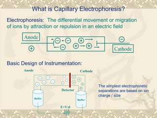
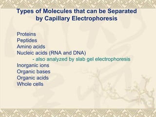
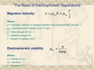
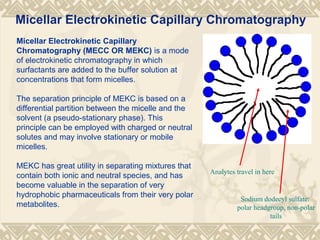
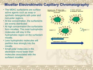
Capillary eleCtrophoresis
Sandhya Talla
M.Pharm (Pharmacology)
What is Capillary Electrophoresis?
Electrophoresis: The differential movement or migration
of ions by attraction or repulsion in an electric field
Anode
Cathode
Basic Design of Instrumentation:
E=V/d
Buffer
Buffer
Anode Cathode
Detector
The simplest electrophoretic
separations are based on ion
charge / size
Proteins
Peptides
Amino acids
Nucleic acids (RNA and DNA)
- also analyzed by slab gel electrophoresis
Inorganic ions
Organic bases
Organic acids
Whole cells
Types of Molecules that can be Separated
by Capillary Electrophoresis
The Basis of Electrophoretic Separations
Migration Velocity:
Where:
v = migration velocity of charged particle in the potential field (cm sec -1
)
µep = electrophoretic mobility (cm2
V-1
sec-1)
E = field strength (V cm -1
)
V = applied voltage (V)
L = length of capillary (cm)
Electrophoretic mobility:
Where:
q = charge on ion
Ī· = viscosity
r = ion radius Frictional retarding forces
L
V
E epep µµν ==
r
q
ep
πη
µ
6
=
Inside the Capillary: The Zeta Potential
• The inside wall of the
capillary is covered
by silanol groups
(SiOH) that are
deprotonated (SiO-
)
at pH > 2
• SiO-
attracts cations
to the inside wall of
the capillary
• The distribution of
charge at the surface
is described by the
Stern double-layer
model and results in
the zeta potential
Top figure: R. N. Zare (Stanford
University), bottom figure: Royal Society
of Chemistry
Note: diffuse
layer rich in +
charges but
still mobile
Electroosmosis
• It would seem that
CE separations would
start in the middle
and separate ions in
two linear directions
• Another effect called
electroosmosis
makes CE like batch
chromatography
• Excess cations in the
diffuse Stern double-
layer flow towards the
cathode, exceeding
the opposite flow
towards the anode
Top figure: R. N. Zare (Stanford
University), bottom figure: Royal Society
of Chemistry
Silanols fully
ionized above
pH = 9
Electroosmotic Flow (EOF)
Where:
v = electroosomotic mobility
εo = dielectric constant of a vacuum
ε = dielectric constant of the buffer
ζ = Zeta potential
Ī· = viscosity
E = electric fieldπη
εζε
µ
4
0
=eo
• Net flow becomes is large at higher pH:
• Key factors that affect electroosmotic mobility: dielectric
constant and viscosity of buffer (controls double-layer
compression)
• EOF can be quenched by protection of silanols or low pH
• Electroosmotic mobility:
EEv eo 





==
πη
εζε
µ
4
0
Electroosmotic Flow Profile
CathodeAnode
Electroosmotic flow profile
Hydrodynamic flow profile
High
Pressure
Low
Pressure
- driving force (charge along
capillary wall)
- no pressure drop is
encountered
- flow velocity is uniform across
the capillary
Frictional forces at the
column walls - cause a
pressure drop across the
column
• Result: electroosmotic flow does not contribute significantly
to band broadening like pressure-driven flow in LC and
related techniques
Electrophoresis and Electroosmosis
• Combining the two effects for migration velocity of an ion
(also applies to neutrals, but with µep = 0):
( ) ( )
L
V
E eoepeoep µµµµν +=+=
• At pH > 2, cations flow to cathode because of positive
contributions from both µep and µeo
• At pH > 2, anions flow to anode because of a negative
contribution from µep, but can be pulled the other way by a
positive contribution from µeo (if EOF is strong enough)
• At pH > 2, neutrals flow to the cathode because of µeo only
Electrophoresis and Electroosmosis
• A pictorial representation of the combined effect in a
capillary, when EO is faster than EP (the common case):
( ) ( )
L
V
E eoepeoep µµµµν +=+=
Figure from R. N. Zare, Stanford
The Electropherogram
• Detectors are placed at the cathode since under common
conditions, all species are driven in this direction by EOF
• Detectors similar to those used in LC, typically UV
absorption, fluorescence, and MS
– Sensitive detectors are needed for small concentrations in CE
• The general layout of an electropherogram:
Figure from Royal Society of Chemistry
CE Theory
The unprecedented resolution of CE is a consequence of
the its extremely high efficiency
Van Deemter Equation:
relates the plate height H to the velocity of the carrier gas
or liquid
CuuBAH ++= /
Where A, B, C are constants, and a lower
value of H corresponds to a higher
separation efficiency
CE Theory
• In CE, a very narrow open-tubular capillary is used
– No A term (multipath) because tube is open
– No C term (mass transfer) because there is no stationary phase
– Only the B term (longitudinal diffusion) remains:
• Cross-section of a capillary:
Figure from R. N. Zare, Stanford
uBH /=
Sample Injection in CE
Hydrodynamic injection
uses a pressure difference between the two ends of the capillary
Vc = āˆ†PĻ€d4
t
128Ī·Lt
Vc, calculated volume of injection
P, pressure difference
d, diameter of the column
t, injection time
Ī·, viscosity
Electrokinetic injection
uses a voltage difference between the two ends of the capillary
Qi = Vapp( kb/ka)tπr2
Ci
Q, moles of analyte
vapp, velocity
t, injection time
kb/ka ratio of conductivities (separation buffer and sample)
r , capillary radius
Ci molar concentration of analyte
Capillary Electrophoresis: Detectors
• LIF (laser-induced fluorescence) is a very popular CE
detector
– These have ~0.01 attomole sensitivity for fluorescent
molecules (e.g. derivatized proteins)
• Direct absorbance (UV-Vis) can be used for organics
• For inorganics, indirect absorbance methods are used
instead, where a absorptive buffer (e.g. chromate) is
displaced by analyte ions
– Detection limits are in the 50-500 ppb range
• Alternative methods involving potentiometric and
conductometric detection are also used
– Potentiometric detection
– Conductometric detection
J. Tanyanyiwa, S. Leuthardt, P. C. Hauser, Conductimetric and potentiometric detection in
conventional and microchip capillary electrophoresis, Electrophoresis 2002, 23, 3659–3666
Capillary Electrophoresis: Applications
• Applications (within analytical chemistry) are broad:
– For example, CE has been heavily studied within the
pharmaceutical industry as an alternative to LC in various
situations
• detecting bacterial/microbial contamination quickly using
CE
– Current methods require several days. Direct innoculation (USP)
requires a sample to be placed in a bacterial growth medium for
several days, during which it is checked under a microscope for
growth or by turbidity measurements
– False positives are common (simply by exposure to air)
– Techniques like ELISA, PCR, hybridization are specific to certain
microorganisms
Advantages
Offers new selectivity, an alternative to HPLC
Easy and predictable selectivity
High separation efficiency (105
to 106
theoretical plates)
Small sample sizes (1-10 ul)
Fast separations (1 to 45 min)
Can be automated
Quantitation (linear)
Easily coupled to MS
Disadvantages
Cannot do preparative scale separations
ā€œStickyā€ compounds
Species that are difficult to dissolve
Reproducibility problems
Advantages and Disadvantages of CE
Capillary Zone electrophoresis (CZE)
Capillary gel electrophoresis (CGE)
Capillary isoelectric focusing (CIEF)
Capillary isotachophoresis (CITP)
Micellar electrokinetic capillary chromatography (MEKC)
Common Modes of CE in Analytical Chemistry
Capillary Zone Electrophoresis
(CZE), also known as free-solution CE
(FSCE), is the simplest form of CE
(what we’ve been talking about).
The separation mechanism is based on
differences in the charge and ionic
radius of the analytes.
Fundamental to CZE are homogeneity
of the buffer solution and constant field
strength throughout the length of the
capillary.
Capillary Zone Electrophoresis (CZE)
Figure from delfin.klte.hu/~agaspar/ce-research.html
Capillary Gel Electrophoresis (CGE) is the adaptation of traditional
gel electrophoresis into the capillary using polymers in solution to
create a molecular sieve also known as replaceable physical gel.
This allows analytes having similar charge-to-mass ratios to also be
resolved by size.
This technique is commonly employed in Gel molecular weight analysis
of proteins and in applications of DNA sequencing and genotyping.
Capillary Gel Electrophoresis (CGE)
Capillary Isoelectric Focusing (CIEF) allows amphoteric molecules,
such as proteins, to be separated by electrophoresis in a pH gradient
generated between the cathode and anode.
A solute will migrate to a point where its net charge is zero. At the
solute’s isoelectric point (pI), migration stops and the sample is focused
into a tight zone.
In CIEF, once a solute has focused at its pI, the zone is mobilized past
the detector by either pressure or chemical means. This technique is
commonly employed in protein characterization as a mechanism to
determine a protein's isoelectric point.
Capillary Isoelectric Focusing (CIEF)
Capillary Isotachophoresis (CITP) is a focusing technique based on
the migration of the sample components between leading and
terminating electrolytes.
(isotach = same speed)
Solutes having mobilities intermediate to those of the leading and
terminating electrolytes stack into sharp, focused zones.
Although it is used as a mode of separation, transient ITP has been
used primarily as a sample concentration technique.
Capillary Isotachophoresis (CITP)
Micellar Electrokinetic Capillary
Chromatography (MECC OR MEKC) is a mode
of electrokinetic chromatography in which
surfactants are added to the buffer solution at
concentrations that form micelles.
The separation principle of MEKC is based on a
differential partition between the micelle and the
solvent (a pseudo-stationary phase). This
principle can be employed with charged or neutral
solutes and may involve stationary or mobile
micelles.
MEKC has great utility in separating mixtures that
contain both ionic and neutral species, and has
become valuable in the separation of very
hydrophobic pharmaceuticals from their very polar
metabolites.
Micellar Electrokinetic Capillary Chromatography
Analytes travel in here
Sodium dodecyl sulfate:
polar headgroup, non-polar
tails
• The MEKC surfactants are surface
active agents such as soap or
synthetic detergents with polar and
non-polar regions.
• At low concentration, the surfactants
are evenly distributed
• At high concentration the surfactants
form micelles. The most hydrophobic
molecules will stay in the
hydrophobic region on the surfactant
micelle.
• Less hydrophobic molecules will
partition less strongly into the
micelle.
• Small polar molecules in the
electrolyte move faster than
molecules associated with the
surfatant micelles.
Micellar Electrokinetic Capillary Chromatography
References
1.Watson G.David,pharmaceutical analysis,2nd
edi.2005,Churchill
Livingstone,Pno.333-353.
2.Frank A. Settle,Handbook of Instrumental Techniques for
Analytical chemistry,1st
edi.,2004,Pearson education,Pno.165.
3. http://www.ceandcec.com/presentation.htm
4.http://www.hbc.ukans.edu/CBAR/Electrochrom.htm

Capillary electrophoresis final ppt.

  • 1.
  • 2.
    What is CapillaryElectrophoresis? Electrophoresis: The differential movement or migration of ions by attraction or repulsion in an electric field Anode Cathode Basic Design of Instrumentation: E=V/d Buffer Buffer Anode Cathode Detector The simplest electrophoretic separations are based on ion charge / size
  • 3.
    Proteins Peptides Amino acids Nucleic acids(RNA and DNA) - also analyzed by slab gel electrophoresis Inorganic ions Organic bases Organic acids Whole cells Types of Molecules that can be Separated by Capillary Electrophoresis
  • 4.
    The Basis ofElectrophoretic Separations Migration Velocity: Where: v = migration velocity of charged particle in the potential field (cm sec -1 ) µep = electrophoretic mobility (cm2 V-1 sec-1) E = field strength (V cm -1 ) V = applied voltage (V) L = length of capillary (cm) Electrophoretic mobility: Where: q = charge on ion Ī· = viscosity r = ion radius Frictional retarding forces L V E epep µµν == r q ep πη µ 6 =
  • 5.
    Inside the Capillary:The Zeta Potential • The inside wall of the capillary is covered by silanol groups (SiOH) that are deprotonated (SiO- ) at pH > 2 • SiO- attracts cations to the inside wall of the capillary • The distribution of charge at the surface is described by the Stern double-layer model and results in the zeta potential Top figure: R. N. Zare (Stanford University), bottom figure: Royal Society of Chemistry Note: diffuse layer rich in + charges but still mobile
  • 6.
    Electroosmosis • It wouldseem that CE separations would start in the middle and separate ions in two linear directions • Another effect called electroosmosis makes CE like batch chromatography • Excess cations in the diffuse Stern double- layer flow towards the cathode, exceeding the opposite flow towards the anode Top figure: R. N. Zare (Stanford University), bottom figure: Royal Society of Chemistry Silanols fully ionized above pH = 9
  • 7.
    Electroosmotic Flow (EOF) Where: v= electroosomotic mobility εo = dielectric constant of a vacuum ε = dielectric constant of the buffer ζ = Zeta potential Ī· = viscosity E = electric fieldπη εζε µ 4 0 =eo • Net flow becomes is large at higher pH: • Key factors that affect electroosmotic mobility: dielectric constant and viscosity of buffer (controls double-layer compression) • EOF can be quenched by protection of silanols or low pH • Electroosmotic mobility: EEv eo       == πη εζε µ 4 0
  • 8.
    Electroosmotic Flow Profile CathodeAnode Electroosmoticflow profile Hydrodynamic flow profile High Pressure Low Pressure - driving force (charge along capillary wall) - no pressure drop is encountered - flow velocity is uniform across the capillary Frictional forces at the column walls - cause a pressure drop across the column • Result: electroosmotic flow does not contribute significantly to band broadening like pressure-driven flow in LC and related techniques
  • 9.
    Electrophoresis and Electroosmosis •Combining the two effects for migration velocity of an ion (also applies to neutrals, but with µep = 0): ( ) ( ) L V E eoepeoep µµµµν +=+= • At pH > 2, cations flow to cathode because of positive contributions from both µep and µeo • At pH > 2, anions flow to anode because of a negative contribution from µep, but can be pulled the other way by a positive contribution from µeo (if EOF is strong enough) • At pH > 2, neutrals flow to the cathode because of µeo only
  • 10.
    Electrophoresis and Electroosmosis •A pictorial representation of the combined effect in a capillary, when EO is faster than EP (the common case): ( ) ( ) L V E eoepeoep µµµµν +=+= Figure from R. N. Zare, Stanford
  • 11.
    The Electropherogram • Detectorsare placed at the cathode since under common conditions, all species are driven in this direction by EOF • Detectors similar to those used in LC, typically UV absorption, fluorescence, and MS – Sensitive detectors are needed for small concentrations in CE • The general layout of an electropherogram: Figure from Royal Society of Chemistry
  • 12.
    CE Theory The unprecedentedresolution of CE is a consequence of the its extremely high efficiency Van Deemter Equation: relates the plate height H to the velocity of the carrier gas or liquid CuuBAH ++= / Where A, B, C are constants, and a lower value of H corresponds to a higher separation efficiency
  • 13.
    CE Theory • InCE, a very narrow open-tubular capillary is used – No A term (multipath) because tube is open – No C term (mass transfer) because there is no stationary phase – Only the B term (longitudinal diffusion) remains: • Cross-section of a capillary: Figure from R. N. Zare, Stanford uBH /=
  • 14.
    Sample Injection inCE Hydrodynamic injection uses a pressure difference between the two ends of the capillary Vc = āˆ†PĻ€d4 t 128Ī·Lt Vc, calculated volume of injection P, pressure difference d, diameter of the column t, injection time Ī·, viscosity Electrokinetic injection uses a voltage difference between the two ends of the capillary Qi = Vapp( kb/ka)tĻ€r2 Ci Q, moles of analyte vapp, velocity t, injection time kb/ka ratio of conductivities (separation buffer and sample) r , capillary radius Ci molar concentration of analyte
  • 15.
    Capillary Electrophoresis: Detectors •LIF (laser-induced fluorescence) is a very popular CE detector – These have ~0.01 attomole sensitivity for fluorescent molecules (e.g. derivatized proteins) • Direct absorbance (UV-Vis) can be used for organics • For inorganics, indirect absorbance methods are used instead, where a absorptive buffer (e.g. chromate) is displaced by analyte ions – Detection limits are in the 50-500 ppb range • Alternative methods involving potentiometric and conductometric detection are also used – Potentiometric detection – Conductometric detection J. Tanyanyiwa, S. Leuthardt, P. C. Hauser, Conductimetric and potentiometric detection in conventional and microchip capillary electrophoresis, Electrophoresis 2002, 23, 3659–3666
  • 16.
    Capillary Electrophoresis: Applications •Applications (within analytical chemistry) are broad: – For example, CE has been heavily studied within the pharmaceutical industry as an alternative to LC in various situations • detecting bacterial/microbial contamination quickly using CE – Current methods require several days. Direct innoculation (USP) requires a sample to be placed in a bacterial growth medium for several days, during which it is checked under a microscope for growth or by turbidity measurements – False positives are common (simply by exposure to air) – Techniques like ELISA, PCR, hybridization are specific to certain microorganisms
  • 17.
    Advantages Offers new selectivity,an alternative to HPLC Easy and predictable selectivity High separation efficiency (105 to 106 theoretical plates) Small sample sizes (1-10 ul) Fast separations (1 to 45 min) Can be automated Quantitation (linear) Easily coupled to MS Disadvantages Cannot do preparative scale separations ā€œStickyā€ compounds Species that are difficult to dissolve Reproducibility problems Advantages and Disadvantages of CE
  • 18.
    Capillary Zone electrophoresis(CZE) Capillary gel electrophoresis (CGE) Capillary isoelectric focusing (CIEF) Capillary isotachophoresis (CITP) Micellar electrokinetic capillary chromatography (MEKC) Common Modes of CE in Analytical Chemistry
  • 19.
    Capillary Zone Electrophoresis (CZE),also known as free-solution CE (FSCE), is the simplest form of CE (what we’ve been talking about). The separation mechanism is based on differences in the charge and ionic radius of the analytes. Fundamental to CZE are homogeneity of the buffer solution and constant field strength throughout the length of the capillary. Capillary Zone Electrophoresis (CZE) Figure from delfin.klte.hu/~agaspar/ce-research.html
  • 20.
    Capillary Gel Electrophoresis(CGE) is the adaptation of traditional gel electrophoresis into the capillary using polymers in solution to create a molecular sieve also known as replaceable physical gel. This allows analytes having similar charge-to-mass ratios to also be resolved by size. This technique is commonly employed in Gel molecular weight analysis of proteins and in applications of DNA sequencing and genotyping. Capillary Gel Electrophoresis (CGE)
  • 21.
    Capillary Isoelectric Focusing(CIEF) allows amphoteric molecules, such as proteins, to be separated by electrophoresis in a pH gradient generated between the cathode and anode. A solute will migrate to a point where its net charge is zero. At the solute’s isoelectric point (pI), migration stops and the sample is focused into a tight zone. In CIEF, once a solute has focused at its pI, the zone is mobilized past the detector by either pressure or chemical means. This technique is commonly employed in protein characterization as a mechanism to determine a protein's isoelectric point. Capillary Isoelectric Focusing (CIEF)
  • 22.
    Capillary Isotachophoresis (CITP)is a focusing technique based on the migration of the sample components between leading and terminating electrolytes. (isotach = same speed) Solutes having mobilities intermediate to those of the leading and terminating electrolytes stack into sharp, focused zones. Although it is used as a mode of separation, transient ITP has been used primarily as a sample concentration technique. Capillary Isotachophoresis (CITP)
  • 23.
    Micellar Electrokinetic Capillary Chromatography(MECC OR MEKC) is a mode of electrokinetic chromatography in which surfactants are added to the buffer solution at concentrations that form micelles. The separation principle of MEKC is based on a differential partition between the micelle and the solvent (a pseudo-stationary phase). This principle can be employed with charged or neutral solutes and may involve stationary or mobile micelles. MEKC has great utility in separating mixtures that contain both ionic and neutral species, and has become valuable in the separation of very hydrophobic pharmaceuticals from their very polar metabolites. Micellar Electrokinetic Capillary Chromatography Analytes travel in here Sodium dodecyl sulfate: polar headgroup, non-polar tails
  • 24.
    • The MEKCsurfactants are surface active agents such as soap or synthetic detergents with polar and non-polar regions. • At low concentration, the surfactants are evenly distributed • At high concentration the surfactants form micelles. The most hydrophobic molecules will stay in the hydrophobic region on the surfactant micelle. • Less hydrophobic molecules will partition less strongly into the micelle. • Small polar molecules in the electrolyte move faster than molecules associated with the surfatant micelles. Micellar Electrokinetic Capillary Chromatography
  • 25.
    References 1.Watson G.David,pharmaceutical analysis,2nd edi.2005,Churchill Livingstone,Pno.333-353. 2.FrankA. Settle,Handbook of Instrumental Techniques for Analytical chemistry,1st edi.,2004,Pearson education,Pno.165. 3. http://www.ceandcec.com/presentation.htm 4.http://www.hbc.ukans.edu/CBAR/Electrochrom.htm

Editor's Notes

  • #3Ā The cathode is defined as the electrode of an electrochemical cell at which reduction occurs (i.e. electrons are added to cations to create neutral atoms). The cathode supplies electrons to the positively charged cations in the cell.
  • #6Ā The Zeta Potential () is the potential at any given point in double layer (decreases with increasing distance from capillary wall). The Stern model (see any good Surface Chemistry textbook for details, or look at pg. 566 in Skoog et al.) is also referred to as the electrical double-layer model.
  • #7Ā Note – electroosmosis is the mobile phase ā€œpumpā€ of capillary zone electrophoresis (CZE) It is the uneven distribution of cations in the diffuse layer that force the net flow towards the cathode. The net flow becomes quite large for high pH situations – a 50 mM pH 8 buffer flows through a 50-cm capillary at 5 cm/min with 25 kV applied potential (see page781 of Skoog et al.)
  • #15Ā Pushing a liquid through a tube may be achieved by simply placing the end of the tube in liquid and applying a pressure at the inlet end, and you get a flow. The Hagen-Poiseuille law (top equation) says that the flow rate is directly proportional to the pressure and to the fourth power of the internal diameter of the tube, and inversely proportional to the length of the tube and the viscosity of the liquid.
  • #16Ā An attomole is 10-18 moles. See page 872, Table 30-1 of Skoog et al. 6th ed for a comparison of CE detectors.
  • #22Ā The isoelectric point (pI) is the pH at which no migration occurs.
  • #26Ā . http://www.ceandcec.com/presentation.htm 3. http://www.hbc.ukans.edu/CBAR/Electrochrom.htm