Download to read offline

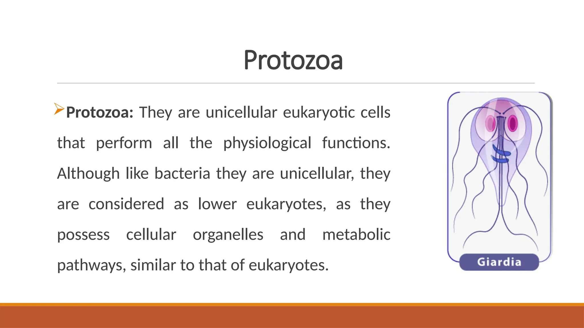
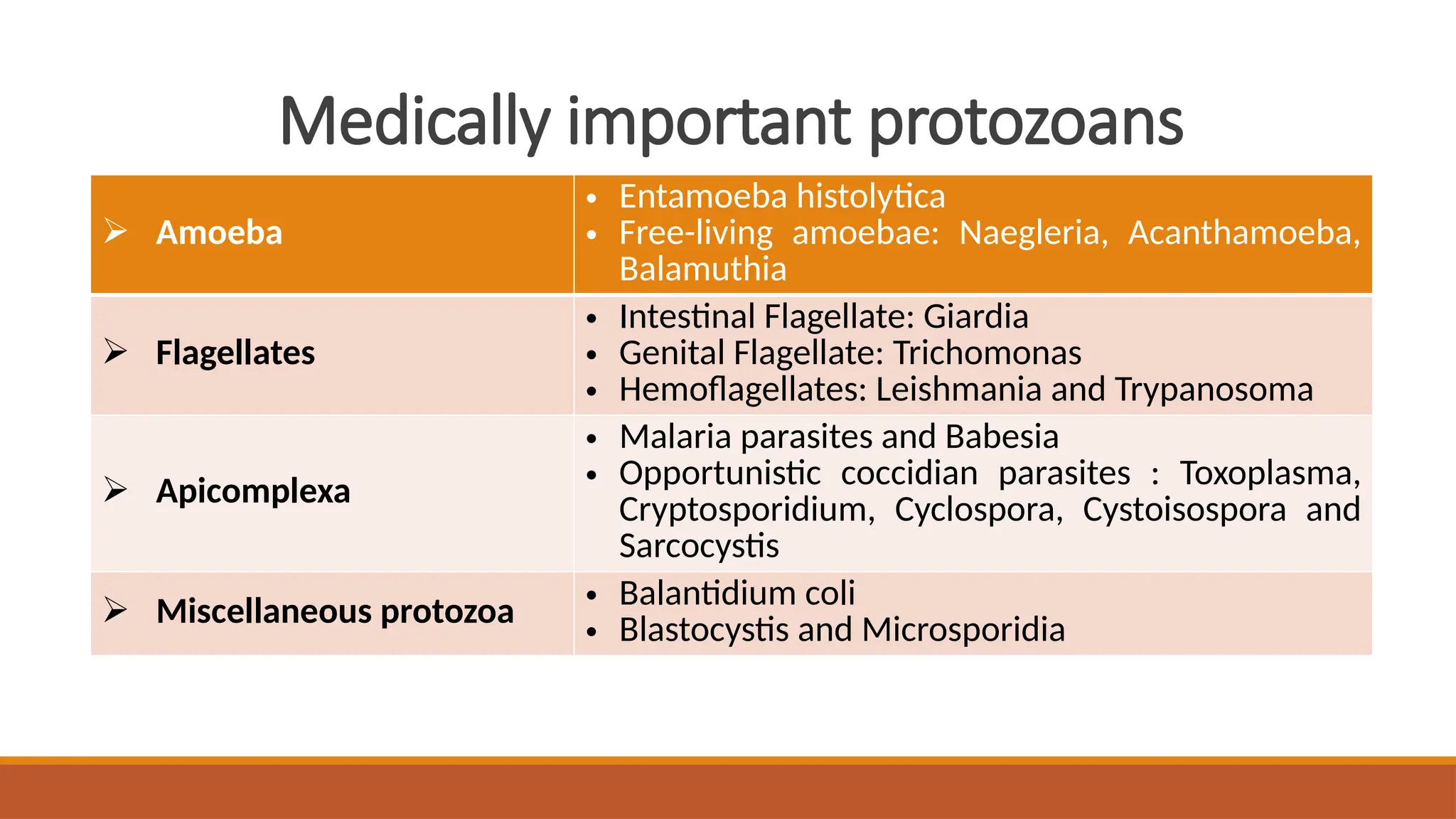
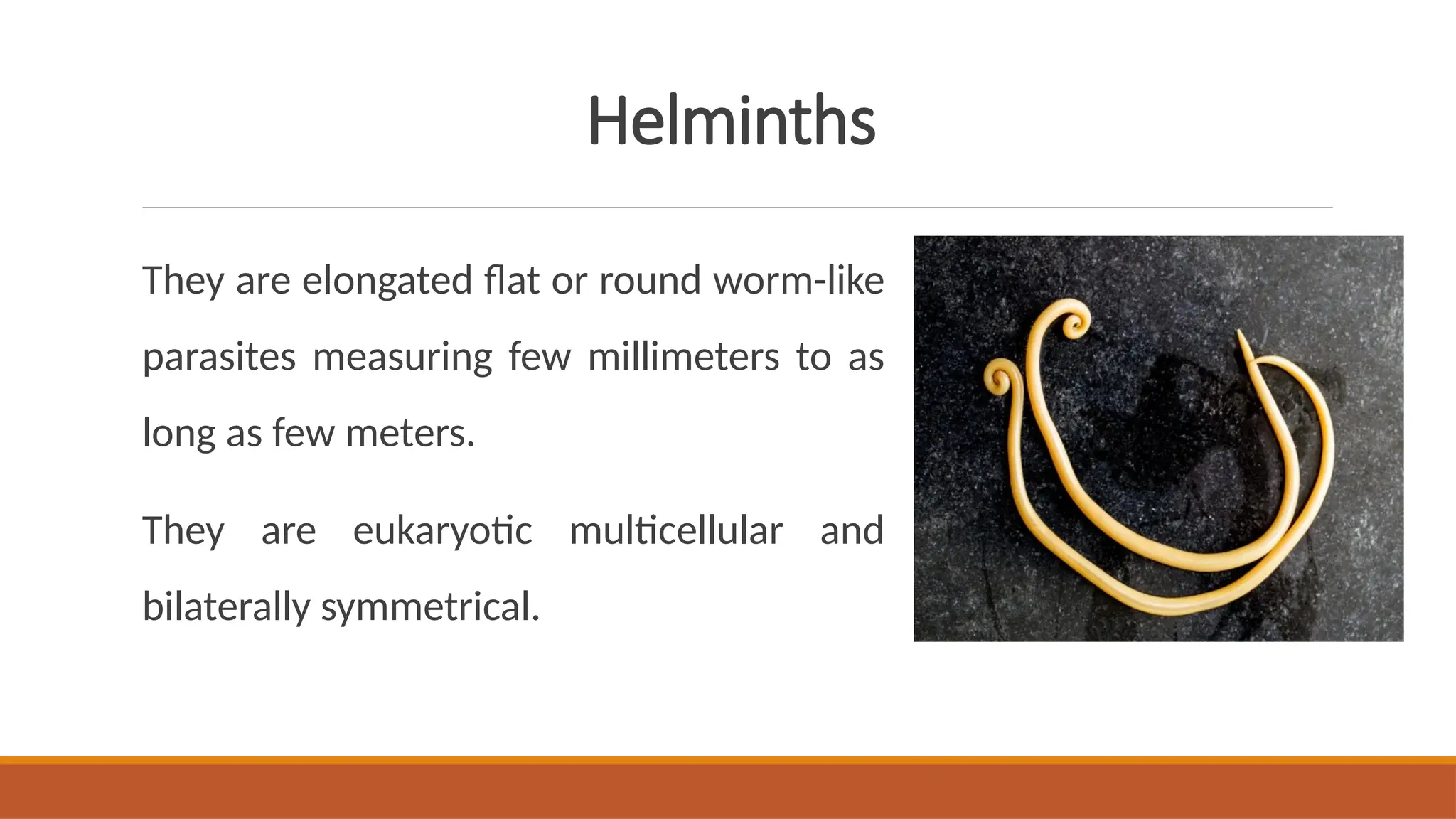
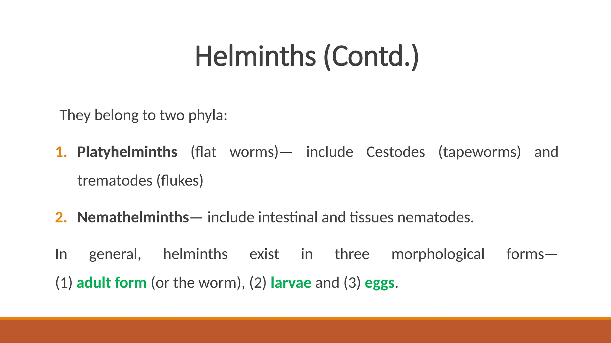
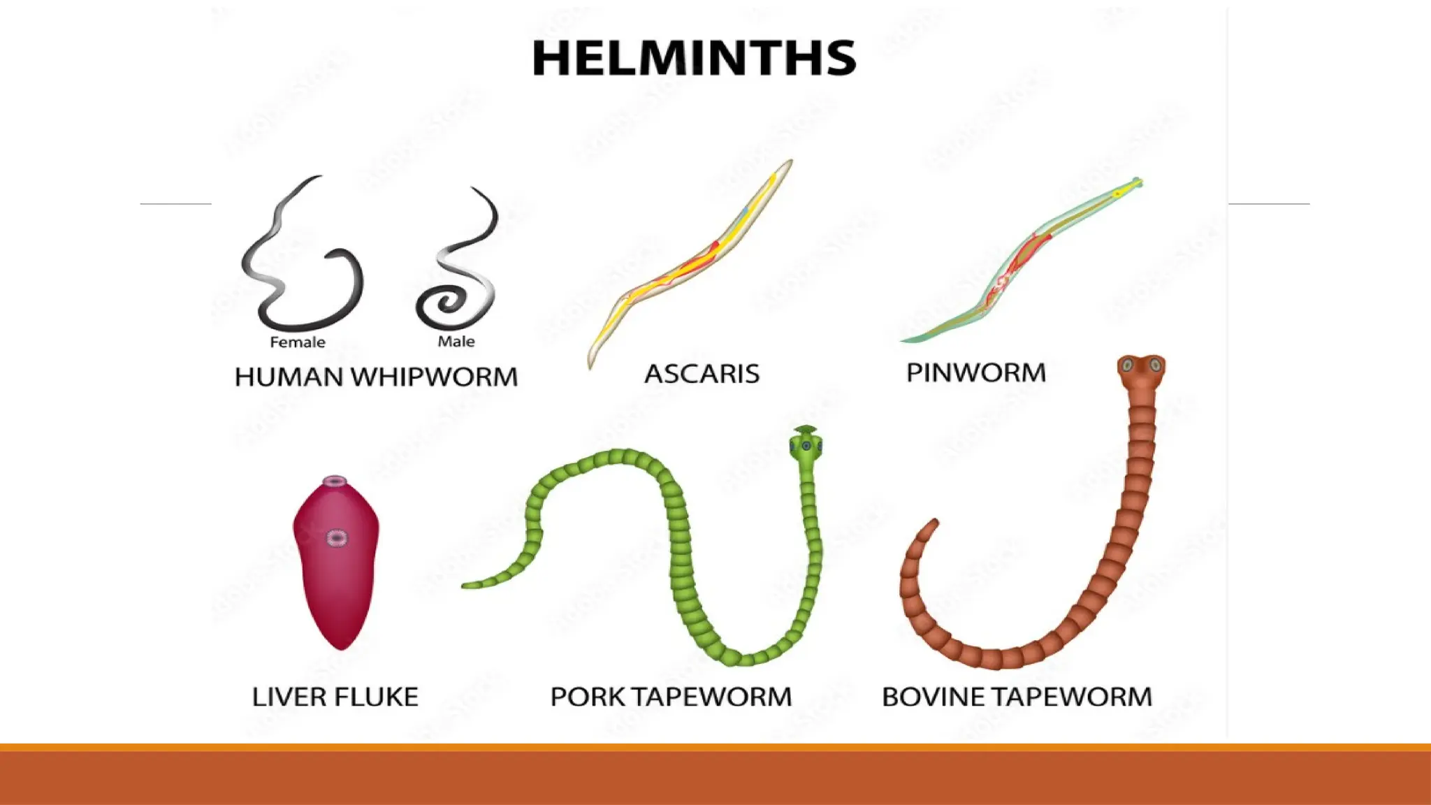


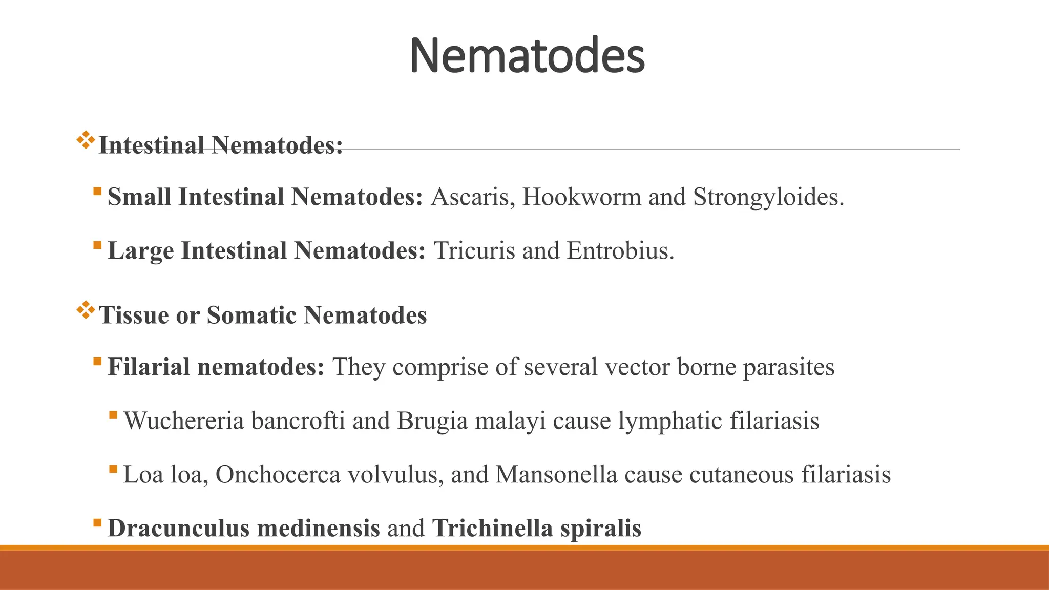
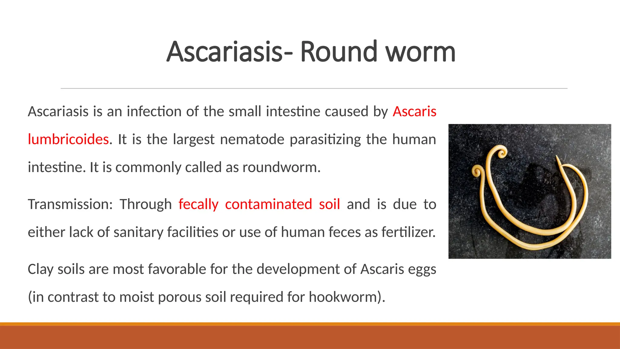

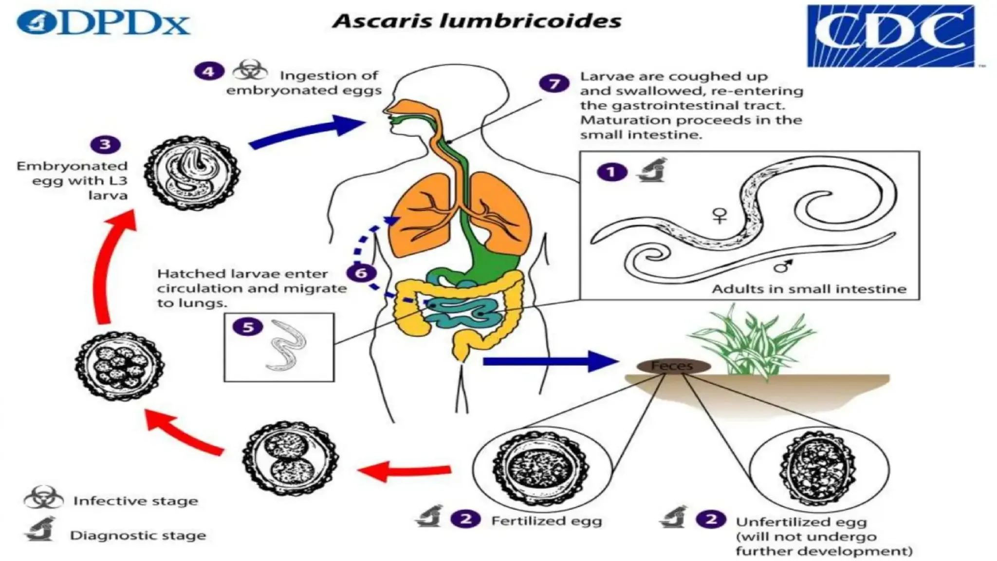


The document provides a comprehensive overview of parasites and parasitism, focusing on protozoans and helminths in medical parasitology. It categorizes parasites into ectoparasites and endoparasites, detailing significant types like protozoa and helminths, their morphological forms, and various medically important species. It additionally covers specific infections caused by helminths such as ascariasis, hookworm, and cystic echinococcosis, including their transmission, clinical features, and life cycles.


