
Recommended
More Related Content
Similar to u,d,ds,.s,d.s,.d,çwleç,,d,nit 5. Tapeworms.ppt
Similar to u,d,ds,.s,d.s,.d,çwleç,,d,nit 5. Tapeworms.ppt (20)
More from Tatiane Fernandes
More from Tatiane Fernandes (15)
Recently uploaded
Recently uploaded (20)
u,d,ds,.s,d.s,.d,çwleç,,d,nit 5. Tapeworms.ppt
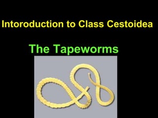
- 1. Intoroduction to Class Cestoidea The Tapeworms
- 2. Otline • General feature of cestodes • General morphology of cestodes – Adult , Larva and Ova • Clarification of cestode • Major differences between order Cyclophyllidea and Pseudophyllidea • Geographical distribution Morphology, differential characteristics, life Cycles of each species • Laboratory diagnosis, control and prevention methods for each species
- 3. Learning objectives • At the end of ths unit the student will be able to: • Explain the general feature of cestode • Explain the general morphology of Adult larvae and ova • Describe the classification of cestode
- 4. Objective… • List the major differences between order Cyclophyllidea and Pseudophyllidea • Explain the geographical distribution Morphology, differential characteristics, life Cycles of each species • Apply the necessary laboratory procedure for detection and identification of cestode parasite
- 5. General characteristics: Adult • Segmented long tape-like worms • Vary from few mm to several meters • No mouth/digestive system. • Obtains its nutrient by absorption through body surface.
- 6. Body is divided into three main regions a. Scolex (head) b. Neck c. Strobila made up of proglottids Morphology
- 7. Scolex – “head” of the organism – Has holdfast organs to keep the tapeworm in place – Three main types of Scolex
- 8. • Three main types of Scolex Cyclophyllidae A-Globular head with 4 Muscular Sucker B- 4 Sucker +Rostellum armed with hooks Pseudohyllidea Bothria-Shallow grooves or pits
- 9. • Area where new segments are created • Give rise to proglottids in strobila. Neck
- 10. Proglottids/segment • Proglottid – Set of reproductive organs – Includes male and female organs and genital pore • Segment – Segments may have one or more proglottids – Body divisisions – More mature as gets farther from neck • Size and shape of segment along with number of proglottids and location of genital pore key to identification many of the tapeworms.
- 11. RED = Male BLUE = Female
- 12. Reproduction • Sex:- Hermaphrodites • Have well developed reproductive system. • Reproduction –Sexual-Oviporous –Asexual-Sometimes multiplication with in larval forms
- 13. Strobila • Is the entire chain of proglottids
- 14. Strobila is divided into three regions: • a/ Immature segment: – near neck, sex organs are immature. • b/ mature segment: – large segment, sex organs are fully mature. • c/ gravid segment: – found at the tail end, uterus is filled with eggs
- 15. Egg: - • Two type – Operculated, immature when voided to the external environment. – Non-operculated ,fully embryonated when voided to the external environment.
- 17. Non-operculated
- 18. Larvae: • Generally two types 1. Solid : – eg. Procercoid, Plerocercoid, cysticercoid 2. Cystic ( true bladder) can be with: – Single scolex eg. Cysticercus; – Many scolexes and/or with daughter cyst eg. hydatid cyst, coenurus cyst, etc
- 20. Classification 1. Order Cyclophyllidea 1. T. saginata, 2. T. solium, 3. Hymenelopis sps 4. Echinococcus granulosus 2. Order Pseudophyllidea 1. Diphyllobothrium latum • Less medically important: – Order Cyclophyllidea • T. multiceps • Dipylidium caninum • Echinococcus multicuralis. – Order Pseudophyllidea • Spirometra species
- 21. Life cycle • Complete in two host (exception H.nana) • Haibtat:- Adult live in small intestine • Man is:- • The only/main DH for T. saginata, T. solium, H. nana and D. latum • IH for E. granulosus and E. multilcolaris • DH & IH for H. nana and T. solium
- 22. Egg Coracidium Oncosphere Cysticercus Cysticercoide, coenuruses, Hydatid Cysts Intermediate Host Adult Definitive Host Cystic larval form Solid larval form procercoid larva Plerocercoid larva Operculated, Non-operculated 1-Pseudohyllidea 2-Cyclophyllidea
- 23. Order- Pseudophillidea Order –Cyclophillidea 1. Scolex Spoonshaped, grooves - globular with 4 suckers 2. Genital pore - venteral - marginal 3. Utrine pore -Present(ventral) - absent 4. Uterus - coiled - sacular tubular or branched 5. Ova - operculated - non-operculated 6. Onchosphere - ciliated - non-ciliated 7. Rostellum - absent - present 8. Progilottids - broader than long - longer than broader 9. Larval forms - solid - cystic
- 24. • Geographical Distribution:- – T. saginata • World wide distribution where cattle are raised and beef is eaten raw or under cooked. • Very common in Ethiopia – T. solium • Not widely distributed as T saginata. • Common in all areas where raw or partially cooked pork is eaten. • Common throughout Mexico, South America and southern Africa & southern Europe. • Not reported from Ethiopia Taenia species
- 25. Morphology: • Adult: – Size: 4-10 m long (can reach up to 20 m) – Colour: ivory white – Strobila : 1000-2000 proglottides – Mature segment: 1-2cm long T. saginata • Adult – Size: 2-3m – Colour: pale blue – Strobila: 800-1000 Proglottides – Mature segment :0.5-1.5 cm T. solium
- 26. Taenia saginata Scolex (head): • Quadrate, with four suckers, no hooks, no rostellum on scolex • Size-2mm across
- 27. Taenia solium Scolex has • Four sucker • two rows of hooks on a prominent rostellum • Size-1mm
- 28. Taenia sp. • Larval stage is cysticercus – Invaginated scolex in fluid filled body – Cysticercus bovis-T saginata – Cysticercus celluless-T solium • Eggs are very round with very thick walls.
- 29. Transmission and life cycle • Transmission – Humans become infected by ingesting raw or undercooked meat infected with cystcerus larvae: • Beef- T saginata • pork meat –T.solium – T. solium can also be transmitted by : • Ingesting ova in food or water • Internal autoinfections
- 31. Taenia solium • Distinct difference with T. saginata is that humans can be infected with egg stage and onocosphere migrates to some site in body and develops into cycticercus • This can be serious, called Cysticercosis
- 33. Clinical manifestation • T saginata –Taeniasis. • Usually asymptomatic but may cause dizziness, abdominal pain, diarrhea, headache and nausea. –Proglottids obvious in feces. – Proglottides have a strong tendency to crawl from the anus during the day when its host is active
- 34. • T. solium –Taeniasis • Major symptoms of taeniasis are as a result of the adult worm. • These include abdominal pain, loss of appetite, and –Cysticercosis • T. solium ( when infected by eggs) cause cysticercosis (larval cysts in lung, liver, eye and brain) resulting in blindness and neurological disorders.
- 35. Laboratory Diagnosis • Detecting eggs in faeces . • Identifying macroscopically – gravid segments in faeces – scolex recovered from clothing or passed in faeces. • In addition • T.saginata- – ova on perianal skin (cellotape slide) • T.solium ( cysticercosis) – Finding calcified larvae in histological or X-rays examination .
- 36. Egg : T.solium &T.saginata • Size: - 33-40 m • Shape: -Round • Colour: - Shell-dark yellowish-brown, • Content: light yellowish gray. • Shell:-Thick, Smooth, brown, radially straited (embryophore) • Content: - A round granular mass enclosed by a fine membrane with six hooklets
- 37. • T. saginata ova stains red (acid fast) in Ziehl- Neelsen stain • this character helps to differentiate it from T.solium which do not have red color in such staining ( not acid fast) Morphologically eggs of T.saginata and T. solium are indistinguishable unless stained by AFB
- 38. Taenia saginata • Gravid proglottide • Detach when fully develop and pass through the anus independently. • Color- white and opaque • Size- 20mmX6mm • Uterus- > 13 main (15- 30) lateral uterine branches.
- 39. Taenia solium • Gravid proglottides- • Grey-blue and transluscent • Size-13mmX8m • 7 to 12, on average 10 lateral compound uterine branches. • Small chains of 3-4 rectangular segments found in the faeces
- 40. • Hymenolepis nana- Dwarf Tape Worm
- 41. Hymenolepis nana • Dwarf Tapeworm – Vampirolepis nana • Definitive Host: Humans, rodents – Most common tapeworm of humans in the world – 1% rate of infection in the southern U.S. – 97.3% rate of infection in Moscow, Russia • Intermediate Host: Larval and adult beetles (but optional) – Larval stage, cysticercoid, can develop in D.H. if it eats the eggs • Probably a recent evolutionary event
- 42. • Small tapeworm • Scolex has rostellum with row of hooks • Proglottids are wider than long with lateral genital pore
- 43. • Geographical Distribution:- – H.nana is widely distributed in countries with warm climates than in cold climates and fairly common in Ethiopia. – Children are more commonly infected than adults.
- 44. • Mode of Transmisssion: - – Ingestion of egg with contaminated food, drink or finger. – Autoinfection. • Life Cycle: H. nana has a direct life cycle with a human host serving as both definitive and intermediate host.
- 46. Hymenolepis dimunata • Slight larger than H. nana but still very small. • Scolex has very small rostellum with no hooks • Proglottid same as H. nana but larger.
- 48. • Egg: H nana – Size: 35-50m – Shape: oval, almost round – Shell: double; thin external membrane and internal membrane often thicker at the poles. Thread like polar filaments coming from both poles – Colour: colour less or very pale gray – Content: Rounded mass (embryo) with six refractile hooklets arranged in fan shaped.
- 49. Egg: H dimunata • Color:-Yellow-brown or bile pigmented. • Size:-70 by 60μm • Shell with double shell and with out thread like polar filaments. • Content: A rounded embryo containing six hooklets arranged in fan shape.
- 50. Echinococcus granulosus • Sheep Tapeworm • Definitive Host: Carnivores including dogs, wolves, and coyotes • Intermediate Host: Herbivores including sheep and mice. • Occasionally infect humans. • The hyatid cysts growin humans very slowly and can overcrowd organs • Geographic Distribution: Most common in sheep raising countries – New Zealand and Australia highest incidence
- 51. MORPHOLOGY • Smallest of all tapeworms – Scolex, neck, 3 segments – Segments look like Taenia sp. • Largest larval stage of all tapeworms – Hydatid cyst
- 53. Clinical feature and Pathology: • The symptoms, depend upon the location of the cyst. • Large abdominal cysts produce increasing discomfort. • Liver cysts cause obstructive jaundice. • Peribronchial cysts may produce pulmonary abscesses. • Brain cysts produce intracranial pressure and Jacksonian epilepsy. • Kidney cysts cause renal dysfunction. • The contents of a cyst may produce anaphylactic responses
- 55. Laboratory Diagnosis • Histological examination to find larvae • X-ray examination to find larvae • Examination of cystic fluid for brood capsules and protoscoleces • Casoni's skin test
- 56. Dipylidium caninum Dog Tapeworm • World wide Distribution • Dogs or cats (humans rarely) as the definitive host • Fleas or lice are the intermediate host. • Habitat: • Adult: mucus membrane of small intestine of carnivores such as dog, cat, Man • Cysticercoid larvae: In the body cavity of insects • Egg: in the faeces of dog, cat, man
- 58. Dipylidium caninum • Flea or louse ingests the eggs in the perianal region of the dog or cat. • The dog or cat (or human) is infected when they ingest a flea or louse infected with the metacestode state (cysticercoid) Dog flea
- 59. Dipylidium caninum Proglottids of Dipylidium caninum compared to a match stick. These are often passed intact in the feces of an infected dog. When the proglottids dry, their appearance is similar to grains of rice.
- 60. Dipylidium caninum • Egg: 5-15 eggs in capsule • 40m in size and yellowish brown in color
- 62. Diphyllobothrium latum Common name: Fish tapeworm • Geographical Distribution:- – Widely distributed in the lake areas of Europe, Asia, Far East, North America, South America and Central Africa .
- 63. Diphyllobothrium latum Fish tapeworm • Important parasite of man. • Definitive hosts can be humans, dogs, foxes, cats, mink, bears, and seals. • Site of attachment : small intestine.
- 64. Diphyllobothrium latum Fish tapeworm • humans are infected with the plerocercoid . • In humans the tapeworm can reach a length of 10 meters (>30 feet) and produce over a million eggs a day! .
- 65. Diphyllobothrium latum • Scolex has bothria – Shallow groove • Segments have one proglottid – Wider than long egg- 58-76µm by 40-51µm Broadly ovoid Light golden yellow, Operculated Thick shell Contains immature
- 66. Morphology Adult: the largest tapeworm Size: 10m or more Grayish-yellow in colour Scolex is elongated, spoon shaped, longitudinal suctorial mangroove/bothria/slits with no rostellum and hooklets. Long and slender neck
- 68. • Clinical feature and Pathology : – Clinical symptoms may be mild, depending on the number of worms. – They include abdominal discomfort, loss of weight, loss of appetite and some malnutrition. – Anemia and neurological problems associated with vitamin B12 deficiency are seen in heavily infected individuals. • Laboratory Diagnosis – Eggs in the faeces – Scolex in the faeces – Adult worms in the faeces
- 69. Egg: • 58-76m by 40-51m • Broadly ovoid • Light golden yellow, Operculated • Thick shell • Contains immature embryo