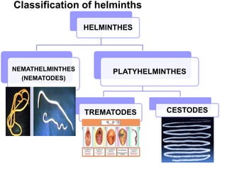
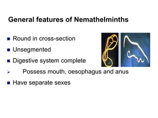
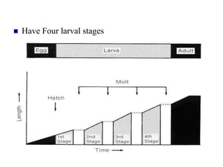
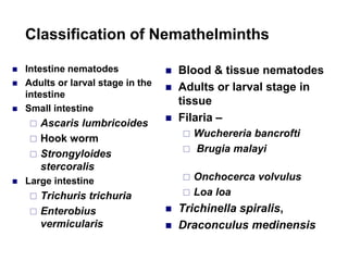
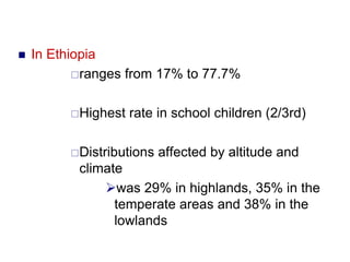
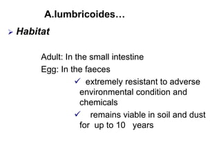
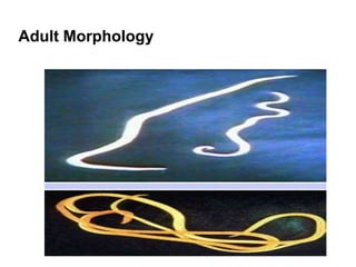
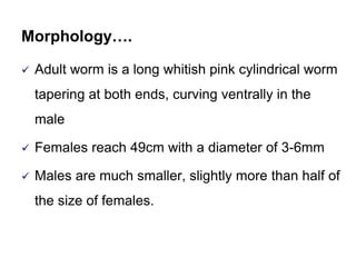
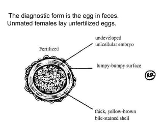
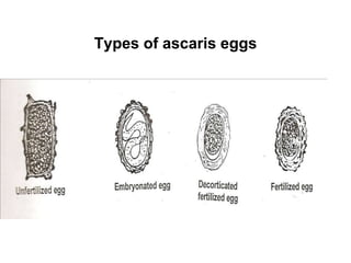
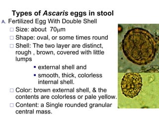
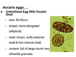
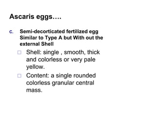
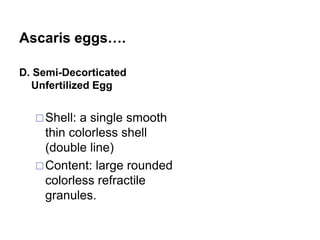
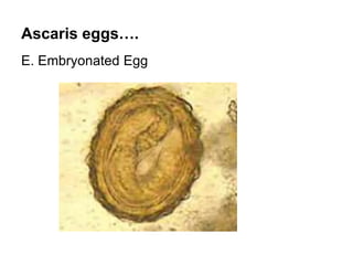
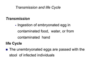
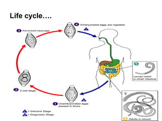
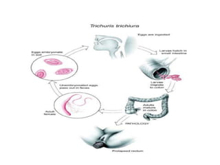
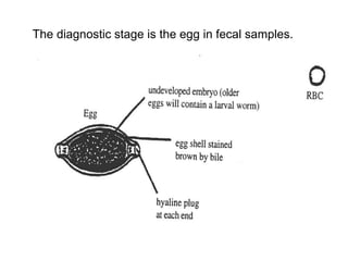
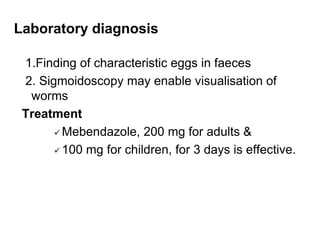
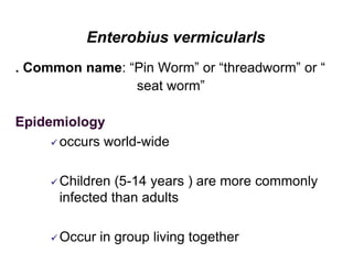
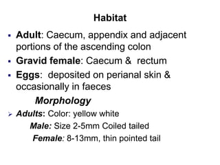
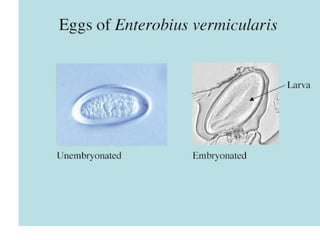
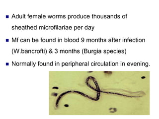
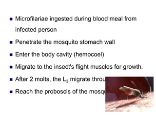
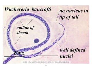
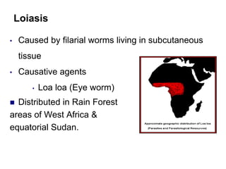
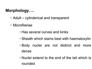
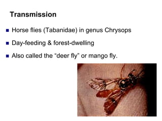
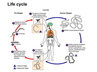
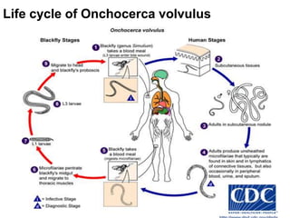
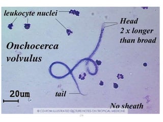
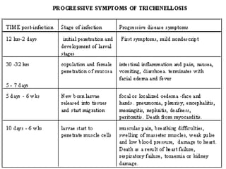
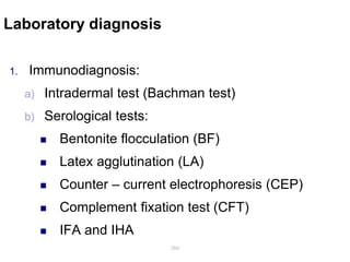
SlideShare a Scribd company logo
UNIT – THREE
HELMINTHS
Learning objective
At the end of this unit the students will be able to:
 Define helminths
 Describe the general features of helminths
 Describe the taxonomic classification of
helminths
Outline
 Introduction to helminths
 General features of helminths
 classification of helminths
3.1. Introduction to Helminths
Medical helminthology: study of these parasitic worms and
their medical consequence
Helminths derived from the Greek word “helminths” or
“helminthose” meaning worm
Int…
 Either free living or parasitic organisms belonging to
phylum:
Nemathelminthes(round worm)
Platyhelminthes(flat worm ),
Aacanthocephala (spinyheaded worms )or
Aannelida (segmented worm )
3.2.General features of helminths
Higher, multicellular forms with specialized organs
 Adult worms vary in size (6mm->10m)
 Their life cycles may be simple or complex
 Pathology, clinical sign and symptoms:
 Depend on the location of the organisms
 May be caused by adults, larva, or egg
Laboratory Diagnosis
 Laboratory diagnosis mainly depends on detection and
identification egg , larva or embryo and rarely adults
HELMINTHES
NEMATHELMINTHES
(NEMATODES)
PLATYHELMINTHES
TREMATODES CESTODES
Classification of helminths
General features of Nemathelminths
 Round in cross-section
 Unsegmented
 Digestive system complete
 Possess mouth, oesophagus and anus
 Have separate sexes
Features…
 Can be oviparous/ovoviviporous/viviparous
Egg (ova) -Larva(L1-L4)-Adult
Possess a shiny cuticle (smooth/spined/ridged)
 Mouth is surrounded by lips or papillae
 Have Four larval stages
Burden and impact on human life
 ≈ 500,000 spp. globally
• Most are free living
 Abundant pathogens in life-stock and pets
 Important pests of many crops
 Cause numerous human diseases
•The warm regions of the world = worm regions.
•The burden of disease is not evenly spread within
developing countries
•The majority of worms are found in the poorest sections of
the community, compounding poverty, and social
deprivation.
•High burden
•In the rural villages
•unsanitary overcrowded cities
•'big three' (Ascaris, Trichuris & Hookworm) is common
•Temperate and cold climates are not spared.
Classification of Nemathelminths
 Intestine nematodes
 Adults or larval stage in the
intestine
 Small intestine
 Ascaris lumbricoides
 Hook worm
 Strongyloides
stercoralis
 Large intestine
 Trichuris trichuria
 Enterobius
vermicularis
 Blood & tissue nematodes
 Adults or larval stage in
tissue
 Filaria –
 Wuchereria bancrofti
 Brugia malayi
 Onchocerca volvulus
 Loa loa
 Trichinella spiralis,
 Draconculus medinensis
Animal nematode of less medically important
or low occurrence
INTESTINAL NEMATODEs
 Toxocara catti &
Toxocara cani
 A. canninum & A.
braziliens
 Cappilaria species
 S. fulliborni
 Trichostrongylus
species
BLOOD & TISSUE
NEMATODEs
 Mansonella ozardi,
 M. peristance,
 M. stereptocerca
INTESTINAL NEMATODES
General features
 Humans are the only or major host of intestinal
nematodes
 Live in gastro-intestinal tract
 Often spread by poor hygiene related to feces
 Most species are geo-helminthes (soil transmitted)
 Female worms are oviparous
Pathogenesis
 Major source of chronic ill health
 Compromising the growth potential, and
intellectual achievements of children all over the
world
Life cycle
 Intestinal nematodes share similar life cycles
that have evolved in response to new ecological
niches
 The core nematode life cycle involves
development from an egg through 5-stages of
growth.
 The 1st 4-stages(L1, L2, L3, L4) are larval
 The 5th & final stage of development is a
sexually mature adult worm.
 At each stage the cuticle of the parasite moults.
Transmission
 Usually only two sites of entry for intestinal
nematodes infecting humans:
 Ingestion of infective egg
 Larva penetrating skin
Laboratory diagnosis:
 Eggs ( most often) and Larvae in faeces
 Recovering egg in the skin around the anus
(perianal area)
Occasional adult worms: A. lumricoudes, E.
vermicularies
Cont…
 Intestinal nematodes infecting humans includes
 Ascaris lumbricoides
 Trichuris trichiura
 Enterobius vermicularis
 Strongyloides stercoralis
 Ancylostoma duodenale
 Nectator amircanus
Ascaris lumbricoides
 Also known as large intestinal round worm
 Is a member of the family Ascoridoidea
 Possess a mouth that has three conspicuous
lips
 Are pathogenic in their adult stage
Epidemiology
 world wide
1.45 billion people are infected annually
WHO estimated it resulted in 60,000 persons death
in 1995
 In Ethiopia
ranges from 17% to 77.7%
Highest rate in school children (2/3rd)
Distributions affected by altitude and
climate
was 29% in highlands, 35% in the
temperate areas and 38% in the
lowlands
A.lumbricoides…
 Habitat
Adult: In the small intestine
Egg: In the faeces
 extremely resistant to adverse
environmental condition and
chemicals
 remains viable in soil and dust
for up to 10 years
Adult Morphology
Morphology….
 Adult worm is a long whitish pink cylindrical worm
tapering at both ends, curving ventrally in the
male
 Females reach 49cm with a diameter of 3-6mm
 Males are much smaller, slightly more than half of
the size of females.
Morphology….
Males:
 have one or two copulatory spicules, but no
bursa copulatrix, Usually no caudal alae
 Female worms produce up to 240, 000eggs/day,
which corresponds to just under 3000eggs/gram
of faeces.
Egg
 Ascaris eggs vary widely in size and appear in
two forms:
I. Fertilized:
golden brown, ovoid and mammilated
about 30-40µm wide & 50-60µm long with a
dense outer irregular shell & a more
translucent regular inner shell.
The thick external mammilated layer is often
lost, giving a decorticate appearance -
Eggs….
 Evidence of segmentation or embryonation is
often seen.
II. Unfertilized: is larger & more elongated with a
length of about 89-95µm and 40-50µm width
 The internal structure of the egg is poorly
differentiated
Transmission and Life Cycle
 Transmission
A. lumbricoides is spread by faecal pollution of
soil
Is favored by conditions that improve the survival
of eggs in the soil, in particular warm moist shady,
conditions
Infective stage: embryonated egg (egg containing
2nd stage larva)
Transmission…
A person acquires infection by
1- Ingestion of food or water contaminated with
embryonated eggs
2-eating soil(geophge) frequently seen in children
3-putting contaminated finger or toys with infective
egg in to mouth
4- rarely by inhalation of eggs carried in air
Transmission….
 The infection is common in areas with
 high density of human population
Poor sanitation
Habit of people to defecate
indiscriminately in and around settlements
Use of infected faeces as fertilizer
Life cycle
 Fully embryonated eggs are swallowed & L2 larvae
hatches in the stomach & penetrate stomach or
duodenal mucosa
 L2 enter blood stream & leave through alveoli into
lung
 Then molt several times in the lungs to L3/L4
 Then move up and get swallowed
Life cycle….
 2-3 months after infection the adult worms start
laying eggs (200,000 daily)
 Eggs are shed with the feces and embryonate
within 2-3 weeks
A.Lumbricoide life cycle
Pathogenesis
1. “Verminous” pneumonia, lung tissue damage
due to migratory larvae.
2. Bowel obstruction - too many adult worms.
3. Parasite secretes trypsin inhibitor, prevents host
from digesting proteins.
4. Aberrant migration of “irritated” adult worms to;
common duct, liver, Pharynx, peritoneum
Pathogenesis…
 With heavier worm
loads a tangled mass
of worms can obstruct
the bowel, or an
individual worm can
block a duct
Laboratory Diagnosis
A. Finding and identification of eggs in the stool.
Direct wet mount
 adequate for detecting moderate to heavy
infections
concentration technique may be used In light
infection, Sodium chloride floatation technique
& Formol-ether concentration technique
B. Adult worms occasionally passed in the stool or
through the mouth or nose
Lab diagnosis….
C. Larvae can be identified in sputum or gastric
aspirate during the pulmonary migration phase
*Examine formalin-fixed organisms for
morphology
The diagnostic form is the egg in feces.
Unmated females lay unfertilized eggs.
Types of ascaris eggs
Types of Ascaris eggs in stool
A. Fertilized Egg With Double Shell
 Size: about 70m
 Shape: oval, or some times round
 Shell: The two layer are distinct,
rough , brown, covered with little
lumps
 external shell and
 smooth, thick, colorless
internal shell.
 Color: brown external shell, & the
contents are colorless or pale yellow.
 Content: a Single rounded granular
central mass.
Ascaris eggs….
B. Unfertilized Egg With Double
Shell
 size: 80-90m
 shape; more elongated
(elliptical)
 shell: brown, puffy external
shell & thin internal shell.
 content: full of large round very
refractile granules
C. Semi-decorticated fertilized egg
Similar to Type A but With out the
external Shell
 Shell: single , smooth, thick
and colorless or very pale
yellow.
 Content: a single rounded
colorless granular central
mass.
Ascaris eggs….
C. Semi-decorticated fertilized egg
Similar to Type A but With out the
external Shell
 Shell: single , smooth, thick
and colorless or very pale
yellow.
 Content: a single rounded
colorless granular central
mass.
Ascaris eggs….
Ascaris eggs….
D. Semi-Decorticated
Unfertilized Egg
Shell: a single smooth
thin colorless shell
(double line)
Content: large rounded
colorless refractile
granules.
Ascaris eggs….
E. Embryonated Egg
Treatment
 Mebendazole, 200 mg, for adults and
 100 mg for children, for 3 days is effective.
Prevention and control
1.Prevention of infection by
 washing hands before eating & trimming
finger nails
 Avoid eating uncooked foods such as
vegetables
Prevention…
2. Preventing soil become faecally polluted by
 sanitary disposal of faeces in latrines
 avoiding the use of night soil as a
fertilizer
3.Treatment and health education
• Mass de-worming programmes repeated at 3-6
month intervals, have been advocated in areas of
high prevalence
Trichuris trichiura
 Common name : whipworm, due to the whip-like
form of the body.
Epidemiology
The third most common round worm of humans
worldwide
Infections more frequent in areas with tropical
weather & poor sanitation practices, and among
children
~ 112 billion cases world-wide
 ~ 1.05 billion people are infected annually
 In Ethiopia
One national survey showed 36.1%
On study in central and northern plateaus:
mean prevalence of 49%
Habitat
 Adult: large intestine (caecum) & appendix
 Eggs: In the faeces, not infective
when passed
Morphology
Adults: whip-like shape, anterior 3/5th of the worm
resembles a whip & the posterior 2/5th
are thick
Male : Size 30-45 mm , coiled tail
Female: 35-50mm, straight thick tail
Morphology….
Eggs
Size: 50-54m
Shape: "tea tray eggs” or
barrel- shaped with a
colorless protruding mucoid
plug at each end
Shell: fairly thick and smooth,
with two layers & bile stained
Color: yellow brown
Content: a central granular
mass which is Unsegmented
ovum
Transmission and life Cycle
Transmission
 Ingestion of embryonated egg in
contaminated food, water, or from
contaminated hand
life Cycle
 The unembryonated eggs are passed with the
stool of infected individuals
Life cycle….
 Mature within three weeks of being deposited in
soil.
require a warm, moist environment with
plenty of oxygen to ensure embryonation
The embryonated eggs are extremely
resistant to environmental conditions
 When embryonated eggs are swallowed larvae are
released into the upper duodenum
 then attach themselves to the villi of small intestine
or invade the intestinal walls
Life cycle…..
 After 3-10 days they move down to the caecum &
ascending colon where they mature into adult
worms
 The adult worms are fixed with the anterior
portions threaded into the mucosa
 The females begin oviposit 60 to 70 days
after infection & shed 3,000 - 20,000 eggs/day
 The life span of the adults is about 1 year
Life cycle….
Clinical features
 Are largely determined by the worm burden:
 <10 worms are asymptomatic (99%
asymptomatic)
 Heavy worm burden mechanical damage to
the intestinal mucosa
 Chronic profuse mucoid & bloody diarrhea
with abdominal pains &
 edematous prolapsed rectum
Clinical features….
Anaemia from blood loss and iron deficiency,
malnutrition, weight loss and sometimes death
Each adult worm sucks about 0.005 ml of blood
per day
Rarely a child will develop congestive cardiac
failure because of anaemia, fluid retention
hypoproteinemia & oedema
The diagnostic stage is the egg in fecal samples.
Laboratory diagnosis
1.Finding of characteristic eggs in faeces
2. Sigmoidoscopy may enable visualisation of
worms
Treatment
 Mebendazole, 200 mg for adults &
 100 mg for children, for 3 days is effective.
Prevention and control
 Sanitary disposal of faeces in latrine
 Avoidance of the use of night soil as a fertilizer
 Treatment of infected individuals and health
education.
Enterobius vermicularls
. Common name: “Pin Worm” or “threadworm” or “
seat worm”
Epidemiology
 occurs world-wide
 Children (5-14 years ) are more commonly
infected than adults
 Occur in group living together
Epidemiology…
 Pinworms eggs can
spread throughout a
house and difficult to
eliminate.
 Small children are
most apt to pick them
up during the
“teething stage.”
Epidemiology….
In Ethiopia :
 5 % school children in rural communities in Gondar
region had E. Vermicularis eggs under their finger
nails and that only 0.5% of them were found to
shed eggs in the stool
 in routine stool examination method, a prevalence
rate up to 1% were reported
Habitat
 Adult: Caecum, appendix and adjacent
portions of the ascending colon
 Gravid female: Caecum & rectum
 Eggs: deposited on perianal skin &
occasionally in faeces
Morphology
 Adults: Color: yellow white
Male: Size 2-5mm Coiled tailed
Female: 8-13mm, thin pointed tail
They are small white worms with
pointed tail
swollen cuticle at anterior end
prominent esophageal end bulb
Morphology…
 Egg
Size: 50-60m
Shape: oval but flattened
on one side, rounded
on the other side
Shell : Smooth and thin
but with double shell
Content: either a small
granular mass or a
small curved up larvae
Transmission and Life cycle
Transmission
 Person –to- person transmission: occur through
handling & sharing of contaminated clothes or
bed linens
 through surfaces in the environment that are
contaminated with pinworm eggs
 Self (Autoinfection) - occurs by transferring
infective eggs to the mouth with hands that have
scratched the perianal area
Transmission …
 Children who suck their fingers are more likely to be
infected
 Exposure to viable eggs on soil (Ingestion)
 Air borne: Some small number of eggs may become
airborne and inhaled
Eggs remain viable 20 days
 Autoinfection
 Retro infection may occur after hatching on the anal
mucosa
Life cycle
 Ingestion of embryonated eggs, usually carried on
fingernails, clothing, bedding or house-dust.
 Eggs hatch in stomach, larvae migrate to caecal
region where they mature into adults
 Copulation takes place in the caecum
 Gravid females migrate nocturnally outside the
anus and oviposit on the perianal area
 1 pin worm lay over 10,000 -15,000 eggs eggs /day
Life cycle…..
 With in 4-6 hours being laid the egg contain
infective larvae
 Perianal itching from the eggs Induces scratching,
& hence the eggs are transferred to the mouth via
fingers
 Retro-infection: the migration of newly hatched
larvae from the anal skin back into the rectum
 interval from ingestion of infective eggs to
oviposition by the adult females is about 1month
Life cycle…
Life cycle…
Pathogenesis
 Mild perianal itching and excoriation
 Appearance of eggs or worms in an ectopic
location
 Atopic worms may result in a granulomatus
response
 In the perianal area causing vaginitis and
postmenopousal bleeding
 Granulomas in the peritoneum
Clinical features
 Nocturnal anal pruritis: The cause of this is unknown,
but may be related to the intensity of the infestation,
and/or an allergic reaction to the parasite
 Sleeplessness, because of the irritation
 Vulvovaginitis, & even urethritis may occur in girls
when migrating worms lay their eggs in these sites
 Abdominal pain or appendicitis resulting from the
worms are considered to be very rare
Adult Pinworms on the perianal skin
Laboratory Diagnosis
1.Finding eggs from perianal skin using adhesive tape
or swab method
Done by pressing transparent adhesive tape
("Scotch test", cellulose-tape) on the perianal skin
and then examining the tape placed on a slide.
Alternatively, anal swabs or "Swube tubes" (a
paddle coated with adhesive material) can also be
used.
Collect sample in the morning, before defecation
and washing,
Lab diagnosis…
 The “Scotch Tape Test”
 place a piece of Scotch
Tape on the anal area.
 The tape is placed on a
slide and examined
under a microscope for
the flat sided eggs.
Lab diagnosis…
2. Finding eggs in the faeces
Less then 10% found in stools, i.e. not a useful
examination;
occasionally eggs can be found in the urine or
vaginal smears
Diagnosis…
3. Finding of female worms from perianal skin
or faeces
Adult worms are also diagnostic, when found in
the perianal area, or during ano-rectal or
vaginal examinations
Treatment
 Two doses (10 mg/kg; maximum of 1g) each of
Pyrental Pamoate two weeks apart give a very
high cure rate.
 Mebendazole is an alternative.
*The whole family should be treated, to avoid
reinfection
Prevention and control
1. Treating all members of a family in which
infection has occurred
2. Wearing tight-fitting cotton pants to infected
children
3. Washing of the anal skin each morning
4. Washing of clothing worn at night
5. washing hands after using toilet and before
eating
6. avoidance of putting fingers in the mouth &
trimming finger
Strongyloides stercoralis
 Common names: Dwarf thread worm
Epidemiology
 Found worldwide
 An estimated 50 to 100 million cases
 Favors warmer tropical and subtropical
climate
Epidemiology…
Epidemiology….
 In Ethiopia
 not highly prevalent in most areas and is found
in the same geographical areas with hookworm
 rates up to 44% reported from 41 of the 50
communities in central and northern Ethiopia
 infection is rare or absent in many arid lowland
areas, including the Ogaden and pastoral areas
in the Awash Valley
Characterstics
1. Parasitic males are absent
2. Parasitic females are present in the submocusa
of small intestine which produce eggs
parthenogenically
3. Can develop in to free living generation in the
soil out side the human host
4. Has internal autoinfection
Habitat
 Has both free living and parasitic generations
 Parasitic Adult females: buried in the mucosal
epithelium of the small intestine of man
 Free living male and female: on external env`t
 Rhabditiform larvae: Passed in the faeces &
external environments
 Filariform larvae infective stage: soil & water.
 Egg : laid in the sub mucosa of small intestine
Morphology
• Free-living females: 1 mm by 60 µm
• Parasitic females: 2.2 mm by 45 µm
• Eggs: 55 µm by 30 µm; as soon as they are laid
in submucosa, the rhabditiform larvae will hatched
Larvae
Rhabditiform larvae
(non-infective form)
Filariform larvae
(infective form)
Morphology…
 Rhabditiform Larvae
Size: 200-300m long
; 15m thick
Motility: very actively
motile in the stool
Tail: Moderately
tapered
Short buccal cavity
and rhabiditiform
esophagus
 Filariforml Larave
About 600-700m
Cylinderical
esophagus
Bifid tail end
Transmission and Life cycle
 Transmission
1. Commonly by penetration of skin by filariform
larva
2. Ingestion of food or water contaminated with
filariform larva( oral route)
3. Rarely: Transmamary & Organ transplantation
4. Autoinfection with rhabidit form larva
Life cycle
Complex , two types of cycles exist:
1.Free-living (indirect) cycle
 Rhabditiform larvae(stool): molt 4x free- living adult
males and females produce eggs rhabditiform larvae
develop to free living adult males or females
 Filariform larvae (this initiate parasitic life cycle)
Parasitic (direct) cycle
Rhabditiform larvae(stool) molt 2x develop
to filarifrom penetrate skin lung Alveolar
space bronchial tree pharynx
swallowed &develop to adult female in small
intestine (molt 2x) produce egg by
parthenogenesis which yield rhabditiform larvae
Life cycle….
 Autoinfection, the rhabditiform larvae become
infective filariform larvae in the host tissue
penetrate intestinal mucosa (internal
autoinfection) or
 perianal area (external autoinfection)
Parasitic life cycle
Free-living cycle
Clinical feature
 It is usually asymptomatic, in symptomatic cases
 People with weaker immune systems such as
elderly people & children are more susceptible.
1.Cutaneous phase
 large number of larva produce itching & erythema
at the site of infection within 24 hours of invasion
Clinical…
2.Pulmonary phase: The migratory larva in the
lung producing bronchopneumonia & full blown
pneumonitis
3. Intestinal phase : Invasion by adult worms may
produce abdominal pain & mucoid diarrhea ,
nausea, vomiting and anemia.
Clinical….
Auto- and hyper-infection syndromes
 characterized by massive larval invasion of the
lung or any other organ including CNS, which is
fatal
 occurs when the immune status of the host
suppressed by either drugs, malnutrition or the
concurrent diseases
Laboratory diagnosis
1. Finding the larvae in faeces or duodenal
aspirates using direct or concentration method
by microscopy
 In hyper-infection syndrome the larva may be
found in sputum, urine and other specimens
 Examination of serial samples may be
necessary because direct stool examination is
relatively insensitive
Diagnosis…
 The stool can be examined in wet mounts:
Directly
After concentration (formalin-ethyl acetate)
After recovery of the larvae by the Baermann
funnel and water emergence semi-concentration
technique
After culture by the Harada-mori filter paper
technique
After culture in agar plates
Diagnosis….
2. Serological tests
 Antibody Detection
 Indicated when the infection is suspected
and the organism cannot be demonstrated
by:
 duodenal aspiration, string tests, or
 repeated examinations of stool
 Use antigens derived from filariform larvae
for the highest sensitivity and specificity
Diagnosis….
 EIA currently recommended because of its
greater sensitivity (90%).
 IFA and IHA tests can be used
Treatment
Ivermectin or thiabendozole
Prevention and control
1. Sanitary disposal of faeces in latrine
2. Avoidance of use of night soil as a fertilizer
3. Wearing protective footwear
4. Treatment of infected individuals and Health
education
Strongyloides fuelleborni
Geographical Distribution
 Widely distributed in Zimbabwe, Zambia, Papua
New Guinea, co-exists with S.stercoralis in
Ethiopia
 It is a common parasite of old world monkeys ,
apes &dog
Transmission and Life cycle
Transmission
 Skin penetration by filariform larvae
 Transmammary
Habitat: Has both free living and parasitic life
Life cycle
 similar to S.stercoralis except it shed eggs in the
faeces
Pathology and treatment
 Similar to S.stercoralis
Laboratory diagnosis
 Finding eggs in fresh stool specimens
Egg: Resembles eggs of hookworms but are
shorter and smaller
Colorless, Oval and 50 by 35µm in size
Contain partially developed larvae
N.B. If there is a delay in examining the faces ,
the larva will hatch.
Prevention and control
The same as described for S. stercoralis
 Are hematophagous nematodes
 Two major species
Ancylomstoma duodenale
Necator americanus
 Less important : A. ceylanicum, A. braziliense
,A. caninum , A.tubaeforme, A. buckleyi
Hook Worms
Epidemiology
widely distributed throughout the tropics and
subtropics
 more than 1 billion people are infected world-
wide
cause daily blood loss of 7 million liters
Most commonly infected are children,
agricultural workers and miners
 In Ethiopia : Necator americanus is more
common than Ancylostoma duodenale
 highest infection rates: Ilubabor, Kefa ,Welega
 A.duodonale is associated with areas of poor
soil coverage and high rate of drainage
 N.americanus is found in red soil areas on flat
plain
Epidemiology….
Epidemiolgy…
 Altitude and moisture are the major factors
affecting their distribution
 Hook worm infection is absent in low ,hot
dry areas of Ethiopia and above 2500m
alttitude
 Adult: Jejunum and less often in the
duodenum of man
 Eggs: In the faeces; not infective to man
 Rhabditiform & filariform larvae: free in soil
and water
Habitat
Adult Adult
A.duodenale N.americanus
Size longer and thicker short and thinner
male 8mm 7-9mm
female 10-13mm 9-11mm
Buccal capsule large &oval small & round
Jaw like teeth cutting plates
Buccal cavity short,10-15m long ,15-16
m length m length
lumen is large lumen is short
Morphology of Adult hookworm
A.duodenale N.americanus
Shape of head slightly conical rounded
Esophagus-
Intestinal junction no gap gap
pathogenecity more pathogenic less
pathogenic
Morphology….
 Head is slightly bend
(hook) and
 the mouth carries
characteristic teeth
(Ancylostoma) or
plates (Necator)
 The posterior end of
the male worm is
elaborated into a
copulatory bursa
 Teeth in their buccal cavity enable their
attachment to intestinal mucosa; from where
they suck their host's blood
 The worm's mean life span Is 1 - 3 years, and
Egg:
 2x egg are produced by A. duodenale
(20,000egg/day) than N. americanus
Size : 65-40m
Shape: oval
Shell: very thin & appears as black line
Color: the cells inside are pale gray
Content: contains an ovum which
appears segmented usually 4-8 blastomeres
Morphology….
Rhabiditiform Larvae Filariform Larvae
1.Size 250-500m 600- 700 m
2.Bucal cavity long short
3.Oesophages 1/3 body length 1/4 body length
4.Tail Pointed end Sharply pointed
end
Morphology of larvae
Hookworm filariform larva
Hookworm rhabditiform larva
Transmission and life cycle
Transmission
 Penetration of the skin by filariform larvae
 Ingestion of the filariform larvae present in the soil
or transmammary
 For A.duodnale, but N.americanus requires
transmammary migration
 Transplacental: rare
Life cycle
 Eggs are passed in the stool, under favorable
conditions (moisture, warmth, shade),
 Rhabditiform larvae hatch in 1 to 2 days in the feces
and/or soil
 After 5 to 10 days (and two molts) they become
filariform (third-stage) larvae that are infective
 larvae can survive 3 to 4 weeks in favorable
environmental conditions.
Life cycle….
 On contact with the human host, the larvae
penetrate the skin & are carried through the veins
to the heart, then to the lungs
 They penetrate into the pulmonary alveoli, ascend
the bronchial tree to the pharynx, and swallowed
 The larvae reach the small intestine, where they
reside & mature into adults
they attach to the intestinal wall with resultant
blood loss by the host
Clinical features
 Arise from a combination of intestinal inflammation &
progressive iron/protein-deficiency anemia
 Most individuals with hookworm infection are
asymptomatic (90%)
 High loads of the parasite(20 - 100 worms) coupled
with poor nutrition (inadequate intake of protein &
iron) eventually lead to anemia
Clinical ….
 Skin penetration and associated secondary
bacterial infection can result in “ground itch”
 Pulmonary phase is usually asymptomatic
 Intestinal phase: adult worms attach to the
mucosa and feed on blood. Worms continuously
move to new places exacerbating bleeding
Clinical….
 Blood loss, 0.03 ml (N.americanus.) to 0.26 ml
(A.duodnale) per worm,
 up to 200 ml per day in heavy infections
 Chronic heavy infections result in anemia & iron
deficiency
 Malnutrition, stunt growth & poor mental dev`t in
children
 Anemia leads to weakness & fatigue in adults
Symptoms of hookworm infection depending on the site at which the
worm is present and the burden of worms
Table 2. Clinical features of hookworm disease
Site Symptoms Pathogenesis
Dermal
Local erythema, macules,
papules (ground itch)
Cutaneous invasion
and subcutaneous
migration of larva
Pulmona
ry
Bronchitis, pneumonitis and,
sometimes, eosinophilia
Migration of larvae
through lung, bronchi,
and trachea
Gastro-
intestinal
Anorexia, epigastric pain
and gastro-intestinal
hemorrhage
Attachment of adult
worms and injury to
upper intestinal
mucosa
Hematol
ogic
Iron deficiency, anemia,
hypoproteinemia, edema,
cardiac failure
Intestinal blood loss
Laboratory diagnosis
1. Finding eggs in faeces
 Microscopic identification of eggs in the stool is the
most common method
• A.duodenale & N.americanus eggs
morphologically indistinguishable
Lab diagnosis…
 The recommended procedure:
1. Collect a stool specimen.
2. Fix the specimen in 10% formalin.
3. Concentrate using the formalin–ethyl acetate
sedimentation technique
4. Examine a wet mount of the sediment for
characteristic eggs.
• Freshly passed faeces should be examined
• If more than 12 hours old ,a larva may be seen
inside the egg
• If more than 24 hours old ,the larva will hatched
and misslead with strongyloides larva
 hookworm : deep buccal cavity
 S. stercoralis : shorter buccal cavity
Diagnosis….
Diagnosis…
2.PCR
 For diagnosis of A.duodenale infection
 Epidemiological studies and monitoring of
success of control programs
3. Serological tests (IgG and IgE)
Treatment
 Pyrantel pamoate, Mebendazole or
Thiabendazole
 if anemic : high protein diet supplemented with
ferrous sulphate, folic acid and vitamin B12
Prevention and control
 As described for Strongyloides stercoralis
summary
1. write the characteristics of nemathelminthes
2. Write the infective and diagnostic stage of medically
important intestinal nematodes
3. List the possible sources of specimen for the
diagnosis of intestinal nematodes
4. Discuss the main differences between rhabiditiform
and filariform larvae of hook worm and S.stercoralis.
Summary…
5. Which intestinal nematodes do not undergo heart
lung migration in their life cycle?
6. What are the differences between adult N.
americanus and A. duodenale ?
7. Write the prevention and control means of
intestinal nematodes.
References
1. Guerrant RL, Walker, DH, Weller PF (2006).
Tropical Infectious Diseases. Principles, pathogens
& practices. 2nd Edition.
2. Gillespie SH & Pearson RD (2001). Principles &
practices of clinical Parasitology.
3. Cheesbrough M (2005). District laboratory practice
in Tropical Countries. 2nd edition updated. Part one.
Cambridge.
4. CDC.
5. Awole M & Cheneke W(2006). Medical Parasitology
for medical laboratory technology students.
Upgraded lecture notes series.
Nematodes
 Blood and Tissue nematodes
Outline
General features
 Classification
 Geographical distribution, morphology, differential
characteristics, life Cycles, laboratory diagnosis, prevention
and control of:
 Wuchereria bancrofti
 Brugia malayi/timori
 Loa loa
 Onchocerca volvulus
 Trichinella spiralis
 Dracunculus medinensis
Learning objective
At the end of this unit the student will be able to:
 Explain the general features of blood and tissue nematodes
 Classify tissue nematodes
 Explain the Geographical distribution, Morphology, differential
characteristics, life Cycles, prevention and control methods of
blood and tissue nematodes
 Collect blood samples in appropriate timing
 Collect skin snip for the diagnosis of O. volvulus
 Detect and identify microfilaria of blood and tissue nematodes
2.1.3.1 General features
Adults:
 are long thread - like worms
 live in the tissues of human lymphatic system,
subcutaneous tissues or muscle
 Requires two host to complete their life cycle.
 Females are viviparous, larvae hatch in the uterus
 The female produce first stage larvae (L1)
 The immature L1 stage larva is called
Microfilariae.
 Microfilariae:
 Small, slender, motile forms
 L1 require blood sucking insects (IH) to
develop to infective form (L3)
 No reproduction in the vector, rather
development
2.1.3.2. Classification:
 Tissue nematodes can be classified based on:
Habitat in the body
Clinical manifestation
Morphology
Three families/ groups
1. FAMILY FILARIDAE( Filarial worm)
Common/pathogenic filaria
 Wuchereria bancrofti
 Brugia malayi
 Brugia timori
 Loa loa
 Onchocerca volvulus
Less/non-pathogenic Filaria
 Mansonella perstance
 Mansonella streptocerca
 Mansonella ozardi
2. Family Trichineloidae
 Trichinella species
3. Family Dracunculidae( guinea worm)
• Dracunculus medinensis
Animal tissue nematodes rarely infect human
 Dirofilaria spps
 Angiostrongylus cantonensis
 Gnathostoma spinigerem
General features:
 Filariae live as adults in various human tissues
 Agents of LF reside in lymphatic vessels &
lymph nodes
• O. Volvulus, Loa loa, M. Ozzardi and M.
Streptocerca in subcutaneous tissues
• M. Streptocerca besides reside in the dermis
• M. Perstans resides in body cavities and
surrounding tissues
160
2.1.3.3. FAMILY FILARIDAE
( Filarial worm)
 Three of these are responsible for most of the
morbidity:
 W. bancrofti & B. malayi cause lymphatic
filariasis,
 O. volvulus causes onchocerciasis (river
blindness).
 Animal reservoirs play no significance role in
most places, except in sub-periodic B. malayi.
161
Cont ….
Diagnosis based on Mf findings:
 Morphologic features:
 Size
 Presence or absence of “ sheath”
 Appearance i.e. curvature, Kinks, coiling etc
 Arrangement of the column of nuclei in the body
 Presence of nuclei at the very tip of the tail
 Other features:
 Periodicity
 Source of specimen
 Factors to be considered when collecting blood
Collect blood at the correct time
Concentration technique recommended
In chronic infection Mf is rarely found in blood
In lymphatic filariasis Mf are higher in capillary
blood than venous blood??
Mf are higher in capillary blood from the ear lob
FAMILY FILARIDAE( Filarial worm)
Morphology
 Adult
The adults are long thread
like worms.
Measure 2 cm – 120 cm
(4 – 10 µm wide)
Morphology…..
Live in body cavities, lymphatic, and
subcutaneous tissues
Release embryos (microfilaria) which live in
blood or dermis (skin)
all require an insect or crusteacian vector as
intermediate host
 Microfilaria
The immature first stage larva of filarial worms
Are motile and live in blood or dermis
Measure, 150-350 µ long
Transparent and colorless with rounded or pointed
tail in unstained smear
Internal structure can be visualized by the use of
fixed stained preparation
Can be sheathed or unsheathed
Microfilaria…
Periodicity:
 Microfilaria of pathogenic filarial worms that
found in the blood which causes lymphatic
filariasis and Loiasis show periodicity.
Mf are found in the blood in greater number in
a certain hours of a day or night.
Corresponds to peak biting times of their
insect vector
Periodicity…
 Nocturnal periodicity: mf is high in blood
during night hrs
 Diurnal periodicity: mf is high in blood during
day hrs
 Nocturnal or diurnal sub-periodicity: mf can
found in blood 24 hrs with slight increase in
number during day or night hrs
170
Filarial worms
(Synonym)
Periodicity Main Vector
(IH)
Reservoir
O. volvulus
(River blindness)
Non Periodic Black fly (Simulium) Human
W. ancrofti (LF) Periodic (N)
22 – 04hr (24hr)
Culex, Anopheles Human
Sub Periodic
20 – 22 (21hr)
14 – 18 (16hr)
Aedes Human
B. Malayi (LF) Periodic (N)
22 – 04hr (24hr)
Anopheles Human
Sub Periodic
20 – 22 (21hr)
Mansonia Human, Monkey, Cat –
Zoonotic
B. Timori (LF) Periodic (N) Anopheles Human
L. Loa (Eye worm) Periodic (D) Deer fly Man, Monkeys
M. streptocerca Non Periodic Midge (Culicoides)
M. perstans Sub Periodic Midge (Culicoides)
M. ozzardi Non periodic Midge (Culicoides)
Black fly
 Filarial worms cause 3 main diseases
lymphatic filariasis (Elephantiasis)
Loiasis
Onchocerciasis (river blindness)
2.1.3.4. Lymphatic Filariasis
Lymphatic Filariasis
• Disease caused by filarial worms living in the
human lymphatic system
• Causative agents
• Wuchereria bancrofti
• Brugia malayi and Burigia timori
• These worms lodge in the lymphatic system
• They live for four to six years, producing millions
of minute larvae that circulate in the blood”
Lymphatic…
 Large numbers are present in the lymphatics of
the:
 Lower extremities (inguinal & obturator
groups),
 Upper extremities (axillary lymph nodes), &
 Male genitalia (epididymis, spermatic cord,
testicle) - particular for W. bancrofti
Social consequences
It is one of the most debilitating and disfiguring
diseases of the world
1. Disfigurement of the limbs and genitals leads
to:
 Stigma
 Anxiety
 Ostracization
 Psychological trauma
 Sexual disability
Social consequences…..
2. The disease impeds
 Mobility
 Travel
 Educational opportunity
 Employment opportunity
 Marriage prospect
Epidemiology of L. Filariasis
 Endemic in 83 countries
 1.2 billion at risk
 > 120 million people infected
 > 25 million men suffer from genital disease,
 > 15 million people suffer from lymphoedema or
elephantiasis of the leg
 ~ 2/3 of infected people live in India and Africa
 Others live in parts of Asia, the Pacific, & in Central
and South America.
Distribution
Distribution
 Wuchereria bancrofti
 affects an estimated 119 million individuals
and disfigures 40 million.
 Wide distribution (Africa, SE Asia, Indonesia,
South Pacific Islands)
 Brugia malayi
 Limited distribution (China, India, SE Asia,
Indonesia, Philippines)
 Brugia timori: Leser sudan, island of Indonesia
Wuchereria bancrofti
Disease: Bancroftian filariasis, Wuchereriasis,
elephantiasis
Distribution: tropical and subtropical countries
Morphology:
1. Adult:
 Thready
 Cylinderical oesophagus
 Creamy white in color
180
W.bancrofti…
Male:
 About 4cm in lentth
 Curved posterior end
 2 unequal spicules and has anal papillae
• Female:
 About 8 cm in length
 2 sets of genitalia
 Vulva opens close to the posterior end
 Viviparous
W. bancrofti...
2. Microfilaria:
250 x 8 
Body forms graceful curves
Body has a column of nuclei separated by free
areas
Rounded anterior & tapered tail ends free of nuclei
182
W. bancrofti
 Loose sheath (stretched vitelline membrane)
closely fits the body but projects beyond the head
& tail ends.
3. Infective larvae: 1500 – 2000 x 20
 Cylinderical oesophagus
183
W. Bancrofti & B. Malayi
184
Transmission and life cycle
Cont ....
• Requires two host
• Human-DH
• Mosquitoes-IH
Transmission
• Bite of female mosquitoes
(Genera Culex, Aedes, Anopheles, Mansonia)
• Infective larvae deposited onto
human skin during the mosquito's
blood meal
• Enter through the mosquito bite
puncture wound or local
abrasions.
 In humans:
 Parasites passes to the lymphatic
system
 Undergo further molts
 Become adult male & female
worms
 Adult female worms produce thousands of
sheathed microfilariae per day
 Mf can be found in blood 9 months after infection
(W.bancrofti) & 3 months (Burgia species)
 Normally found in peripheral circulation in evening.
 Microfilariae ingested during blood meal from
infected person
 Penetrate the mosquito stomach wall
 Enter the body cavity (hemocoel)
 Migrate to the insect's flight muscles for growth.
 After 2 molts, the L3 migrate through the head,
 Reach the proboscis of the mosquito.
Clinical manifestation
 Depends on:
Site occupied by adult
Number of worms,
Length of infection and
Immune response of the host
Clinical manifestation.
1. Many infections are asymptomatic
2. Circulating Mf probably do not cause pathology
3. The main pathological lesions are:
a) Inflammatory manifestations: due to toxic
products of living or dead adult worms
Clinical…
 There may be:
 Recurrent attacks of lymphangitis
 Funiculits
 Epididymitis
 Orchitis, etc...
 Lymphadenitis
 Swelling and redness of affected parts
 Fever, chills, headache, vomiting & malais
b) Obstructive manifestations
• Fibrosis following the inflammatory process
• Coiled worms inside lymphatics.
• This may result in:
 Dilatation
 Rupture of distended lymphatics in the
 urinary passage – chyluria
 pleura – chylothorax
 peritoneal cavity – chylous ascitis
 testis – chylocoele
 Elephantiasis:
 Hard and thick, rough and fissured skin
 Frequently legs & genitalia (scrotum, penis &
vulva)
 Less often arms and breasts.
Elephanthiasis…
Clinical ......
4. Tropical pulmonary eosinophilia
 Pulmonary symptoms: cough, dyspnoea,
"asthmatic syndrome".
 Chest X-rays: patchy infiltrates
 Splenomegaly
 High ESR & marked eosinophilia
 No microfilariae in the peripheral blood.
 Serological tests strongly positive
 Responds very well to therapy with DEC
196
Diagnosis of W. bancrofti
1. BF (taken at night)
 Thin and thick smears
 Concentration methods
 Lyzed capillary blood technique
 Tube centrifugation lyzed blood technique
 Microhematocrit tube technique
 Membrane filtration technique
 Counting chamber technique
 DEC provocation test
197
Diagnosis...
 Mf in:
 Aspirates of hydrocele
 Lymph gland fluid
 Chylous urine
2. Intradermal test (antigen from Dirofilaria immitis)
3. Serological tests as IHA, IFA
4. Antigen detection: ICT filariasis test
198
200
201
Adult female worm of W. bancrofti
202
Adult male worm of W. bancrofti
Differential diagnosis
 Podoconosis (syn. lymphatic siderosilicosis or
lymphoconosis):
Very slow onset of edema
Lymphoedema
Elephantiasis (mostly limited to below the knee)
 Caused by immune response to certain minerals.
• No hydrocoele, eosinophilia, nocturnal
microfilaraemia 203
Treatment of W. bancrofti
 Diethyl carbamazine (DEC)
 General measures:
 Rest, antibiotics, antihistamines, and bandaging
 Surgical measures for elephantiasis
204
Prevention and control
• Control of mosquitoes
 Avoid mosquito bite
 Treat patients
 Health education
 Global LF elimination program strategy:
 Mass drug administration
 Care for chronic cases
205
2.1.3.5. Loiasis
Loiasis
• Caused by filarial worms living in subcutaneous
tissue
• Causative agents
• Loa loa (Eye worm)
 Distributed in Rain Forest
areas of West Africa &
equatorial Sudan.
Loa loa (Eye worm)
 Habitat:
 Adults live in:
 Connective tissues under the skin
 Mesentry
 Parietal peritoneum
 Subconjunctival tissue of the eye or thin
skinned areas

208
Loa loa Habitat..
 Microfilaria in peripheral blood of man during day
time
 Infective larvae in the gut, mouth parts and
muscles of chrysops
209
Morphology….
 Adult – cylinderical and transparent
 Microfilariae
 Has several curves and kinks
 Sheath which stains best with haematoxylin
 Body nuclei are not distinct and more
dense
 Nuclei extend to the end of the tail which is
rounded
211
Transmission
 Horse flies (Tabanidae) in genus Chrysops
 Day-feeding & forest-dwelling
 Also called the “deer fly” or mango fly.
Life cycle
Life Cycle
 Adult worms continuously migrate through tissue at
a rate of about 1 cm per minute.
 Found in back, chest, axilla, groin, penis, scalp and
eyes.
Clinical manifastation
 Loiasis is often asymptomatic.
 Calabar swellings (episodic angioedema)
Itchy, red, and nonpitting swollen areas in the skin
2-10 cm in diameter, Often painful/may be painless
In any portion of the skin/wrists & ankles most
frequent
Clinical manifestations
 Adult worms also migrate in sub-conjuctival tissues.
 They can cause inflammation & irritation but not
blindness
Laboratory diagnosis
 Mf in stained BF taken during the daytime
 Occasionally the Mf can be found in joint fluid
 Differentiation from mansonella required
(hematoxylin staining)
217
 Loa loa:
 Sheathed,
 Relatively dense nuclear column
 Tail tapers & is frequently coiled, and
 Nuclei extend to the end of the tail.
 Mansonella perstans:
 Smaller than L. loa
 No sheath
 Blunt tail with nuclei extending to the end of
the tail
219
220
Mf of Loa loa
Mf of M. pestans
2.1.3.6. Onchocerciasis
Onchocerciasis
 Is a filarial disease caused by O. Volvulus
 Commonly known as river blindness
 The world’s second leading infectious cause of
blindness
 WHO estimates the global prevalence is 17.7 million,
of whom about 270,000 are blind
222
Onchocerciasis
DISTRUBUTION MAP
Tropical Africa between
the 15° north and the 13°
south
Foci are present in Southern
Arabia, Yemen and in S. & C.
America
 Occurs most widely along
the courses of fast running
rivers in the forests &
Savannah areas of west and
central Africa
 Also occurs in the Yemen,
Arab Republic, Central and
South America
Onchocerca volvulus
 Habitat:
Adult:
 Subcutaneous nodules and in the skin
 Adults can live ~ 8 – 10 years in nodules
Microfilariae:
 Skin, eye and other organs of the body
Infective larvae in:
 Gut, mouth parts and muscles of black fly
226
Onchocerca volvulus
Morphology
 Microfilariae:
220 to 360 µm by 5 to 9 µm
No sheath
Head end is slightly enlarged
Anterior nuclei are positioned side by side
No nuclei in the end of the tail
Tail is long and pointed
227
Onchocerca volvulus
 Adult:
Females - 33 to 50 cm in length and 270 to 400
μm
Males - 19 to 42 mm by 130 to 210 μm.
228
Onchocerca volvulus
Transmission:
 By the bite of infected vector (simulium species)
229
Life cycle
 During a blood meal, infected black fly introduces L3
(infective stage) larvae onto the skin of the human
 L3 penetrate into the bite wound
 In subcutaneous tissues the larvae develop into adult
filariae
 Adult commonly reside in nodules in subcutaneous
connective tissues
230
Life cycle
 The female worms produce Mf for ~ 9 years
 Mf have a life span ~ 2 years
 Mf found:
Typically in the skin and in the lymphatics of
connective tissues
Occasionally in peripheral blood, urine and sputum
231
Life cycle…..
 A black fly ingests the Mf during a blood meal
 Mf migrate from the black fly's midgut through the
hemocoel to the thoracic muscles
 Mf develop into L1 larvae and then to L3
 L3 migrate to the black fly's proboscis
 Infection occurs when the fly takes a blood meal
232
Life cycle of Onchocerca volvulus
233
Clinical feature
Onchocerciasis
 Acute onchocerciasis:
Itchy (pruritic)
Erythematous
Papular rash with thickening of the skin
234
Clinical feature
 Chronic onchocerciasis:
 Elephant or lizard skin Hanging groin
 Leopard skin River blindness
235
Clinical feature
 Onchocercomata:
Upper part of the body
(American onchocerciasis)
Pelvic region (African form)
• Nodules surrounded by
concentric bands of fibrous
tissue
236
Laboratory diagnosis
 Mf in skin snips
 Mf in urine, blood & most body fluids (in heavy
infection)
 Wet mount preparation Staining
237
Skin biopsy Skin fragment
238
 Mf must be differentiated from Mf of M.
Streptocerca and M. Ozzardi.
Mf of O. Volvulus are longer and do not have
nuclei to the end of the tail
239
240
241
Prevention and control
 Destruction of vector (Simulium)
 Avoiding Simulium bites
 Treatment of communities (~APOC)
242
Treatment
 Ivermectin:
Paralysis of worms
Reduces the microfilarial load
 Surgical Care:
Nodulectomy
Removes adult worms
243
2.1.3.7. Trichinellosis
Trichinella spiralis
 A tissue nematode caused by Trichinella spiralis
 Zoonotic disease
 Disease in humans: Trichinosis, Trichiniasis,
Trichinelliasis, Trichinellosis
 Distribution: Temperate regions where pork is eaten
1. T. Spiralis spiralis – found in temperate regions
2. T. Spiralis nativa – found in the Arctic
3. T. Spiralis nelsoni – found in Africa and S.
Europe 245
Trichinella spiralis
 Habitat:
 Adults in the small intestine of man and animals
especially pigs and rats (reservoir hosts)
 Larvae : encysted in muscles
246
Trichinella spiralis
 Morphology;
1. Adults:
 Attenuated anterior end
 Cellular oesophagus
 Anus or cloaca terminal
Male: 1.5 mm in length
 Posterior end curved ventrally
 2 caudal papillae
 One set of genitalia
247
Morph ....
 Female: 3.5 mm in length
 Posterior end bluntly rounded
 One set of genitalia
 Vulva opens at the junction of the anterior 5th
with the rest of the body
 Larviparous (viviparous)
248
Morph ....
2. Encycted larva: in cyst wall formed by tissue
reaction
 Larva (1mm) coiled inside the cyst (0.5 x 0.2 mm)
 Larva grows from 0.1 to 1mm (~ 2 weeks to
become infective)
 Lies along the longitudinal axis of muscle fibres
 Cyst usually become calcified
249
Transmission
 Eating flesh of infected pork (raw/undercooked)
250
Life cycle
251
Life cycle...
 The same host (animal/man) act as DH & IH
 After fertilization, males die and are expelled.
 Females penetrate deeply in the mucosa and lay
 Female lays ~ 1500 larvae in its life span (~ 2
months)
 Larvae to the circulation
 Passes through pulmonary filter
252
 Life cycle......
 Larvae coil & encyst in the long axis of muscles
 Pigs become infected by eating infected flesh
from other pigs or ingestion of infected dead
pigs and rats
 Rats are infected by eating flesh of dead pigs or
rats and by canibalism
253
Life cycle
 Larvae liberated from the cysts in small intestine
and mature to adults
 Larvae start to be deposited by the female
254
Pathogenicity
 Intestinal invasion by adult worms
 Abdominal pain, nausea, vomiting, diarrhoea and
colic.
255
 Migration of larvae
256
 Encystment of larvae
 Manifestations
depend up on
organs affected.
 > 50 – 100
larvae/gm of
muscle are
symptomatic
 < 10 larvae are
often asymptomatic
257
Clinical signs & syptoms
 The main findings are:
 Oedema chiefly orbital
 Muscle pain and tenderness
 Headache, fever, rash, dyspnoea, general
weakness
 Death occurs in severe cases from exhaustion,
heart failure (myocarditis), pneumonia, etc.
258
259
Laboratory diagnosis
1. Immunodiagnosis:
a) Intradermal test (Bachman test)
b) Serological tests:
 Bentonite flocculation (BF)
 Latex agglutination (LA)
 Counter – current electrophoresis (CEP)
 Complement fixation test (CFT)
 IFA and IHA
260
Diagnosis .....
2. Muscle biopsy:
 Direct examination
 After digestion in a pepsin hydrochloric acid
medium
3. Eosinophilic leucocytosis in the acute stage
261
262
263 Adult worm from intestine wall
Prevention & control
 Thorough cooking of pork
 770c or freezing at – 150c for 20 days
 – 180c for 24 hours
 Proper breeding of pigs
 Sterilizing garbage
 Antirat campaign
 Inspection of pork in slaughter houses
 Trichinoscope.
264
Treatment
 Non specific symptomatic treatment:
 Sedatives
 Cortisone and ACTH
 Supportive treatment:
 Rest, fluids, smooth diet and vitamins
 Thiabendazole
 Mebendazole
265
Dracontiasis
2.1.3.8. Dracunculus medinenis
“Guinea worm, ”
Dracunculosis
 Synonyms: Dracontiasis, Dracunculosis,
Dracunculiasis
 Causative agent
 Scientific name: Dracunculus medinensis
 Common name: Medina worm or Guinea worm
268
Epidemiology
 Most common in areas of limited water
supply where individuals acquire water by
physically entering water sources.
“Walk-in well”
Water holes in parts of Africa
Distribution of
Dracunculus medinensis
Global: Nile valley,
India and areas
where wells are
used for water
supply
Ethiopia:
Dracunculosis
 Habitat:
 Adults in subcutaneous tissues of man/reservoir
animals
271
Morphology
I. Adult :thread like,
cylinderical oesophagus
II. Male: About 3 cm in
length
 Posterior end coiled
 2 unequal spicules
I. Female: About
30 to 100 cm in
length
 Swollen
anterior end
 Hooked
posterior end
 Inconspicuous
vulva near
anterior end
272
D. medinensis
2. Larva (or embryo):
 600 x 20 
 Rhabditiform
oesophagus
 Anterior end rounded
 Tapering and long tail
(1/3 body)
273
Life Cycle of Dracunculus
medinensis
 A blister is formed from the female worm's
production of embryos released beneath the skin,
due to a burning pain that comes with this, the
victims often immerses their legs in water for relief.
 With the sudden drop in temperature that follows, the
blisters usually rupture, releasing the worms.
 These worms may release thousands of infective
juveniles at this time, which enter the water.
Before
After
 The cephalic end of the
fertilized female
pressing on the skin,
produces a papule that
becomes a blister and
then ruptures forming
an ulcer
Life Cycle of Dracunculus medinensis
Infective larvae
In water, larvae Must be eaten by Copepod
(Crustacean), the IH,
Life Cycle of Dracunculus medinensis
 Once within the
copepod, the infective
juvenile larvae moves
into the hemocoel where
they develop into 3rd
stage juveniles.
 These get consumed
when humans drink
water with infected
copepods.
Life cycle of D. medinensis
 Man is infected on drinking water containing cyclops
 In the small intestine, the cyclops is digested , larvae
liberated and penetrate through the duodenal wall
and migrate to the subcutaneous tissues probably
via lymphatics.
 At this point the females are fertilized by the males,
and the males die. The females then migrate to the
skin, reach sexual maturity, and produce juveniles.
278
Life cycle
 They tend to go to parts most likely to come in
contact with water as the lower extremities
(positive hygrotropism and geotropism)
 Several months (9 or more) elapse between
infection and appearance of the gravid female at
the skin surface
 Male dies after copulation
Life cycle of D. medinensis
 The cephalic end of the fertilized female pressing on
the skin, produces a papule that becomes a blister
and then ruptures forming an ulcer
 When the ulcer contacts with water, a loop of the
uterus prolapses through a rupture in the anterior
end of the worm and larvae are discharged.
 larvae penetrate the intestine and settle in the body
cavity to become infective in about 3 weeks
280
Copepod
281
Life Cycle of D. medinensis
282
Pathogencity of D. medinensis
 Early manifestatiosn when the female worm
approaches the skin. It liberates a toxic substance
that results in local erythema, tenderness and pain.
 Formation of a blister that turns into a cesicle &
ultimately ulcerates
 Local or systemic symptoms as urticaria, pruritus,
pain, dyspnoea, nausea and vomiting, which
subside with rupture of the blister
 The ulcer may be secondarily infected producing
cellulitis and induration
 Eosinophilia
283
Adult worm of D. medinensis
284
Adult worm of D. medinensis
285
D. medinensis
286
Blister containing the worm Ruptured blister with filamentous worm
A B
D. medinensis
287
D. medinensis
288
Adult Dracunculus in foot
B
Diagnosis of D. medinensis
 Laboratory tests to investigate dracunculiasis are
limited because the larvae are normally washed
into water
 A diagnosis is usually made when the blister has
ruptured and the anterior end of the female worm
can be seen
289
Diagnosis of D. medinensis
 If required, laboratory confirmation of the diagnosis
can be made as follows:
1. Place a few drops of water on the ulcer to
encourage discharge of the larvae
2. After a few minutes collect the water in a plastic
bulb pipette or pasteur pipette
3. Transfer the water to a slide and examine
microscopically using 10x objective – motile
larvae will be observed
290
Adult D. medinensis
291
Prevention & Treatment
 People with an open Guinea worm wound should
not enter ponds or wells used for drinking water.
 Water can be boiled, filtered through tightly woven
nylon cloth, or treated with a larvae-killing chemical.
 No medication is available to end or prevent
infection.
Prevention and control
 The only treatment is to remove the worm over
many weeks by winding it around a small stick
and pulling it out a tiny bit at a time.
 Sometimes the worm can be pulled out
completely within a few days, but the process
usually takes weeks or months.
 The worm can be surgically removed before the
wound begins to swell.
 Antihistamines and antibiotics can reduce
swelling and ease removal of the worm.
Summary
 Write the general characteristics of blood and
tissue nematodes
 What are the basis for classification of tissue
nematodes
 Write the diagnostic and differential
characteristics of microfilariae of tissue
nematodes
 What are the factors to be considered in
collection of samples for diagnosis
 Write the vectors responsible for transmission of
blood and tissue nematodes
References
1. Guerrant RL, Walker, DH, Weller PF (2006). Tropical
Infectious Diseases. Principles, pathogens & practices.
2nd Edition. Elsevier Inc
2. Gillespie SH & Pearson RD (2001). Principles &
practices of clinical Parasitology. John Wiley & sons
LTD
3. Cheesbrough M (2005). District laboratory practice in
Tropical Countries. 2nd edition updated. Part one.
Cambridge.
4. www.CDC.gov.
5. Awole M & Cheneke W(2006). Medical Parasitology for
medical laboratory technology students. Upgraded
lecture notes series.
6. Jaffeey & Leach. Atlas of Medical Helminthology &
protozology. 2nd edition.

More Related Content

What's hot

Trematodes:FLUKES
Trematodes:FLUKESTrematodes:FLUKES
Trematodes:FLUKES
Dayachandran
 
Lung And Reproductive Trematodes
Lung And  Reproductive  TrematodesLung And  Reproductive  Trematodes
Lung And Reproductive Trematodesraj kumar
 
Taenia saginata
Taenia saginataTaenia saginata
Taenia saginata
Usman Ayub Awan
 
ascaris lumbricoids
ascaris lumbricoidsascaris lumbricoids
ascaris lumbricoids
iyumva aimable
 
Liver fluke
Liver flukeLiver fluke
Liver fluke
mah neem mah
 
Taenia solium and taenia saginata
Taenia solium and taenia saginataTaenia solium and taenia saginata
Taenia solium and taenia saginata
Saad Ijaz
 
Schistosoma Mansoni and Schistosoma japonicum
Schistosoma Mansoni and Schistosoma japonicumSchistosoma Mansoni and Schistosoma japonicum
Schistosoma Mansoni and Schistosoma japonicum
Adib Dilshad
 
Giardia lamblia
Giardia lamblia Giardia lamblia
Giardia lamblia
azizkhan1995
 
Ascaris lumbricoides
Ascaris lumbricoidesAscaris lumbricoides
Ascaris lumbricoides
RaNa MB
 
Trichinella spiralis
Trichinella spiralisTrichinella spiralis
Trichinella spiralis
Dr.Jatheesh Mohan
 
Basic Introduction to Parasitology
Basic Introduction to ParasitologyBasic Introduction to Parasitology
Basic Introduction to Parasitology
Dr Shifa Ul Haq
 
Trypanosomiasis
TrypanosomiasisTrypanosomiasis
Trypanosomiasis
harpreet kaur
 
Trichuris trichiura
Trichuris trichiuraTrichuris trichiura
Trichuris trichiura
MahaGamalAldein
 
Introduction To Tapeworms
Introduction To  TapewormsIntroduction To  Tapeworms
Introduction To Tapewormsraj kumar
 
Introduction To Parasitology
Introduction To ParasitologyIntroduction To Parasitology
Introduction To Parasitologyraj kumar
 
Trichinella spiralis
Trichinella  spiralisTrichinella  spiralis
Trichinella spiralis
Prabhat Kumar Shantanu
 
Trematodes
TrematodesTrematodes
Trematodes
Jessabeth Aluba
 
SCHISTONOMA MANSONI
SCHISTONOMA MANSONISCHISTONOMA MANSONI
SCHISTONOMA MANSONI
Balamurugan r
 
Parasitology
ParasitologyParasitology
Parasitology
Ashish Jawarkar
 

What's hot (20)

Trematodes:FLUKES
Trematodes:FLUKESTrematodes:FLUKES
Trematodes:FLUKES
 
Lung And Reproductive Trematodes
Lung And  Reproductive  TrematodesLung And  Reproductive  Trematodes
Lung And Reproductive Trematodes
 
Taenia saginata
Taenia saginataTaenia saginata
Taenia saginata
 
ascaris lumbricoids
ascaris lumbricoidsascaris lumbricoids
ascaris lumbricoids
 
Liver fluke
Liver flukeLiver fluke
Liver fluke
 
Trematodes
TrematodesTrematodes
Trematodes
 
Taenia solium and taenia saginata
Taenia solium and taenia saginataTaenia solium and taenia saginata
Taenia solium and taenia saginata
 
Schistosoma Mansoni and Schistosoma japonicum
Schistosoma Mansoni and Schistosoma japonicumSchistosoma Mansoni and Schistosoma japonicum
Schistosoma Mansoni and Schistosoma japonicum
 
Giardia lamblia
Giardia lamblia Giardia lamblia
Giardia lamblia
 
Ascaris lumbricoides
Ascaris lumbricoidesAscaris lumbricoides
Ascaris lumbricoides
 
Trichinella spiralis
Trichinella spiralisTrichinella spiralis
Trichinella spiralis
 
Basic Introduction to Parasitology
Basic Introduction to ParasitologyBasic Introduction to Parasitology
Basic Introduction to Parasitology
 
Trypanosomiasis
TrypanosomiasisTrypanosomiasis
Trypanosomiasis
 
Trichuris trichiura
Trichuris trichiuraTrichuris trichiura
Trichuris trichiura
 
Introduction To Tapeworms
Introduction To  TapewormsIntroduction To  Tapeworms
Introduction To Tapeworms
 
Introduction To Parasitology
Introduction To ParasitologyIntroduction To Parasitology
Introduction To Parasitology
 
Trichinella spiralis
Trichinella  spiralisTrichinella  spiralis
Trichinella spiralis
 
Trematodes
TrematodesTrematodes
Trematodes
 
SCHISTONOMA MANSONI
SCHISTONOMA MANSONISCHISTONOMA MANSONI
SCHISTONOMA MANSONI
 
Parasitology
ParasitologyParasitology
Parasitology
 

Similar to Helminthes

Cdc health and parasitology
Cdc health and parasitologyCdc health and parasitology
Cdc health and parasitology
Sylvania_12
 
CHAPTER THREE.pptx
CHAPTER THREE.pptxCHAPTER THREE.pptx
CHAPTER THREE.pptx
AdugnaWari
 
Ascaris lumbricoides
Ascaris lumbricoidesAscaris lumbricoides
Ascaris lumbricoides
Aruna Rani Behera
 
Ascariasis
AscariasisAscariasis
Ascariasis
raghunathp
 
Nematode
NematodeNematode
Nematode
Dr.Naresh Yadav
 
Cestodes and trematodes
Cestodes and trematodesCestodes and trematodes
Cestodes and trematodes
Kim Sonido
 
Entomology Louse, Bedbugs & Sand fly
Entomology Louse, Bedbugs & Sand flyEntomology Louse, Bedbugs & Sand fly
Entomology Louse, Bedbugs & Sand fly
utpal sharma
 
nematodes.pptx
nematodes.pptxnematodes.pptx
nematodes.pptx
Emerito Angeles
 
Cestodes and trematodes
Cestodes and trematodesCestodes and trematodes
Cestodes and trematodes
Kim Sonido
 
Chapter 15-NEMATODES(SHAYNE SUNICO).pptx
Chapter 15-NEMATODES(SHAYNE SUNICO).pptxChapter 15-NEMATODES(SHAYNE SUNICO).pptx
Chapter 15-NEMATODES(SHAYNE SUNICO).pptx
princessmarieaujero6
 
Chapter 15-NEMATODES(SHAYNE SUNICO).pptx
Chapter 15-NEMATODES(SHAYNE SUNICO).pptxChapter 15-NEMATODES(SHAYNE SUNICO).pptx
Chapter 15-NEMATODES(SHAYNE SUNICO).pptx
princessmarieaujero6
 
Parasitology Helmin Ascaris & Enterobius.pptx
Parasitology Helmin Ascaris & Enterobius.pptxParasitology Helmin Ascaris & Enterobius.pptx
Parasitology Helmin Ascaris & Enterobius.pptx
mohamederrmali1
 
Ascariasis
Ascariasis Ascariasis
Ascariasis
10gupta
 
1. Medical Helminthology.ppt
1. Medical Helminthology.ppt1. Medical Helminthology.ppt
1. Medical Helminthology.ppt
NimonaAAyele
 
HELMINTHOLOGY.pptx
HELMINTHOLOGY.pptxHELMINTHOLOGY.pptx
HELMINTHOLOGY.pptx
Happychifunda
 
V E T E R I N A R Y H E L M I N T H O L O G Y
V E T E R I N A R Y  H E L M I N T H O L O G YV E T E R I N A R Y  H E L M I N T H O L O G Y
V E T E R I N A R Y H E L M I N T H O L O G Yhumanupgrade velasquez
 

Similar to Helminthes (20)

Cdc health and parasitology
Cdc health and parasitologyCdc health and parasitology
Cdc health and parasitology
 
CHAPTER THREE.pptx
CHAPTER THREE.pptxCHAPTER THREE.pptx
CHAPTER THREE.pptx
 
Ascaris lumbricoides
Ascaris lumbricoidesAscaris lumbricoides
Ascaris lumbricoides
 
Ascariasis
AscariasisAscariasis
Ascariasis
 
Nematode
NematodeNematode
Nematode
 
Cestodes and trematodes
Cestodes and trematodesCestodes and trematodes
Cestodes and trematodes
 
Entomology Louse, Bedbugs & Sand fly
Entomology Louse, Bedbugs & Sand flyEntomology Louse, Bedbugs & Sand fly
Entomology Louse, Bedbugs & Sand fly
 
nematodes.pptx
nematodes.pptxnematodes.pptx
nematodes.pptx
 
2.a.lumbricoides and hookworms
2.a.lumbricoides and hookworms2.a.lumbricoides and hookworms
2.a.lumbricoides and hookworms
 
Cestodes and trematodes
Cestodes and trematodesCestodes and trematodes
Cestodes and trematodes
 
Chapter 15-NEMATODES(SHAYNE SUNICO).pptx
Chapter 15-NEMATODES(SHAYNE SUNICO).pptxChapter 15-NEMATODES(SHAYNE SUNICO).pptx
Chapter 15-NEMATODES(SHAYNE SUNICO).pptx
 
Chapter 15-NEMATODES(SHAYNE SUNICO).pptx
Chapter 15-NEMATODES(SHAYNE SUNICO).pptxChapter 15-NEMATODES(SHAYNE SUNICO).pptx
Chapter 15-NEMATODES(SHAYNE SUNICO).pptx
 
Ascaris lumbricoides
Ascaris lumbricoidesAscaris lumbricoides
Ascaris lumbricoides
 
Parasitology Helmin Ascaris & Enterobius.pptx
Parasitology Helmin Ascaris & Enterobius.pptxParasitology Helmin Ascaris & Enterobius.pptx
Parasitology Helmin Ascaris & Enterobius.pptx
 
Ascariasis
Ascariasis Ascariasis
Ascariasis
 
Ascaris
AscarisAscaris
Ascaris
 
1. Medical Helminthology.ppt
1. Medical Helminthology.ppt1. Medical Helminthology.ppt
1. Medical Helminthology.ppt
 
HELMINTHOLOGY.pptx
HELMINTHOLOGY.pptxHELMINTHOLOGY.pptx
HELMINTHOLOGY.pptx
 
V E T E R I N A R Y H E L M I N T H O L O G Y
V E T E R I N A R Y  H E L M I N T H O L O G YV E T E R I N A R Y  H E L M I N T H O L O G Y
V E T E R I N A R Y H E L M I N T H O L O G Y
 
Veterinary Helminthology
Veterinary HelminthologyVeterinary Helminthology
Veterinary Helminthology
 

Recently uploaded

Physiology of Special Chemical Sensation of Taste
Physiology of Special Chemical Sensation of TastePhysiology of Special Chemical Sensation of Taste
Physiology of Special Chemical Sensation of Taste
MedicoseAcademics
 
Knee anatomy and clinical tests 2024.pdf
Knee anatomy and clinical tests 2024.pdfKnee anatomy and clinical tests 2024.pdf
Knee anatomy and clinical tests 2024.pdf
vimalpl1234
 
POST OPERATIVE OLIGURIA and its management
POST OPERATIVE OLIGURIA and its managementPOST OPERATIVE OLIGURIA and its management
POST OPERATIVE OLIGURIA and its management
touseefaziz1
 
Report Back from SGO 2024: What’s the Latest in Cervical Cancer?
Report Back from SGO 2024: What’s the Latest in Cervical Cancer?Report Back from SGO 2024: What’s the Latest in Cervical Cancer?
Report Back from SGO 2024: What’s the Latest in Cervical Cancer?
bkling
 
Couples presenting to the infertility clinic- Do they really have infertility...
Couples presenting to the infertility clinic- Do they really have infertility...Couples presenting to the infertility clinic- Do they really have infertility...
Couples presenting to the infertility clinic- Do they really have infertility...
Sujoy Dasgupta
 
HOT NEW PRODUCT! BIG SALES FAST SHIPPING NOW FROM CHINA!! EU KU DB BK substit...
HOT NEW PRODUCT! BIG SALES FAST SHIPPING NOW FROM CHINA!! EU KU DB BK substit...HOT NEW PRODUCT! BIG SALES FAST SHIPPING NOW FROM CHINA!! EU KU DB BK substit...
HOT NEW PRODUCT! BIG SALES FAST SHIPPING NOW FROM CHINA!! EU KU DB BK substit...
GL Anaacs
 
Maxilla, Mandible & Hyoid Bone & Clinical Correlations by Dr. RIG.pptx
Maxilla, Mandible & Hyoid Bone & Clinical Correlations by Dr. RIG.pptxMaxilla, Mandible & Hyoid Bone & Clinical Correlations by Dr. RIG.pptx
Maxilla, Mandible & Hyoid Bone & Clinical Correlations by Dr. RIG.pptx
Dr. Rabia Inam Gandapore
 
TEST BANK for Operations Management, 14th Edition by William J. Stevenson, Ve...
TEST BANK for Operations Management, 14th Edition by William J. Stevenson, Ve...TEST BANK for Operations Management, 14th Edition by William J. Stevenson, Ve...
TEST BANK for Operations Management, 14th Edition by William J. Stevenson, Ve...
kevinkariuki227
 
Triangles of Neck and Clinical Correlation by Dr. RIG.pptx
Triangles of Neck and Clinical Correlation by Dr. RIG.pptxTriangles of Neck and Clinical Correlation by Dr. RIG.pptx
Triangles of Neck and Clinical Correlation by Dr. RIG.pptx
Dr. Rabia Inam Gandapore
 
263778731218 Abortion Clinic /Pills In Harare ,
263778731218 Abortion Clinic /Pills In Harare ,263778731218 Abortion Clinic /Pills In Harare ,
263778731218 Abortion Clinic /Pills In Harare ,
sisternakatoto
 
Evaluation of antidepressant activity of clitoris ternatea in animals
Evaluation of antidepressant activity of clitoris ternatea in animalsEvaluation of antidepressant activity of clitoris ternatea in animals
Evaluation of antidepressant activity of clitoris ternatea in animals
Shweta
 
heat stroke and heat exhaustion in children
heat stroke and heat exhaustion in childrenheat stroke and heat exhaustion in children
heat stroke and heat exhaustion in children
SumeraAhmad5
 
BRACHYTHERAPY OVERVIEW AND APPLICATORS
BRACHYTHERAPY OVERVIEW  AND  APPLICATORSBRACHYTHERAPY OVERVIEW  AND  APPLICATORS
BRACHYTHERAPY OVERVIEW AND APPLICATORS
Krishan Murari
 
Superficial & Deep Fascia of the NECK.pptx
Superficial & Deep Fascia of the NECK.pptxSuperficial & Deep Fascia of the NECK.pptx
Superficial & Deep Fascia of the NECK.pptx
Dr. Rabia Inam Gandapore
 
basicmodesofventilation2022-220313203758.pdf
basicmodesofventilation2022-220313203758.pdfbasicmodesofventilation2022-220313203758.pdf
basicmodesofventilation2022-220313203758.pdf
aljamhori teaching hospital
 
Charaka Samhita Sutra sthana Chapter 15 Upakalpaniyaadhyaya
Charaka Samhita Sutra sthana Chapter 15 UpakalpaniyaadhyayaCharaka Samhita Sutra sthana Chapter 15 Upakalpaniyaadhyaya
Charaka Samhita Sutra sthana Chapter 15 Upakalpaniyaadhyaya
Dr KHALID B.M
 
ACUTE SCROTUM.....pdf. ACUTE SCROTAL CONDITIOND
ACUTE SCROTUM.....pdf. ACUTE SCROTAL CONDITIONDACUTE SCROTUM.....pdf. ACUTE SCROTAL CONDITIOND
ACUTE SCROTUM.....pdf. ACUTE SCROTAL CONDITIOND
DR SETH JOTHAM
 
Flu Vaccine Alert in Bangalore Karnataka
Flu Vaccine Alert in Bangalore KarnatakaFlu Vaccine Alert in Bangalore Karnataka
Flu Vaccine Alert in Bangalore Karnataka
addon Scans
 
ARTIFICIAL INTELLIGENCE IN HEALTHCARE.pdf
ARTIFICIAL INTELLIGENCE IN  HEALTHCARE.pdfARTIFICIAL INTELLIGENCE IN  HEALTHCARE.pdf
ARTIFICIAL INTELLIGENCE IN HEALTHCARE.pdf
Anujkumaranit
 
Lung Cancer: Artificial Intelligence, Synergetics, Complex System Analysis, S...
Lung Cancer: Artificial Intelligence, Synergetics, Complex System Analysis, S...Lung Cancer: Artificial Intelligence, Synergetics, Complex System Analysis, S...
Lung Cancer: Artificial Intelligence, Synergetics, Complex System Analysis, S...
Oleg Kshivets
 

Recently uploaded (20)

Physiology of Special Chemical Sensation of Taste
Physiology of Special Chemical Sensation of TastePhysiology of Special Chemical Sensation of Taste
Physiology of Special Chemical Sensation of Taste
 
Knee anatomy and clinical tests 2024.pdf
Knee anatomy and clinical tests 2024.pdfKnee anatomy and clinical tests 2024.pdf
Knee anatomy and clinical tests 2024.pdf
 
POST OPERATIVE OLIGURIA and its management
POST OPERATIVE OLIGURIA and its managementPOST OPERATIVE OLIGURIA and its management
POST OPERATIVE OLIGURIA and its management
 
Report Back from SGO 2024: What’s the Latest in Cervical Cancer?
Report Back from SGO 2024: What’s the Latest in Cervical Cancer?Report Back from SGO 2024: What’s the Latest in Cervical Cancer?
Report Back from SGO 2024: What’s the Latest in Cervical Cancer?
 
Couples presenting to the infertility clinic- Do they really have infertility...
Couples presenting to the infertility clinic- Do they really have infertility...Couples presenting to the infertility clinic- Do they really have infertility...
Couples presenting to the infertility clinic- Do they really have infertility...
 
HOT NEW PRODUCT! BIG SALES FAST SHIPPING NOW FROM CHINA!! EU KU DB BK substit...
HOT NEW PRODUCT! BIG SALES FAST SHIPPING NOW FROM CHINA!! EU KU DB BK substit...HOT NEW PRODUCT! BIG SALES FAST SHIPPING NOW FROM CHINA!! EU KU DB BK substit...
HOT NEW PRODUCT! BIG SALES FAST SHIPPING NOW FROM CHINA!! EU KU DB BK substit...
 
Maxilla, Mandible & Hyoid Bone & Clinical Correlations by Dr. RIG.pptx
Maxilla, Mandible & Hyoid Bone & Clinical Correlations by Dr. RIG.pptxMaxilla, Mandible & Hyoid Bone & Clinical Correlations by Dr. RIG.pptx
Maxilla, Mandible & Hyoid Bone & Clinical Correlations by Dr. RIG.pptx
 
TEST BANK for Operations Management, 14th Edition by William J. Stevenson, Ve...
TEST BANK for Operations Management, 14th Edition by William J. Stevenson, Ve...TEST BANK for Operations Management, 14th Edition by William J. Stevenson, Ve...
TEST BANK for Operations Management, 14th Edition by William J. Stevenson, Ve...
 
Triangles of Neck and Clinical Correlation by Dr. RIG.pptx
Triangles of Neck and Clinical Correlation by Dr. RIG.pptxTriangles of Neck and Clinical Correlation by Dr. RIG.pptx
Triangles of Neck and Clinical Correlation by Dr. RIG.pptx
 
263778731218 Abortion Clinic /Pills In Harare ,
263778731218 Abortion Clinic /Pills In Harare ,263778731218 Abortion Clinic /Pills In Harare ,
263778731218 Abortion Clinic /Pills In Harare ,
 
Evaluation of antidepressant activity of clitoris ternatea in animals
Evaluation of antidepressant activity of clitoris ternatea in animalsEvaluation of antidepressant activity of clitoris ternatea in animals
Evaluation of antidepressant activity of clitoris ternatea in animals
 
heat stroke and heat exhaustion in children
heat stroke and heat exhaustion in childrenheat stroke and heat exhaustion in children
heat stroke and heat exhaustion in children
 
BRACHYTHERAPY OVERVIEW AND APPLICATORS
BRACHYTHERAPY OVERVIEW  AND  APPLICATORSBRACHYTHERAPY OVERVIEW  AND  APPLICATORS
BRACHYTHERAPY OVERVIEW AND APPLICATORS
 
Superficial & Deep Fascia of the NECK.pptx
Superficial & Deep Fascia of the NECK.pptxSuperficial & Deep Fascia of the NECK.pptx
Superficial & Deep Fascia of the NECK.pptx
 
basicmodesofventilation2022-220313203758.pdf
basicmodesofventilation2022-220313203758.pdfbasicmodesofventilation2022-220313203758.pdf
basicmodesofventilation2022-220313203758.pdf
 
Charaka Samhita Sutra sthana Chapter 15 Upakalpaniyaadhyaya
Charaka Samhita Sutra sthana Chapter 15 UpakalpaniyaadhyayaCharaka Samhita Sutra sthana Chapter 15 Upakalpaniyaadhyaya
Charaka Samhita Sutra sthana Chapter 15 Upakalpaniyaadhyaya
 
ACUTE SCROTUM.....pdf. ACUTE SCROTAL CONDITIOND
ACUTE SCROTUM.....pdf. ACUTE SCROTAL CONDITIONDACUTE SCROTUM.....pdf. ACUTE SCROTAL CONDITIOND
ACUTE SCROTUM.....pdf. ACUTE SCROTAL CONDITIOND
 
Flu Vaccine Alert in Bangalore Karnataka
Flu Vaccine Alert in Bangalore KarnatakaFlu Vaccine Alert in Bangalore Karnataka
Flu Vaccine Alert in Bangalore Karnataka
 
ARTIFICIAL INTELLIGENCE IN HEALTHCARE.pdf
ARTIFICIAL INTELLIGENCE IN  HEALTHCARE.pdfARTIFICIAL INTELLIGENCE IN  HEALTHCARE.pdf
ARTIFICIAL INTELLIGENCE IN HEALTHCARE.pdf
 
Lung Cancer: Artificial Intelligence, Synergetics, Complex System Analysis, S...
Lung Cancer: Artificial Intelligence, Synergetics, Complex System Analysis, S...Lung Cancer: Artificial Intelligence, Synergetics, Complex System Analysis, S...
Lung Cancer: Artificial Intelligence, Synergetics, Complex System Analysis, S...
 

Helminthes

  • 2. Learning objective At the end of this unit the students will be able to:  Define helminths  Describe the general features of helminths  Describe the taxonomic classification of helminths
  • 3. Outline  Introduction to helminths  General features of helminths  classification of helminths
  • 4. 3.1. Introduction to Helminths Medical helminthology: study of these parasitic worms and their medical consequence Helminths derived from the Greek word “helminths” or “helminthose” meaning worm
  • 5. Int…  Either free living or parasitic organisms belonging to phylum: Nemathelminthes(round worm) Platyhelminthes(flat worm ), Aacanthocephala (spinyheaded worms )or Aannelida (segmented worm )
  • 6. 3.2.General features of helminths Higher, multicellular forms with specialized organs  Adult worms vary in size (6mm->10m)  Their life cycles may be simple or complex  Pathology, clinical sign and symptoms:  Depend on the location of the organisms  May be caused by adults, larva, or egg
  • 7. Laboratory Diagnosis  Laboratory diagnosis mainly depends on detection and identification egg , larva or embryo and rarely adults
  • 9. General features of Nemathelminths  Round in cross-section  Unsegmented  Digestive system complete  Possess mouth, oesophagus and anus  Have separate sexes
  • 10. Features…  Can be oviparous/ovoviviporous/viviparous Egg (ova) -Larva(L1-L4)-Adult Possess a shiny cuticle (smooth/spined/ridged)  Mouth is surrounded by lips or papillae
  • 11.  Have Four larval stages
  • 12. Burden and impact on human life  ≈ 500,000 spp. globally • Most are free living  Abundant pathogens in life-stock and pets  Important pests of many crops  Cause numerous human diseases
  • 13. •The warm regions of the world = worm regions. •The burden of disease is not evenly spread within developing countries •The majority of worms are found in the poorest sections of the community, compounding poverty, and social deprivation. •High burden •In the rural villages •unsanitary overcrowded cities •'big three' (Ascaris, Trichuris & Hookworm) is common •Temperate and cold climates are not spared.
  • 14. Classification of Nemathelminths  Intestine nematodes  Adults or larval stage in the intestine  Small intestine  Ascaris lumbricoides  Hook worm  Strongyloides stercoralis  Large intestine  Trichuris trichuria  Enterobius vermicularis  Blood & tissue nematodes  Adults or larval stage in tissue  Filaria –  Wuchereria bancrofti  Brugia malayi  Onchocerca volvulus  Loa loa  Trichinella spiralis,  Draconculus medinensis
  • 15. Animal nematode of less medically important or low occurrence INTESTINAL NEMATODEs  Toxocara catti & Toxocara cani  A. canninum & A. braziliens  Cappilaria species  S. fulliborni  Trichostrongylus species BLOOD & TISSUE NEMATODEs  Mansonella ozardi,  M. peristance,  M. stereptocerca
  • 16. INTESTINAL NEMATODES General features  Humans are the only or major host of intestinal nematodes  Live in gastro-intestinal tract  Often spread by poor hygiene related to feces  Most species are geo-helminthes (soil transmitted)  Female worms are oviparous
  • 17. Pathogenesis  Major source of chronic ill health  Compromising the growth potential, and intellectual achievements of children all over the world
  • 18. Life cycle  Intestinal nematodes share similar life cycles that have evolved in response to new ecological niches  The core nematode life cycle involves development from an egg through 5-stages of growth.  The 1st 4-stages(L1, L2, L3, L4) are larval  The 5th & final stage of development is a sexually mature adult worm.  At each stage the cuticle of the parasite moults.
  • 19. Transmission  Usually only two sites of entry for intestinal nematodes infecting humans:  Ingestion of infective egg  Larva penetrating skin Laboratory diagnosis:  Eggs ( most often) and Larvae in faeces  Recovering egg in the skin around the anus (perianal area) Occasional adult worms: A. lumricoudes, E. vermicularies
  • 20. Cont…  Intestinal nematodes infecting humans includes  Ascaris lumbricoides  Trichuris trichiura  Enterobius vermicularis  Strongyloides stercoralis  Ancylostoma duodenale  Nectator amircanus
  • 21. Ascaris lumbricoides  Also known as large intestinal round worm  Is a member of the family Ascoridoidea  Possess a mouth that has three conspicuous lips  Are pathogenic in their adult stage Epidemiology  world wide
  • 22. 1.45 billion people are infected annually WHO estimated it resulted in 60,000 persons death in 1995
  • 23.  In Ethiopia ranges from 17% to 77.7% Highest rate in school children (2/3rd) Distributions affected by altitude and climate was 29% in highlands, 35% in the temperate areas and 38% in the lowlands
  • 24. A.lumbricoides…  Habitat Adult: In the small intestine Egg: In the faeces  extremely resistant to adverse environmental condition and chemicals  remains viable in soil and dust for up to 10 years
  • 26. Morphology….  Adult worm is a long whitish pink cylindrical worm tapering at both ends, curving ventrally in the male  Females reach 49cm with a diameter of 3-6mm  Males are much smaller, slightly more than half of the size of females.
  • 27. Morphology…. Males:  have one or two copulatory spicules, but no bursa copulatrix, Usually no caudal alae  Female worms produce up to 240, 000eggs/day, which corresponds to just under 3000eggs/gram of faeces.
  • 28. Egg  Ascaris eggs vary widely in size and appear in two forms: I. Fertilized: golden brown, ovoid and mammilated about 30-40µm wide & 50-60µm long with a dense outer irregular shell & a more translucent regular inner shell. The thick external mammilated layer is often lost, giving a decorticate appearance -
  • 29. Eggs….  Evidence of segmentation or embryonation is often seen. II. Unfertilized: is larger & more elongated with a length of about 89-95µm and 40-50µm width  The internal structure of the egg is poorly differentiated
  • 30. Transmission and Life Cycle  Transmission A. lumbricoides is spread by faecal pollution of soil Is favored by conditions that improve the survival of eggs in the soil, in particular warm moist shady, conditions Infective stage: embryonated egg (egg containing 2nd stage larva)
  • 31. Transmission… A person acquires infection by 1- Ingestion of food or water contaminated with embryonated eggs 2-eating soil(geophge) frequently seen in children 3-putting contaminated finger or toys with infective egg in to mouth 4- rarely by inhalation of eggs carried in air
  • 32. Transmission….  The infection is common in areas with  high density of human population Poor sanitation Habit of people to defecate indiscriminately in and around settlements Use of infected faeces as fertilizer
  • 33. Life cycle  Fully embryonated eggs are swallowed & L2 larvae hatches in the stomach & penetrate stomach or duodenal mucosa  L2 enter blood stream & leave through alveoli into lung  Then molt several times in the lungs to L3/L4  Then move up and get swallowed
  • 34. Life cycle….  2-3 months after infection the adult worms start laying eggs (200,000 daily)  Eggs are shed with the feces and embryonate within 2-3 weeks
  • 36. Pathogenesis 1. “Verminous” pneumonia, lung tissue damage due to migratory larvae. 2. Bowel obstruction - too many adult worms. 3. Parasite secretes trypsin inhibitor, prevents host from digesting proteins. 4. Aberrant migration of “irritated” adult worms to; common duct, liver, Pharynx, peritoneum
  • 37. Pathogenesis…  With heavier worm loads a tangled mass of worms can obstruct the bowel, or an individual worm can block a duct
  • 38.
  • 39.
  • 40. Laboratory Diagnosis A. Finding and identification of eggs in the stool. Direct wet mount  adequate for detecting moderate to heavy infections concentration technique may be used In light infection, Sodium chloride floatation technique & Formol-ether concentration technique B. Adult worms occasionally passed in the stool or through the mouth or nose
  • 41. Lab diagnosis…. C. Larvae can be identified in sputum or gastric aspirate during the pulmonary migration phase *Examine formalin-fixed organisms for morphology
  • 42. The diagnostic form is the egg in feces. Unmated females lay unfertilized eggs.
  • 44. Types of Ascaris eggs in stool A. Fertilized Egg With Double Shell  Size: about 70m  Shape: oval, or some times round  Shell: The two layer are distinct, rough , brown, covered with little lumps  external shell and  smooth, thick, colorless internal shell.  Color: brown external shell, & the contents are colorless or pale yellow.  Content: a Single rounded granular central mass.
  • 45. Ascaris eggs…. B. Unfertilized Egg With Double Shell  size: 80-90m  shape; more elongated (elliptical)  shell: brown, puffy external shell & thin internal shell.  content: full of large round very refractile granules
  • 46. C. Semi-decorticated fertilized egg Similar to Type A but With out the external Shell  Shell: single , smooth, thick and colorless or very pale yellow.  Content: a single rounded colorless granular central mass. Ascaris eggs….
  • 47. C. Semi-decorticated fertilized egg Similar to Type A but With out the external Shell  Shell: single , smooth, thick and colorless or very pale yellow.  Content: a single rounded colorless granular central mass. Ascaris eggs….
  • 48. Ascaris eggs…. D. Semi-Decorticated Unfertilized Egg Shell: a single smooth thin colorless shell (double line) Content: large rounded colorless refractile granules.
  • 50. Treatment  Mebendazole, 200 mg, for adults and  100 mg for children, for 3 days is effective.
  • 51. Prevention and control 1.Prevention of infection by  washing hands before eating & trimming finger nails  Avoid eating uncooked foods such as vegetables
  • 52. Prevention… 2. Preventing soil become faecally polluted by  sanitary disposal of faeces in latrines  avoiding the use of night soil as a fertilizer 3.Treatment and health education • Mass de-worming programmes repeated at 3-6 month intervals, have been advocated in areas of high prevalence
  • 53. Trichuris trichiura  Common name : whipworm, due to the whip-like form of the body. Epidemiology The third most common round worm of humans worldwide Infections more frequent in areas with tropical weather & poor sanitation practices, and among children
  • 54. ~ 112 billion cases world-wide  ~ 1.05 billion people are infected annually  In Ethiopia One national survey showed 36.1% On study in central and northern plateaus: mean prevalence of 49%
  • 55. Habitat  Adult: large intestine (caecum) & appendix  Eggs: In the faeces, not infective when passed Morphology Adults: whip-like shape, anterior 3/5th of the worm resembles a whip & the posterior 2/5th are thick Male : Size 30-45 mm , coiled tail Female: 35-50mm, straight thick tail
  • 57. Eggs Size: 50-54m Shape: "tea tray eggs” or barrel- shaped with a colorless protruding mucoid plug at each end Shell: fairly thick and smooth, with two layers & bile stained Color: yellow brown Content: a central granular mass which is Unsegmented ovum
  • 58. Transmission and life Cycle Transmission  Ingestion of embryonated egg in contaminated food, water, or from contaminated hand life Cycle  The unembryonated eggs are passed with the stool of infected individuals
  • 59. Life cycle….  Mature within three weeks of being deposited in soil. require a warm, moist environment with plenty of oxygen to ensure embryonation The embryonated eggs are extremely resistant to environmental conditions  When embryonated eggs are swallowed larvae are released into the upper duodenum  then attach themselves to the villi of small intestine or invade the intestinal walls
  • 60. Life cycle…..  After 3-10 days they move down to the caecum & ascending colon where they mature into adult worms  The adult worms are fixed with the anterior portions threaded into the mucosa  The females begin oviposit 60 to 70 days after infection & shed 3,000 - 20,000 eggs/day  The life span of the adults is about 1 year
  • 62.
  • 63. Clinical features  Are largely determined by the worm burden:  <10 worms are asymptomatic (99% asymptomatic)  Heavy worm burden mechanical damage to the intestinal mucosa  Chronic profuse mucoid & bloody diarrhea with abdominal pains &  edematous prolapsed rectum
  • 64. Clinical features…. Anaemia from blood loss and iron deficiency, malnutrition, weight loss and sometimes death Each adult worm sucks about 0.005 ml of blood per day Rarely a child will develop congestive cardiac failure because of anaemia, fluid retention hypoproteinemia & oedema
  • 65.
  • 66. The diagnostic stage is the egg in fecal samples.
  • 67. Laboratory diagnosis 1.Finding of characteristic eggs in faeces 2. Sigmoidoscopy may enable visualisation of worms Treatment  Mebendazole, 200 mg for adults &  100 mg for children, for 3 days is effective.
  • 68. Prevention and control  Sanitary disposal of faeces in latrine  Avoidance of the use of night soil as a fertilizer  Treatment of infected individuals and health education.
  • 69. Enterobius vermicularls . Common name: “Pin Worm” or “threadworm” or “ seat worm” Epidemiology  occurs world-wide  Children (5-14 years ) are more commonly infected than adults  Occur in group living together
  • 70. Epidemiology…  Pinworms eggs can spread throughout a house and difficult to eliminate.  Small children are most apt to pick them up during the “teething stage.”
  • 71. Epidemiology…. In Ethiopia :  5 % school children in rural communities in Gondar region had E. Vermicularis eggs under their finger nails and that only 0.5% of them were found to shed eggs in the stool  in routine stool examination method, a prevalence rate up to 1% were reported
  • 72. Habitat  Adult: Caecum, appendix and adjacent portions of the ascending colon  Gravid female: Caecum & rectum  Eggs: deposited on perianal skin & occasionally in faeces Morphology  Adults: Color: yellow white Male: Size 2-5mm Coiled tailed Female: 8-13mm, thin pointed tail
  • 73. They are small white worms with pointed tail swollen cuticle at anterior end prominent esophageal end bulb
  • 74. Morphology…  Egg Size: 50-60m Shape: oval but flattened on one side, rounded on the other side Shell : Smooth and thin but with double shell Content: either a small granular mass or a small curved up larvae
  • 75.
  • 76. Transmission and Life cycle Transmission  Person –to- person transmission: occur through handling & sharing of contaminated clothes or bed linens  through surfaces in the environment that are contaminated with pinworm eggs  Self (Autoinfection) - occurs by transferring infective eggs to the mouth with hands that have scratched the perianal area
  • 77. Transmission …  Children who suck their fingers are more likely to be infected  Exposure to viable eggs on soil (Ingestion)  Air borne: Some small number of eggs may become airborne and inhaled Eggs remain viable 20 days  Autoinfection  Retro infection may occur after hatching on the anal mucosa
  • 78. Life cycle  Ingestion of embryonated eggs, usually carried on fingernails, clothing, bedding or house-dust.  Eggs hatch in stomach, larvae migrate to caecal region where they mature into adults  Copulation takes place in the caecum  Gravid females migrate nocturnally outside the anus and oviposit on the perianal area  1 pin worm lay over 10,000 -15,000 eggs eggs /day
  • 79. Life cycle…..  With in 4-6 hours being laid the egg contain infective larvae  Perianal itching from the eggs Induces scratching, & hence the eggs are transferred to the mouth via fingers  Retro-infection: the migration of newly hatched larvae from the anal skin back into the rectum  interval from ingestion of infective eggs to oviposition by the adult females is about 1month
  • 82. Pathogenesis  Mild perianal itching and excoriation  Appearance of eggs or worms in an ectopic location  Atopic worms may result in a granulomatus response  In the perianal area causing vaginitis and postmenopousal bleeding  Granulomas in the peritoneum
  • 83. Clinical features  Nocturnal anal pruritis: The cause of this is unknown, but may be related to the intensity of the infestation, and/or an allergic reaction to the parasite  Sleeplessness, because of the irritation  Vulvovaginitis, & even urethritis may occur in girls when migrating worms lay their eggs in these sites  Abdominal pain or appendicitis resulting from the worms are considered to be very rare
  • 84. Adult Pinworms on the perianal skin
  • 85. Laboratory Diagnosis 1.Finding eggs from perianal skin using adhesive tape or swab method Done by pressing transparent adhesive tape ("Scotch test", cellulose-tape) on the perianal skin and then examining the tape placed on a slide. Alternatively, anal swabs or "Swube tubes" (a paddle coated with adhesive material) can also be used. Collect sample in the morning, before defecation and washing,
  • 86.
  • 87. Lab diagnosis…  The “Scotch Tape Test”  place a piece of Scotch Tape on the anal area.  The tape is placed on a slide and examined under a microscope for the flat sided eggs.
  • 88. Lab diagnosis… 2. Finding eggs in the faeces Less then 10% found in stools, i.e. not a useful examination; occasionally eggs can be found in the urine or vaginal smears
  • 89. Diagnosis… 3. Finding of female worms from perianal skin or faeces Adult worms are also diagnostic, when found in the perianal area, or during ano-rectal or vaginal examinations
  • 90. Treatment  Two doses (10 mg/kg; maximum of 1g) each of Pyrental Pamoate two weeks apart give a very high cure rate.  Mebendazole is an alternative. *The whole family should be treated, to avoid reinfection
  • 91. Prevention and control 1. Treating all members of a family in which infection has occurred 2. Wearing tight-fitting cotton pants to infected children 3. Washing of the anal skin each morning 4. Washing of clothing worn at night 5. washing hands after using toilet and before eating 6. avoidance of putting fingers in the mouth & trimming finger
  • 92. Strongyloides stercoralis  Common names: Dwarf thread worm Epidemiology  Found worldwide  An estimated 50 to 100 million cases  Favors warmer tropical and subtropical climate
  • 94. Epidemiology….  In Ethiopia  not highly prevalent in most areas and is found in the same geographical areas with hookworm  rates up to 44% reported from 41 of the 50 communities in central and northern Ethiopia  infection is rare or absent in many arid lowland areas, including the Ogaden and pastoral areas in the Awash Valley
  • 95. Characterstics 1. Parasitic males are absent 2. Parasitic females are present in the submocusa of small intestine which produce eggs parthenogenically 3. Can develop in to free living generation in the soil out side the human host 4. Has internal autoinfection
  • 96. Habitat  Has both free living and parasitic generations  Parasitic Adult females: buried in the mucosal epithelium of the small intestine of man  Free living male and female: on external env`t  Rhabditiform larvae: Passed in the faeces & external environments  Filariform larvae infective stage: soil & water.  Egg : laid in the sub mucosa of small intestine
  • 97. Morphology • Free-living females: 1 mm by 60 µm • Parasitic females: 2.2 mm by 45 µm • Eggs: 55 µm by 30 µm; as soon as they are laid in submucosa, the rhabditiform larvae will hatched
  • 99.
  • 100. Morphology…  Rhabditiform Larvae Size: 200-300m long ; 15m thick Motility: very actively motile in the stool Tail: Moderately tapered Short buccal cavity and rhabiditiform esophagus  Filariforml Larave About 600-700m Cylinderical esophagus Bifid tail end
  • 101. Transmission and Life cycle  Transmission 1. Commonly by penetration of skin by filariform larva 2. Ingestion of food or water contaminated with filariform larva( oral route) 3. Rarely: Transmamary & Organ transplantation 4. Autoinfection with rhabidit form larva
  • 102. Life cycle Complex , two types of cycles exist: 1.Free-living (indirect) cycle  Rhabditiform larvae(stool): molt 4x free- living adult males and females produce eggs rhabditiform larvae develop to free living adult males or females  Filariform larvae (this initiate parasitic life cycle)
  • 103. Parasitic (direct) cycle Rhabditiform larvae(stool) molt 2x develop to filarifrom penetrate skin lung Alveolar space bronchial tree pharynx swallowed &develop to adult female in small intestine (molt 2x) produce egg by parthenogenesis which yield rhabditiform larvae
  • 104. Life cycle….  Autoinfection, the rhabditiform larvae become infective filariform larvae in the host tissue penetrate intestinal mucosa (internal autoinfection) or  perianal area (external autoinfection)
  • 106. Clinical feature  It is usually asymptomatic, in symptomatic cases  People with weaker immune systems such as elderly people & children are more susceptible. 1.Cutaneous phase  large number of larva produce itching & erythema at the site of infection within 24 hours of invasion
  • 107. Clinical… 2.Pulmonary phase: The migratory larva in the lung producing bronchopneumonia & full blown pneumonitis 3. Intestinal phase : Invasion by adult worms may produce abdominal pain & mucoid diarrhea , nausea, vomiting and anemia.
  • 108. Clinical…. Auto- and hyper-infection syndromes  characterized by massive larval invasion of the lung or any other organ including CNS, which is fatal  occurs when the immune status of the host suppressed by either drugs, malnutrition or the concurrent diseases
  • 109. Laboratory diagnosis 1. Finding the larvae in faeces or duodenal aspirates using direct or concentration method by microscopy  In hyper-infection syndrome the larva may be found in sputum, urine and other specimens  Examination of serial samples may be necessary because direct stool examination is relatively insensitive
  • 110. Diagnosis…  The stool can be examined in wet mounts: Directly After concentration (formalin-ethyl acetate) After recovery of the larvae by the Baermann funnel and water emergence semi-concentration technique After culture by the Harada-mori filter paper technique After culture in agar plates
  • 111. Diagnosis…. 2. Serological tests  Antibody Detection  Indicated when the infection is suspected and the organism cannot be demonstrated by:  duodenal aspiration, string tests, or  repeated examinations of stool  Use antigens derived from filariform larvae for the highest sensitivity and specificity
  • 112. Diagnosis….  EIA currently recommended because of its greater sensitivity (90%).  IFA and IHA tests can be used
  • 114. Prevention and control 1. Sanitary disposal of faeces in latrine 2. Avoidance of use of night soil as a fertilizer 3. Wearing protective footwear 4. Treatment of infected individuals and Health education
  • 115. Strongyloides fuelleborni Geographical Distribution  Widely distributed in Zimbabwe, Zambia, Papua New Guinea, co-exists with S.stercoralis in Ethiopia  It is a common parasite of old world monkeys , apes &dog
  • 116. Transmission and Life cycle Transmission  Skin penetration by filariform larvae  Transmammary Habitat: Has both free living and parasitic life Life cycle  similar to S.stercoralis except it shed eggs in the faeces
  • 117. Pathology and treatment  Similar to S.stercoralis Laboratory diagnosis  Finding eggs in fresh stool specimens Egg: Resembles eggs of hookworms but are shorter and smaller Colorless, Oval and 50 by 35µm in size Contain partially developed larvae N.B. If there is a delay in examining the faces , the larva will hatch.
  • 118. Prevention and control The same as described for S. stercoralis
  • 119.  Are hematophagous nematodes  Two major species Ancylomstoma duodenale Necator americanus  Less important : A. ceylanicum, A. braziliense ,A. caninum , A.tubaeforme, A. buckleyi Hook Worms
  • 120. Epidemiology widely distributed throughout the tropics and subtropics  more than 1 billion people are infected world- wide cause daily blood loss of 7 million liters Most commonly infected are children, agricultural workers and miners
  • 121.
  • 122.  In Ethiopia : Necator americanus is more common than Ancylostoma duodenale  highest infection rates: Ilubabor, Kefa ,Welega  A.duodonale is associated with areas of poor soil coverage and high rate of drainage  N.americanus is found in red soil areas on flat plain Epidemiology….
  • 123. Epidemiolgy…  Altitude and moisture are the major factors affecting their distribution  Hook worm infection is absent in low ,hot dry areas of Ethiopia and above 2500m alttitude
  • 124.  Adult: Jejunum and less often in the duodenum of man  Eggs: In the faeces; not infective to man  Rhabditiform & filariform larvae: free in soil and water Habitat
  • 125. Adult Adult A.duodenale N.americanus Size longer and thicker short and thinner male 8mm 7-9mm female 10-13mm 9-11mm Buccal capsule large &oval small & round Jaw like teeth cutting plates Buccal cavity short,10-15m long ,15-16 m length m length lumen is large lumen is short Morphology of Adult hookworm
  • 126. A.duodenale N.americanus Shape of head slightly conical rounded Esophagus- Intestinal junction no gap gap pathogenecity more pathogenic less pathogenic Morphology….
  • 127.  Head is slightly bend (hook) and  the mouth carries characteristic teeth (Ancylostoma) or plates (Necator)  The posterior end of the male worm is elaborated into a copulatory bursa
  • 128.
  • 129.  Teeth in their buccal cavity enable their attachment to intestinal mucosa; from where they suck their host's blood  The worm's mean life span Is 1 - 3 years, and
  • 130. Egg:  2x egg are produced by A. duodenale (20,000egg/day) than N. americanus Size : 65-40m Shape: oval Shell: very thin & appears as black line Color: the cells inside are pale gray Content: contains an ovum which appears segmented usually 4-8 blastomeres Morphology….
  • 131. Rhabiditiform Larvae Filariform Larvae 1.Size 250-500m 600- 700 m 2.Bucal cavity long short 3.Oesophages 1/3 body length 1/4 body length 4.Tail Pointed end Sharply pointed end Morphology of larvae
  • 132.
  • 133. Hookworm filariform larva Hookworm rhabditiform larva
  • 134. Transmission and life cycle Transmission  Penetration of the skin by filariform larvae  Ingestion of the filariform larvae present in the soil or transmammary  For A.duodnale, but N.americanus requires transmammary migration  Transplacental: rare
  • 135. Life cycle  Eggs are passed in the stool, under favorable conditions (moisture, warmth, shade),  Rhabditiform larvae hatch in 1 to 2 days in the feces and/or soil  After 5 to 10 days (and two molts) they become filariform (third-stage) larvae that are infective  larvae can survive 3 to 4 weeks in favorable environmental conditions.
  • 136. Life cycle….  On contact with the human host, the larvae penetrate the skin & are carried through the veins to the heart, then to the lungs  They penetrate into the pulmonary alveoli, ascend the bronchial tree to the pharynx, and swallowed  The larvae reach the small intestine, where they reside & mature into adults they attach to the intestinal wall with resultant blood loss by the host
  • 137.
  • 138. Clinical features  Arise from a combination of intestinal inflammation & progressive iron/protein-deficiency anemia  Most individuals with hookworm infection are asymptomatic (90%)  High loads of the parasite(20 - 100 worms) coupled with poor nutrition (inadequate intake of protein & iron) eventually lead to anemia
  • 139. Clinical ….  Skin penetration and associated secondary bacterial infection can result in “ground itch”  Pulmonary phase is usually asymptomatic  Intestinal phase: adult worms attach to the mucosa and feed on blood. Worms continuously move to new places exacerbating bleeding
  • 140. Clinical….  Blood loss, 0.03 ml (N.americanus.) to 0.26 ml (A.duodnale) per worm,  up to 200 ml per day in heavy infections  Chronic heavy infections result in anemia & iron deficiency  Malnutrition, stunt growth & poor mental dev`t in children  Anemia leads to weakness & fatigue in adults
  • 141. Symptoms of hookworm infection depending on the site at which the worm is present and the burden of worms Table 2. Clinical features of hookworm disease Site Symptoms Pathogenesis Dermal Local erythema, macules, papules (ground itch) Cutaneous invasion and subcutaneous migration of larva Pulmona ry Bronchitis, pneumonitis and, sometimes, eosinophilia Migration of larvae through lung, bronchi, and trachea Gastro- intestinal Anorexia, epigastric pain and gastro-intestinal hemorrhage Attachment of adult worms and injury to upper intestinal mucosa Hematol ogic Iron deficiency, anemia, hypoproteinemia, edema, cardiac failure Intestinal blood loss
  • 142. Laboratory diagnosis 1. Finding eggs in faeces  Microscopic identification of eggs in the stool is the most common method • A.duodenale & N.americanus eggs morphologically indistinguishable
  • 143. Lab diagnosis…  The recommended procedure: 1. Collect a stool specimen. 2. Fix the specimen in 10% formalin. 3. Concentrate using the formalin–ethyl acetate sedimentation technique 4. Examine a wet mount of the sediment for characteristic eggs.
  • 144.
  • 145. • Freshly passed faeces should be examined • If more than 12 hours old ,a larva may be seen inside the egg • If more than 24 hours old ,the larva will hatched and misslead with strongyloides larva  hookworm : deep buccal cavity  S. stercoralis : shorter buccal cavity Diagnosis….
  • 146.
  • 147. Diagnosis… 2.PCR  For diagnosis of A.duodenale infection  Epidemiological studies and monitoring of success of control programs 3. Serological tests (IgG and IgE)
  • 148. Treatment  Pyrantel pamoate, Mebendazole or Thiabendazole  if anemic : high protein diet supplemented with ferrous sulphate, folic acid and vitamin B12 Prevention and control  As described for Strongyloides stercoralis
  • 149. summary 1. write the characteristics of nemathelminthes 2. Write the infective and diagnostic stage of medically important intestinal nematodes 3. List the possible sources of specimen for the diagnosis of intestinal nematodes 4. Discuss the main differences between rhabiditiform and filariform larvae of hook worm and S.stercoralis.
  • 150. Summary… 5. Which intestinal nematodes do not undergo heart lung migration in their life cycle? 6. What are the differences between adult N. americanus and A. duodenale ? 7. Write the prevention and control means of intestinal nematodes.
  • 151. References 1. Guerrant RL, Walker, DH, Weller PF (2006). Tropical Infectious Diseases. Principles, pathogens & practices. 2nd Edition. 2. Gillespie SH & Pearson RD (2001). Principles & practices of clinical Parasitology. 3. Cheesbrough M (2005). District laboratory practice in Tropical Countries. 2nd edition updated. Part one. Cambridge. 4. CDC. 5. Awole M & Cheneke W(2006). Medical Parasitology for medical laboratory technology students. Upgraded lecture notes series.
  • 152. Nematodes  Blood and Tissue nematodes
  • 153. Outline General features  Classification  Geographical distribution, morphology, differential characteristics, life Cycles, laboratory diagnosis, prevention and control of:  Wuchereria bancrofti  Brugia malayi/timori  Loa loa  Onchocerca volvulus  Trichinella spiralis  Dracunculus medinensis
  • 154. Learning objective At the end of this unit the student will be able to:  Explain the general features of blood and tissue nematodes  Classify tissue nematodes  Explain the Geographical distribution, Morphology, differential characteristics, life Cycles, prevention and control methods of blood and tissue nematodes  Collect blood samples in appropriate timing  Collect skin snip for the diagnosis of O. volvulus  Detect and identify microfilaria of blood and tissue nematodes
  • 155. 2.1.3.1 General features Adults:  are long thread - like worms  live in the tissues of human lymphatic system, subcutaneous tissues or muscle  Requires two host to complete their life cycle.  Females are viviparous, larvae hatch in the uterus  The female produce first stage larvae (L1)
  • 156.  The immature L1 stage larva is called Microfilariae.  Microfilariae:  Small, slender, motile forms  L1 require blood sucking insects (IH) to develop to infective form (L3)  No reproduction in the vector, rather development
  • 157. 2.1.3.2. Classification:  Tissue nematodes can be classified based on: Habitat in the body Clinical manifestation Morphology
  • 158. Three families/ groups 1. FAMILY FILARIDAE( Filarial worm) Common/pathogenic filaria  Wuchereria bancrofti  Brugia malayi  Brugia timori  Loa loa  Onchocerca volvulus Less/non-pathogenic Filaria  Mansonella perstance  Mansonella streptocerca  Mansonella ozardi
  • 159. 2. Family Trichineloidae  Trichinella species 3. Family Dracunculidae( guinea worm) • Dracunculus medinensis Animal tissue nematodes rarely infect human  Dirofilaria spps  Angiostrongylus cantonensis  Gnathostoma spinigerem
  • 160. General features:  Filariae live as adults in various human tissues  Agents of LF reside in lymphatic vessels & lymph nodes • O. Volvulus, Loa loa, M. Ozzardi and M. Streptocerca in subcutaneous tissues • M. Streptocerca besides reside in the dermis • M. Perstans resides in body cavities and surrounding tissues 160 2.1.3.3. FAMILY FILARIDAE ( Filarial worm)
  • 161.  Three of these are responsible for most of the morbidity:  W. bancrofti & B. malayi cause lymphatic filariasis,  O. volvulus causes onchocerciasis (river blindness).  Animal reservoirs play no significance role in most places, except in sub-periodic B. malayi. 161 Cont ….
  • 162. Diagnosis based on Mf findings:  Morphologic features:  Size  Presence or absence of “ sheath”  Appearance i.e. curvature, Kinks, coiling etc  Arrangement of the column of nuclei in the body  Presence of nuclei at the very tip of the tail  Other features:  Periodicity  Source of specimen
  • 163.  Factors to be considered when collecting blood Collect blood at the correct time Concentration technique recommended In chronic infection Mf is rarely found in blood In lymphatic filariasis Mf are higher in capillary blood than venous blood?? Mf are higher in capillary blood from the ear lob
  • 164. FAMILY FILARIDAE( Filarial worm) Morphology  Adult The adults are long thread like worms. Measure 2 cm – 120 cm (4 – 10 µm wide)
  • 165. Morphology….. Live in body cavities, lymphatic, and subcutaneous tissues Release embryos (microfilaria) which live in blood or dermis (skin) all require an insect or crusteacian vector as intermediate host
  • 166.  Microfilaria The immature first stage larva of filarial worms Are motile and live in blood or dermis Measure, 150-350 µ long Transparent and colorless with rounded or pointed tail in unstained smear Internal structure can be visualized by the use of fixed stained preparation Can be sheathed or unsheathed
  • 168. Periodicity:  Microfilaria of pathogenic filarial worms that found in the blood which causes lymphatic filariasis and Loiasis show periodicity. Mf are found in the blood in greater number in a certain hours of a day or night. Corresponds to peak biting times of their insect vector
  • 169. Periodicity…  Nocturnal periodicity: mf is high in blood during night hrs  Diurnal periodicity: mf is high in blood during day hrs  Nocturnal or diurnal sub-periodicity: mf can found in blood 24 hrs with slight increase in number during day or night hrs
  • 170. 170 Filarial worms (Synonym) Periodicity Main Vector (IH) Reservoir O. volvulus (River blindness) Non Periodic Black fly (Simulium) Human W. ancrofti (LF) Periodic (N) 22 – 04hr (24hr) Culex, Anopheles Human Sub Periodic 20 – 22 (21hr) 14 – 18 (16hr) Aedes Human B. Malayi (LF) Periodic (N) 22 – 04hr (24hr) Anopheles Human Sub Periodic 20 – 22 (21hr) Mansonia Human, Monkey, Cat – Zoonotic B. Timori (LF) Periodic (N) Anopheles Human L. Loa (Eye worm) Periodic (D) Deer fly Man, Monkeys M. streptocerca Non Periodic Midge (Culicoides) M. perstans Sub Periodic Midge (Culicoides) M. ozzardi Non periodic Midge (Culicoides) Black fly
  • 171.  Filarial worms cause 3 main diseases lymphatic filariasis (Elephantiasis) Loiasis Onchocerciasis (river blindness)
  • 173. Lymphatic Filariasis • Disease caused by filarial worms living in the human lymphatic system • Causative agents • Wuchereria bancrofti • Brugia malayi and Burigia timori • These worms lodge in the lymphatic system • They live for four to six years, producing millions of minute larvae that circulate in the blood”
  • 174. Lymphatic…  Large numbers are present in the lymphatics of the:  Lower extremities (inguinal & obturator groups),  Upper extremities (axillary lymph nodes), &  Male genitalia (epididymis, spermatic cord, testicle) - particular for W. bancrofti
  • 175. Social consequences It is one of the most debilitating and disfiguring diseases of the world 1. Disfigurement of the limbs and genitals leads to:  Stigma  Anxiety  Ostracization  Psychological trauma  Sexual disability
  • 176. Social consequences….. 2. The disease impeds  Mobility  Travel  Educational opportunity  Employment opportunity  Marriage prospect
  • 177. Epidemiology of L. Filariasis  Endemic in 83 countries  1.2 billion at risk  > 120 million people infected  > 25 million men suffer from genital disease,  > 15 million people suffer from lymphoedema or elephantiasis of the leg  ~ 2/3 of infected people live in India and Africa  Others live in parts of Asia, the Pacific, & in Central and South America.
  • 179. Distribution  Wuchereria bancrofti  affects an estimated 119 million individuals and disfigures 40 million.  Wide distribution (Africa, SE Asia, Indonesia, South Pacific Islands)  Brugia malayi  Limited distribution (China, India, SE Asia, Indonesia, Philippines)  Brugia timori: Leser sudan, island of Indonesia
  • 180. Wuchereria bancrofti Disease: Bancroftian filariasis, Wuchereriasis, elephantiasis Distribution: tropical and subtropical countries Morphology: 1. Adult:  Thready  Cylinderical oesophagus  Creamy white in color 180
  • 181. W.bancrofti… Male:  About 4cm in lentth  Curved posterior end  2 unequal spicules and has anal papillae • Female:  About 8 cm in length  2 sets of genitalia  Vulva opens close to the posterior end  Viviparous
  • 182. W. bancrofti... 2. Microfilaria: 250 x 8  Body forms graceful curves Body has a column of nuclei separated by free areas Rounded anterior & tapered tail ends free of nuclei 182
  • 183. W. bancrofti  Loose sheath (stretched vitelline membrane) closely fits the body but projects beyond the head & tail ends. 3. Infective larvae: 1500 – 2000 x 20  Cylinderical oesophagus 183
  • 184. W. Bancrofti & B. Malayi 184
  • 186. Cont .... • Requires two host • Human-DH • Mosquitoes-IH Transmission • Bite of female mosquitoes (Genera Culex, Aedes, Anopheles, Mansonia)
  • 187. • Infective larvae deposited onto human skin during the mosquito's blood meal • Enter through the mosquito bite puncture wound or local abrasions.  In humans:  Parasites passes to the lymphatic system  Undergo further molts  Become adult male & female worms
  • 188.  Adult female worms produce thousands of sheathed microfilariae per day  Mf can be found in blood 9 months after infection (W.bancrofti) & 3 months (Burgia species)  Normally found in peripheral circulation in evening.
  • 189.  Microfilariae ingested during blood meal from infected person  Penetrate the mosquito stomach wall  Enter the body cavity (hemocoel)  Migrate to the insect's flight muscles for growth.  After 2 molts, the L3 migrate through the head,  Reach the proboscis of the mosquito.
  • 190. Clinical manifestation  Depends on: Site occupied by adult Number of worms, Length of infection and Immune response of the host
  • 191. Clinical manifestation. 1. Many infections are asymptomatic 2. Circulating Mf probably do not cause pathology 3. The main pathological lesions are: a) Inflammatory manifestations: due to toxic products of living or dead adult worms
  • 192. Clinical…  There may be:  Recurrent attacks of lymphangitis  Funiculits  Epididymitis  Orchitis, etc...  Lymphadenitis  Swelling and redness of affected parts  Fever, chills, headache, vomiting & malais
  • 193. b) Obstructive manifestations • Fibrosis following the inflammatory process • Coiled worms inside lymphatics. • This may result in:  Dilatation  Rupture of distended lymphatics in the  urinary passage – chyluria  pleura – chylothorax  peritoneal cavity – chylous ascitis  testis – chylocoele
  • 194.  Elephantiasis:  Hard and thick, rough and fissured skin  Frequently legs & genitalia (scrotum, penis & vulva)  Less often arms and breasts.
  • 196. Clinical ...... 4. Tropical pulmonary eosinophilia  Pulmonary symptoms: cough, dyspnoea, "asthmatic syndrome".  Chest X-rays: patchy infiltrates  Splenomegaly  High ESR & marked eosinophilia  No microfilariae in the peripheral blood.  Serological tests strongly positive  Responds very well to therapy with DEC 196
  • 197. Diagnosis of W. bancrofti 1. BF (taken at night)  Thin and thick smears  Concentration methods  Lyzed capillary blood technique  Tube centrifugation lyzed blood technique  Microhematocrit tube technique  Membrane filtration technique  Counting chamber technique  DEC provocation test 197
  • 198. Diagnosis...  Mf in:  Aspirates of hydrocele  Lymph gland fluid  Chylous urine 2. Intradermal test (antigen from Dirofilaria immitis) 3. Serological tests as IHA, IFA 4. Antigen detection: ICT filariasis test 198
  • 199.
  • 200. 200
  • 201. 201
  • 202. Adult female worm of W. bancrofti 202 Adult male worm of W. bancrofti
  • 203. Differential diagnosis  Podoconosis (syn. lymphatic siderosilicosis or lymphoconosis): Very slow onset of edema Lymphoedema Elephantiasis (mostly limited to below the knee)  Caused by immune response to certain minerals. • No hydrocoele, eosinophilia, nocturnal microfilaraemia 203
  • 204. Treatment of W. bancrofti  Diethyl carbamazine (DEC)  General measures:  Rest, antibiotics, antihistamines, and bandaging  Surgical measures for elephantiasis 204
  • 205. Prevention and control • Control of mosquitoes  Avoid mosquito bite  Treat patients  Health education  Global LF elimination program strategy:  Mass drug administration  Care for chronic cases 205
  • 207. Loiasis • Caused by filarial worms living in subcutaneous tissue • Causative agents • Loa loa (Eye worm)  Distributed in Rain Forest areas of West Africa & equatorial Sudan.
  • 208. Loa loa (Eye worm)  Habitat:  Adults live in:  Connective tissues under the skin  Mesentry  Parietal peritoneum  Subconjunctival tissue of the eye or thin skinned areas  208
  • 209. Loa loa Habitat..  Microfilaria in peripheral blood of man during day time  Infective larvae in the gut, mouth parts and muscles of chrysops 209
  • 210. Morphology….  Adult – cylinderical and transparent  Microfilariae  Has several curves and kinks  Sheath which stains best with haematoxylin  Body nuclei are not distinct and more dense  Nuclei extend to the end of the tail which is rounded
  • 211. 211
  • 212. Transmission  Horse flies (Tabanidae) in genus Chrysops  Day-feeding & forest-dwelling  Also called the “deer fly” or mango fly.
  • 214. Life Cycle  Adult worms continuously migrate through tissue at a rate of about 1 cm per minute.  Found in back, chest, axilla, groin, penis, scalp and eyes.
  • 215. Clinical manifastation  Loiasis is often asymptomatic.  Calabar swellings (episodic angioedema) Itchy, red, and nonpitting swollen areas in the skin 2-10 cm in diameter, Often painful/may be painless In any portion of the skin/wrists & ankles most frequent
  • 216. Clinical manifestations  Adult worms also migrate in sub-conjuctival tissues.  They can cause inflammation & irritation but not blindness
  • 217. Laboratory diagnosis  Mf in stained BF taken during the daytime  Occasionally the Mf can be found in joint fluid  Differentiation from mansonella required (hematoxylin staining) 217
  • 218.
  • 219.  Loa loa:  Sheathed,  Relatively dense nuclear column  Tail tapers & is frequently coiled, and  Nuclei extend to the end of the tail.  Mansonella perstans:  Smaller than L. loa  No sheath  Blunt tail with nuclei extending to the end of the tail 219
  • 220. 220 Mf of Loa loa Mf of M. pestans
  • 222. Onchocerciasis  Is a filarial disease caused by O. Volvulus  Commonly known as river blindness  The world’s second leading infectious cause of blindness  WHO estimates the global prevalence is 17.7 million, of whom about 270,000 are blind 222
  • 224. DISTRUBUTION MAP Tropical Africa between the 15° north and the 13° south Foci are present in Southern Arabia, Yemen and in S. & C. America
  • 225.  Occurs most widely along the courses of fast running rivers in the forests & Savannah areas of west and central Africa  Also occurs in the Yemen, Arab Republic, Central and South America
  • 226. Onchocerca volvulus  Habitat: Adult:  Subcutaneous nodules and in the skin  Adults can live ~ 8 – 10 years in nodules Microfilariae:  Skin, eye and other organs of the body Infective larvae in:  Gut, mouth parts and muscles of black fly 226
  • 227. Onchocerca volvulus Morphology  Microfilariae: 220 to 360 µm by 5 to 9 µm No sheath Head end is slightly enlarged Anterior nuclei are positioned side by side No nuclei in the end of the tail Tail is long and pointed 227
  • 228. Onchocerca volvulus  Adult: Females - 33 to 50 cm in length and 270 to 400 μm Males - 19 to 42 mm by 130 to 210 μm. 228
  • 229. Onchocerca volvulus Transmission:  By the bite of infected vector (simulium species) 229
  • 230. Life cycle  During a blood meal, infected black fly introduces L3 (infective stage) larvae onto the skin of the human  L3 penetrate into the bite wound  In subcutaneous tissues the larvae develop into adult filariae  Adult commonly reside in nodules in subcutaneous connective tissues 230
  • 231. Life cycle  The female worms produce Mf for ~ 9 years  Mf have a life span ~ 2 years  Mf found: Typically in the skin and in the lymphatics of connective tissues Occasionally in peripheral blood, urine and sputum 231
  • 232. Life cycle…..  A black fly ingests the Mf during a blood meal  Mf migrate from the black fly's midgut through the hemocoel to the thoracic muscles  Mf develop into L1 larvae and then to L3  L3 migrate to the black fly's proboscis  Infection occurs when the fly takes a blood meal 232
  • 233. Life cycle of Onchocerca volvulus 233
  • 234. Clinical feature Onchocerciasis  Acute onchocerciasis: Itchy (pruritic) Erythematous Papular rash with thickening of the skin 234
  • 235. Clinical feature  Chronic onchocerciasis:  Elephant or lizard skin Hanging groin  Leopard skin River blindness 235
  • 236. Clinical feature  Onchocercomata: Upper part of the body (American onchocerciasis) Pelvic region (African form) • Nodules surrounded by concentric bands of fibrous tissue 236
  • 237. Laboratory diagnosis  Mf in skin snips  Mf in urine, blood & most body fluids (in heavy infection)  Wet mount preparation Staining 237 Skin biopsy Skin fragment
  • 238. 238
  • 239.  Mf must be differentiated from Mf of M. Streptocerca and M. Ozzardi. Mf of O. Volvulus are longer and do not have nuclei to the end of the tail 239
  • 240. 240
  • 241. 241
  • 242. Prevention and control  Destruction of vector (Simulium)  Avoiding Simulium bites  Treatment of communities (~APOC) 242
  • 243. Treatment  Ivermectin: Paralysis of worms Reduces the microfilarial load  Surgical Care: Nodulectomy Removes adult worms 243
  • 245. Trichinella spiralis  A tissue nematode caused by Trichinella spiralis  Zoonotic disease  Disease in humans: Trichinosis, Trichiniasis, Trichinelliasis, Trichinellosis  Distribution: Temperate regions where pork is eaten 1. T. Spiralis spiralis – found in temperate regions 2. T. Spiralis nativa – found in the Arctic 3. T. Spiralis nelsoni – found in Africa and S. Europe 245
  • 246. Trichinella spiralis  Habitat:  Adults in the small intestine of man and animals especially pigs and rats (reservoir hosts)  Larvae : encysted in muscles 246
  • 247. Trichinella spiralis  Morphology; 1. Adults:  Attenuated anterior end  Cellular oesophagus  Anus or cloaca terminal Male: 1.5 mm in length  Posterior end curved ventrally  2 caudal papillae  One set of genitalia 247
  • 248. Morph ....  Female: 3.5 mm in length  Posterior end bluntly rounded  One set of genitalia  Vulva opens at the junction of the anterior 5th with the rest of the body  Larviparous (viviparous) 248
  • 249. Morph .... 2. Encycted larva: in cyst wall formed by tissue reaction  Larva (1mm) coiled inside the cyst (0.5 x 0.2 mm)  Larva grows from 0.1 to 1mm (~ 2 weeks to become infective)  Lies along the longitudinal axis of muscle fibres  Cyst usually become calcified 249
  • 250. Transmission  Eating flesh of infected pork (raw/undercooked) 250
  • 252. Life cycle...  The same host (animal/man) act as DH & IH  After fertilization, males die and are expelled.  Females penetrate deeply in the mucosa and lay  Female lays ~ 1500 larvae in its life span (~ 2 months)  Larvae to the circulation  Passes through pulmonary filter 252
  • 253.  Life cycle......  Larvae coil & encyst in the long axis of muscles  Pigs become infected by eating infected flesh from other pigs or ingestion of infected dead pigs and rats  Rats are infected by eating flesh of dead pigs or rats and by canibalism 253
  • 254. Life cycle  Larvae liberated from the cysts in small intestine and mature to adults  Larvae start to be deposited by the female 254
  • 255. Pathogenicity  Intestinal invasion by adult worms  Abdominal pain, nausea, vomiting, diarrhoea and colic. 255
  • 256.  Migration of larvae 256
  • 257.  Encystment of larvae  Manifestations depend up on organs affected.  > 50 – 100 larvae/gm of muscle are symptomatic  < 10 larvae are often asymptomatic 257
  • 258. Clinical signs & syptoms  The main findings are:  Oedema chiefly orbital  Muscle pain and tenderness  Headache, fever, rash, dyspnoea, general weakness  Death occurs in severe cases from exhaustion, heart failure (myocarditis), pneumonia, etc. 258
  • 259. 259
  • 260. Laboratory diagnosis 1. Immunodiagnosis: a) Intradermal test (Bachman test) b) Serological tests:  Bentonite flocculation (BF)  Latex agglutination (LA)  Counter – current electrophoresis (CEP)  Complement fixation test (CFT)  IFA and IHA 260
  • 261. Diagnosis ..... 2. Muscle biopsy:  Direct examination  After digestion in a pepsin hydrochloric acid medium 3. Eosinophilic leucocytosis in the acute stage 261
  • 262. 262
  • 263. 263 Adult worm from intestine wall
  • 264. Prevention & control  Thorough cooking of pork  770c or freezing at – 150c for 20 days  – 180c for 24 hours  Proper breeding of pigs  Sterilizing garbage  Antirat campaign  Inspection of pork in slaughter houses  Trichinoscope. 264
  • 265. Treatment  Non specific symptomatic treatment:  Sedatives  Cortisone and ACTH  Supportive treatment:  Rest, fluids, smooth diet and vitamins  Thiabendazole  Mebendazole 265
  • 268. Dracunculosis  Synonyms: Dracontiasis, Dracunculosis, Dracunculiasis  Causative agent  Scientific name: Dracunculus medinensis  Common name: Medina worm or Guinea worm 268
  • 269. Epidemiology  Most common in areas of limited water supply where individuals acquire water by physically entering water sources. “Walk-in well” Water holes in parts of Africa
  • 270. Distribution of Dracunculus medinensis Global: Nile valley, India and areas where wells are used for water supply Ethiopia:
  • 271. Dracunculosis  Habitat:  Adults in subcutaneous tissues of man/reservoir animals 271
  • 272. Morphology I. Adult :thread like, cylinderical oesophagus II. Male: About 3 cm in length  Posterior end coiled  2 unequal spicules I. Female: About 30 to 100 cm in length  Swollen anterior end  Hooked posterior end  Inconspicuous vulva near anterior end 272
  • 273. D. medinensis 2. Larva (or embryo):  600 x 20   Rhabditiform oesophagus  Anterior end rounded  Tapering and long tail (1/3 body) 273
  • 274. Life Cycle of Dracunculus medinensis  A blister is formed from the female worm's production of embryos released beneath the skin, due to a burning pain that comes with this, the victims often immerses their legs in water for relief.  With the sudden drop in temperature that follows, the blisters usually rupture, releasing the worms.  These worms may release thousands of infective juveniles at this time, which enter the water.
  • 275. Before After  The cephalic end of the fertilized female pressing on the skin, produces a papule that becomes a blister and then ruptures forming an ulcer
  • 276. Life Cycle of Dracunculus medinensis Infective larvae In water, larvae Must be eaten by Copepod (Crustacean), the IH,
  • 277. Life Cycle of Dracunculus medinensis  Once within the copepod, the infective juvenile larvae moves into the hemocoel where they develop into 3rd stage juveniles.  These get consumed when humans drink water with infected copepods.
  • 278. Life cycle of D. medinensis  Man is infected on drinking water containing cyclops  In the small intestine, the cyclops is digested , larvae liberated and penetrate through the duodenal wall and migrate to the subcutaneous tissues probably via lymphatics.  At this point the females are fertilized by the males, and the males die. The females then migrate to the skin, reach sexual maturity, and produce juveniles. 278
  • 279. Life cycle  They tend to go to parts most likely to come in contact with water as the lower extremities (positive hygrotropism and geotropism)  Several months (9 or more) elapse between infection and appearance of the gravid female at the skin surface  Male dies after copulation
  • 280. Life cycle of D. medinensis  The cephalic end of the fertilized female pressing on the skin, produces a papule that becomes a blister and then ruptures forming an ulcer  When the ulcer contacts with water, a loop of the uterus prolapses through a rupture in the anterior end of the worm and larvae are discharged.  larvae penetrate the intestine and settle in the body cavity to become infective in about 3 weeks 280
  • 282. Life Cycle of D. medinensis 282
  • 283. Pathogencity of D. medinensis  Early manifestatiosn when the female worm approaches the skin. It liberates a toxic substance that results in local erythema, tenderness and pain.  Formation of a blister that turns into a cesicle & ultimately ulcerates  Local or systemic symptoms as urticaria, pruritus, pain, dyspnoea, nausea and vomiting, which subside with rupture of the blister  The ulcer may be secondarily infected producing cellulitis and induration  Eosinophilia 283
  • 284. Adult worm of D. medinensis 284
  • 285. Adult worm of D. medinensis 285
  • 286. D. medinensis 286 Blister containing the worm Ruptured blister with filamentous worm A B
  • 289. Diagnosis of D. medinensis  Laboratory tests to investigate dracunculiasis are limited because the larvae are normally washed into water  A diagnosis is usually made when the blister has ruptured and the anterior end of the female worm can be seen 289
  • 290. Diagnosis of D. medinensis  If required, laboratory confirmation of the diagnosis can be made as follows: 1. Place a few drops of water on the ulcer to encourage discharge of the larvae 2. After a few minutes collect the water in a plastic bulb pipette or pasteur pipette 3. Transfer the water to a slide and examine microscopically using 10x objective – motile larvae will be observed 290
  • 292. Prevention & Treatment  People with an open Guinea worm wound should not enter ponds or wells used for drinking water.  Water can be boiled, filtered through tightly woven nylon cloth, or treated with a larvae-killing chemical.  No medication is available to end or prevent infection.
  • 293. Prevention and control  The only treatment is to remove the worm over many weeks by winding it around a small stick and pulling it out a tiny bit at a time.  Sometimes the worm can be pulled out completely within a few days, but the process usually takes weeks or months.  The worm can be surgically removed before the wound begins to swell.  Antihistamines and antibiotics can reduce swelling and ease removal of the worm.
  • 294. Summary  Write the general characteristics of blood and tissue nematodes  What are the basis for classification of tissue nematodes  Write the diagnostic and differential characteristics of microfilariae of tissue nematodes  What are the factors to be considered in collection of samples for diagnosis  Write the vectors responsible for transmission of blood and tissue nematodes
  • 295. References 1. Guerrant RL, Walker, DH, Weller PF (2006). Tropical Infectious Diseases. Principles, pathogens & practices. 2nd Edition. Elsevier Inc 2. Gillespie SH & Pearson RD (2001). Principles & practices of clinical Parasitology. John Wiley & sons LTD 3. Cheesbrough M (2005). District laboratory practice in Tropical Countries. 2nd edition updated. Part one. Cambridge. 4. www.CDC.gov. 5. Awole M & Cheneke W(2006). Medical Parasitology for medical laboratory technology students. Upgraded lecture notes series. 6. Jaffeey & Leach. Atlas of Medical Helminthology & protozology. 2nd edition.