Download to read offline
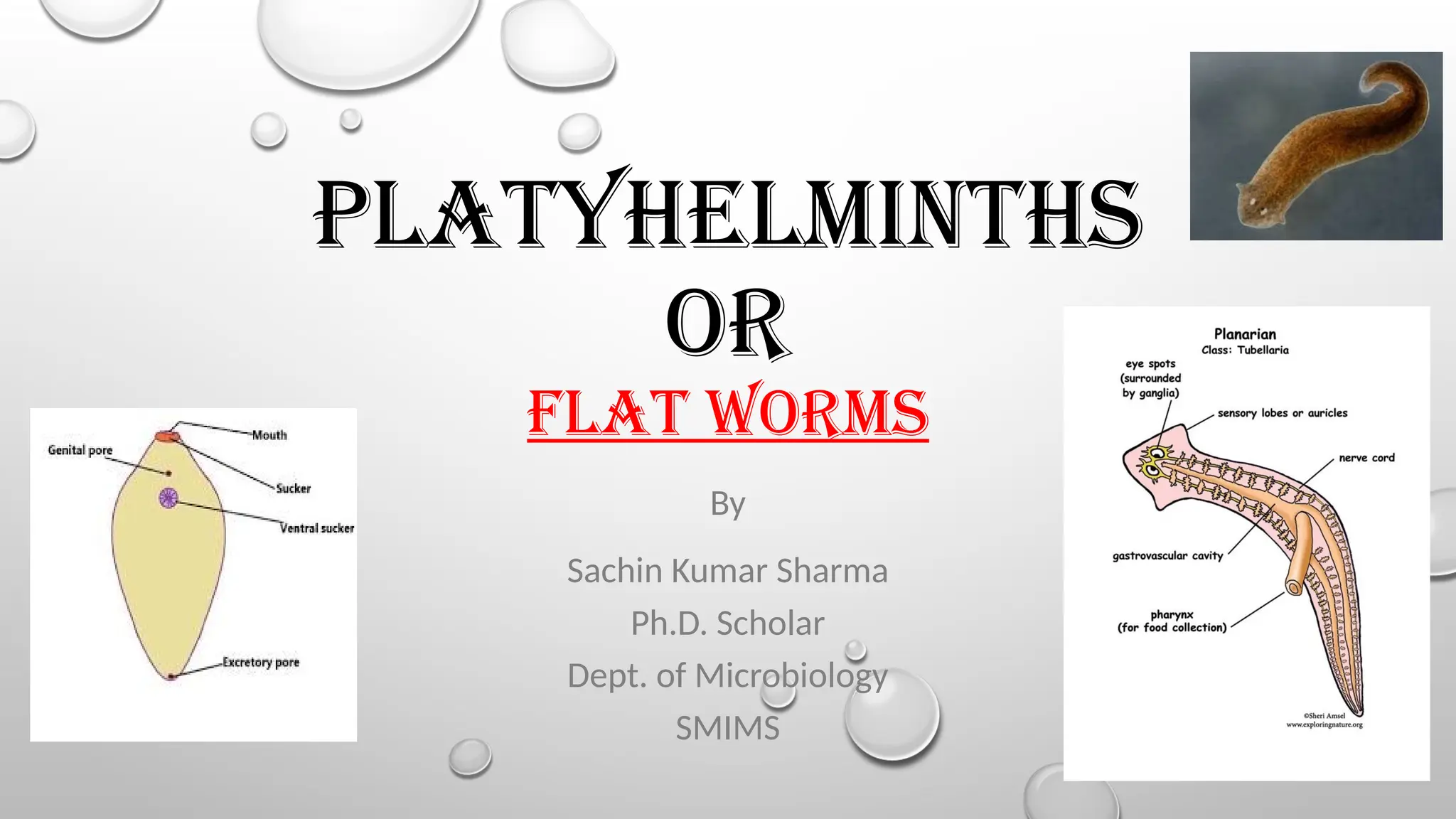

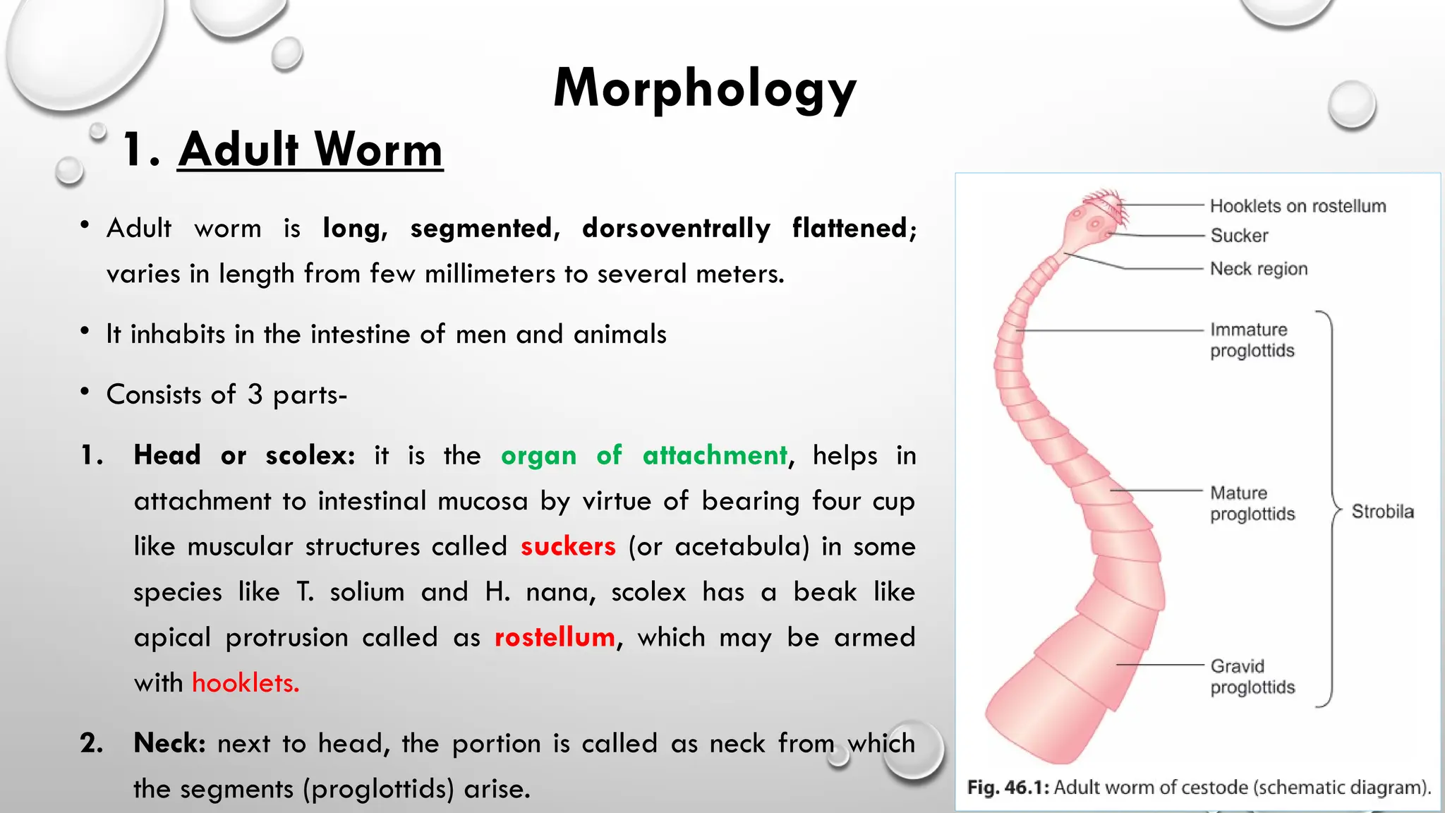
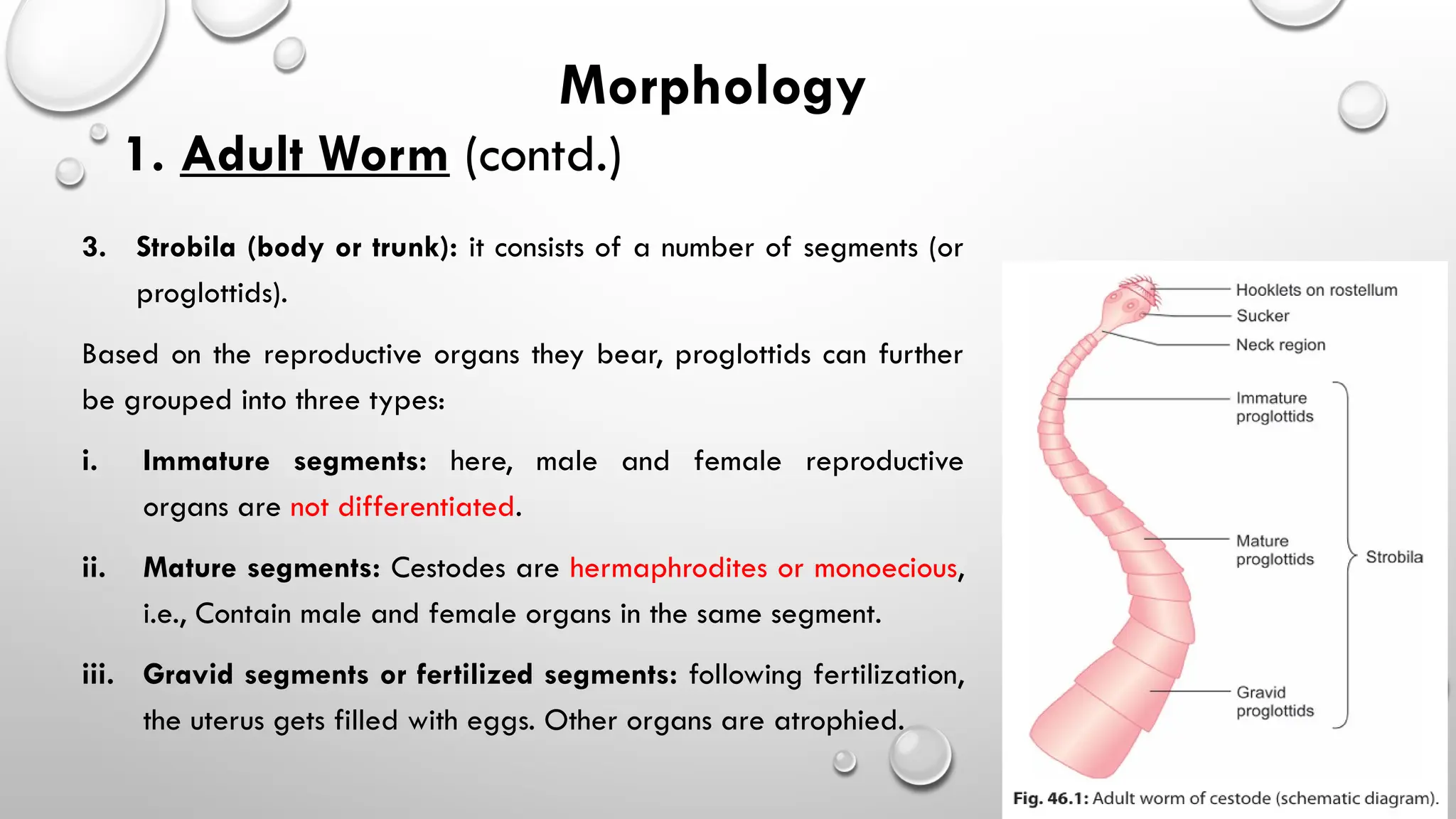

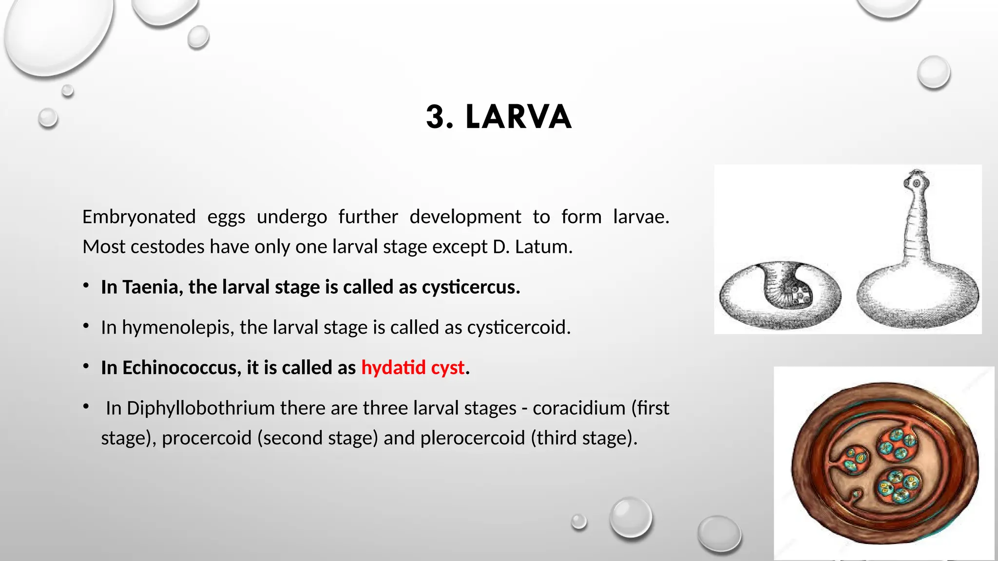
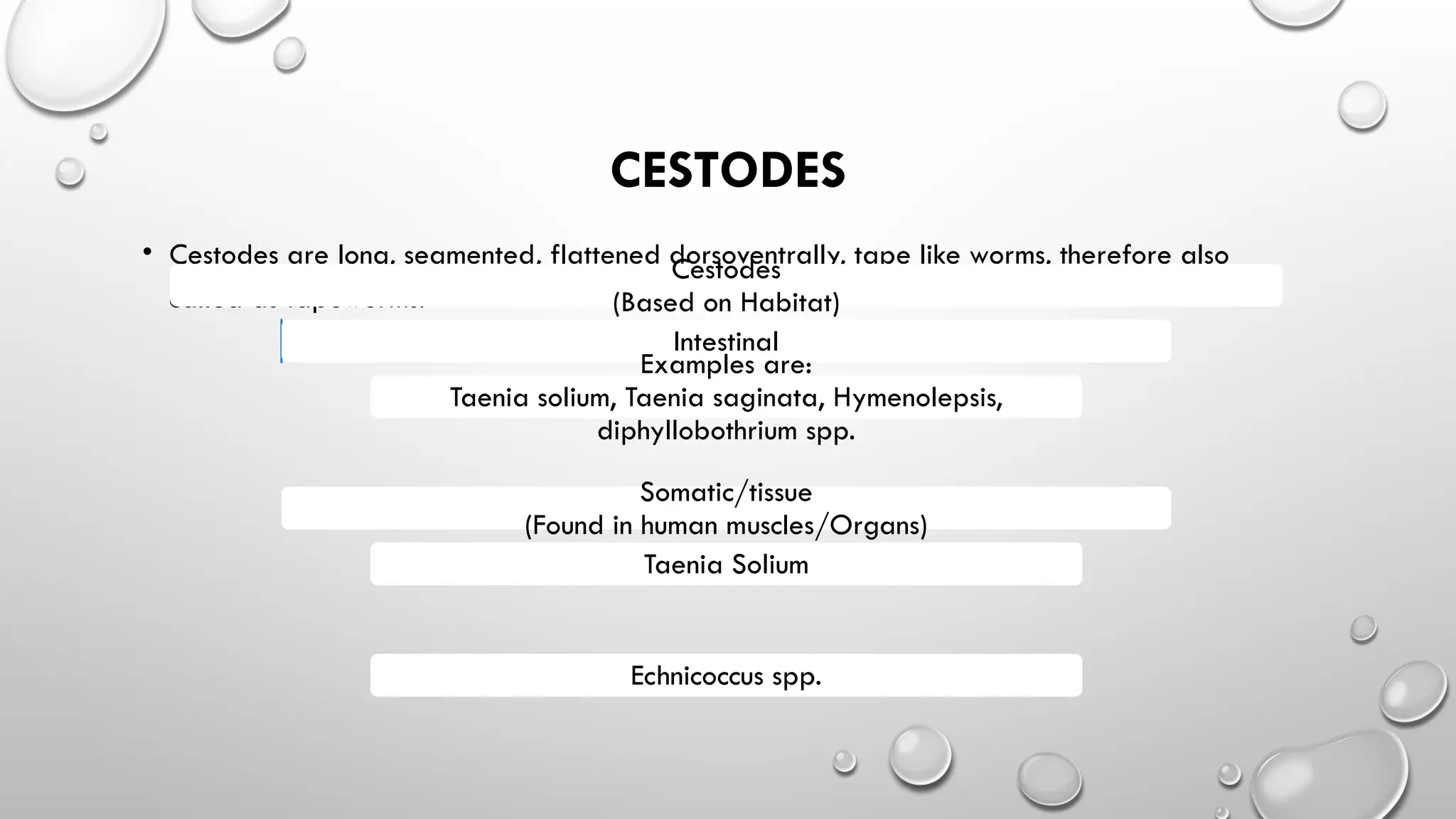

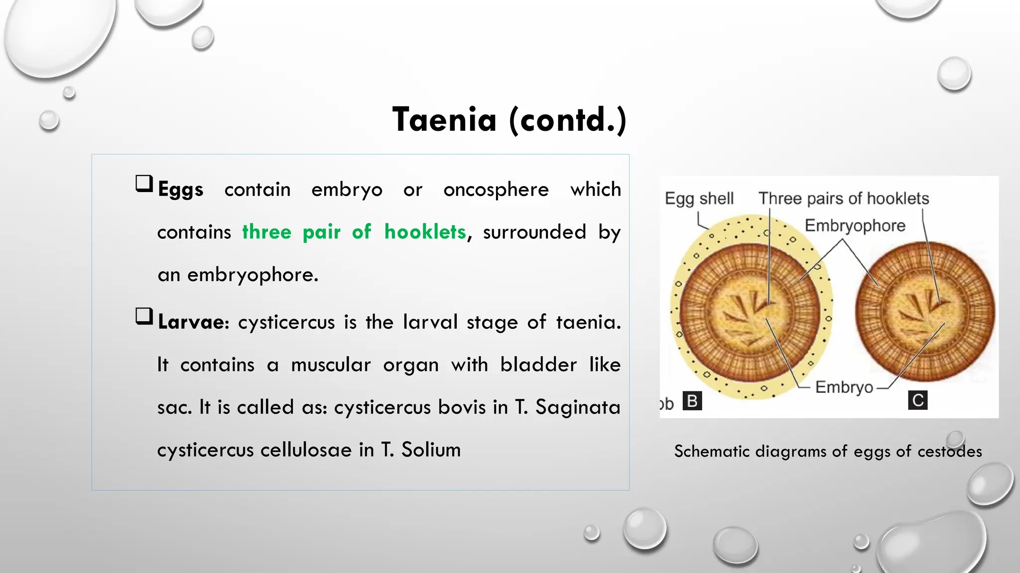


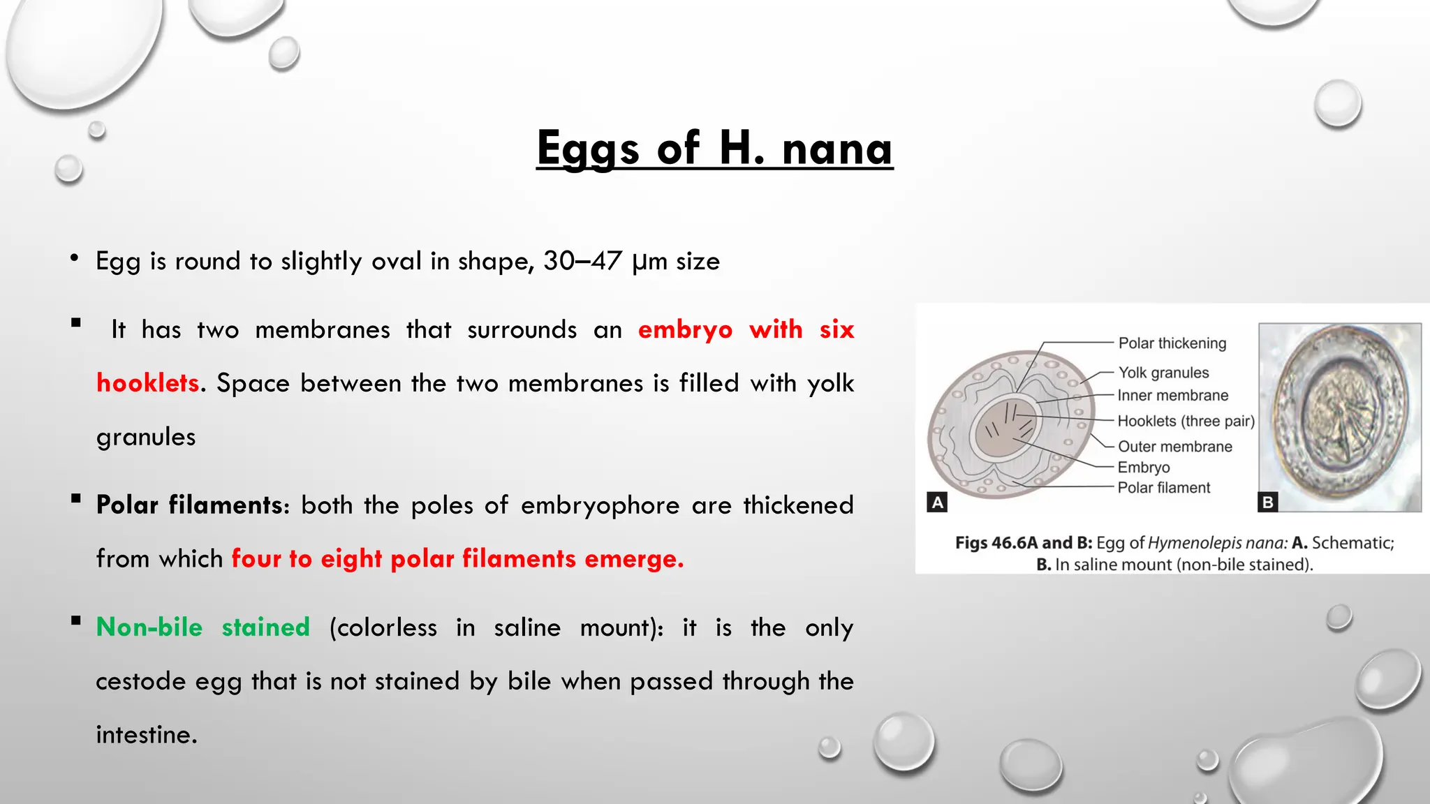
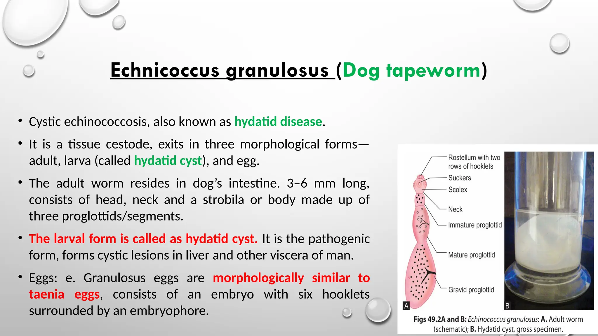
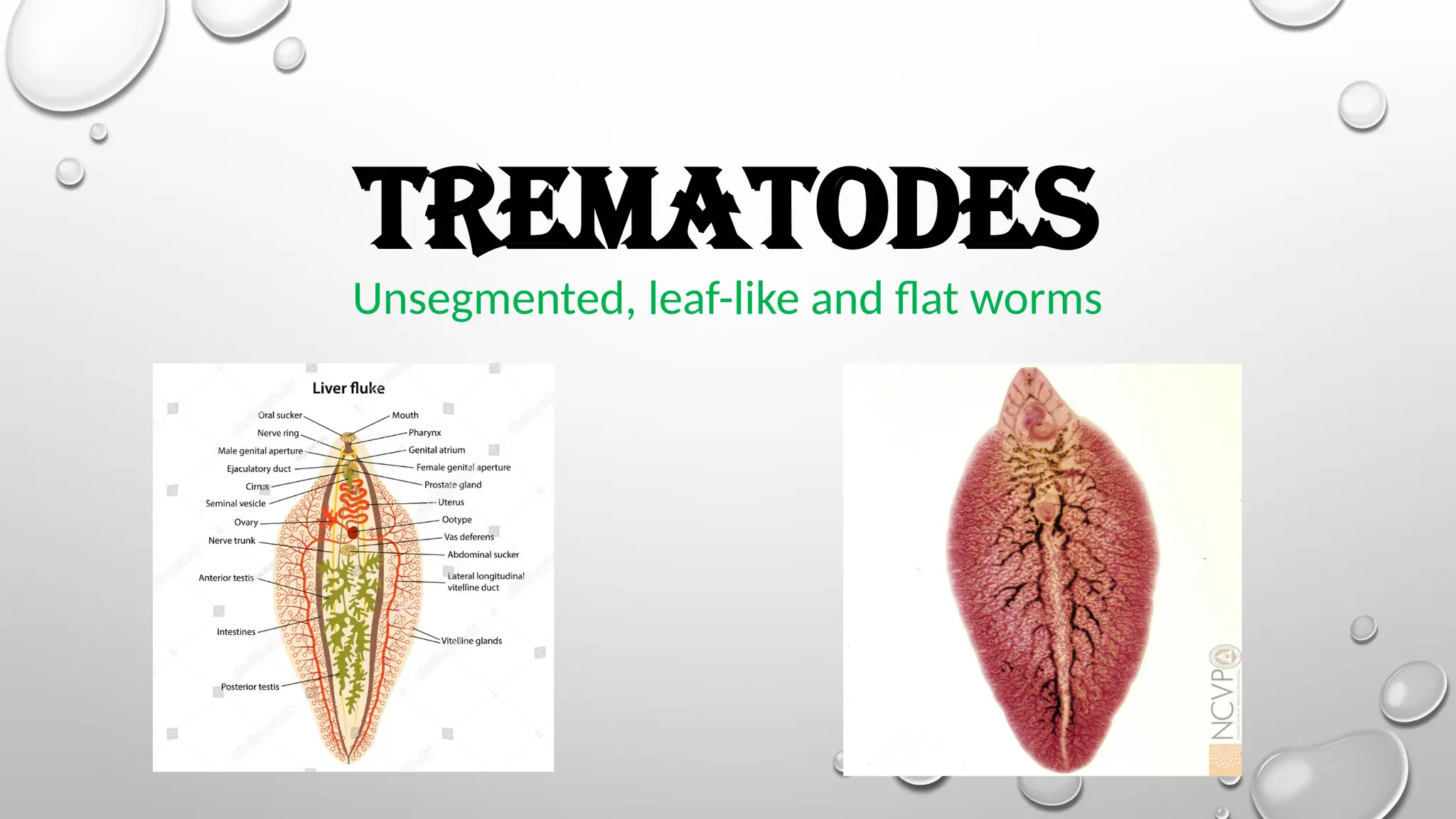

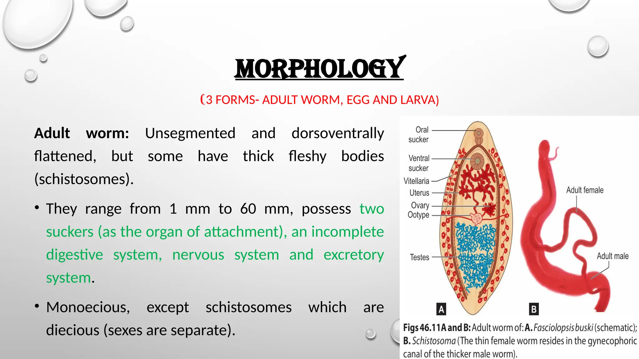
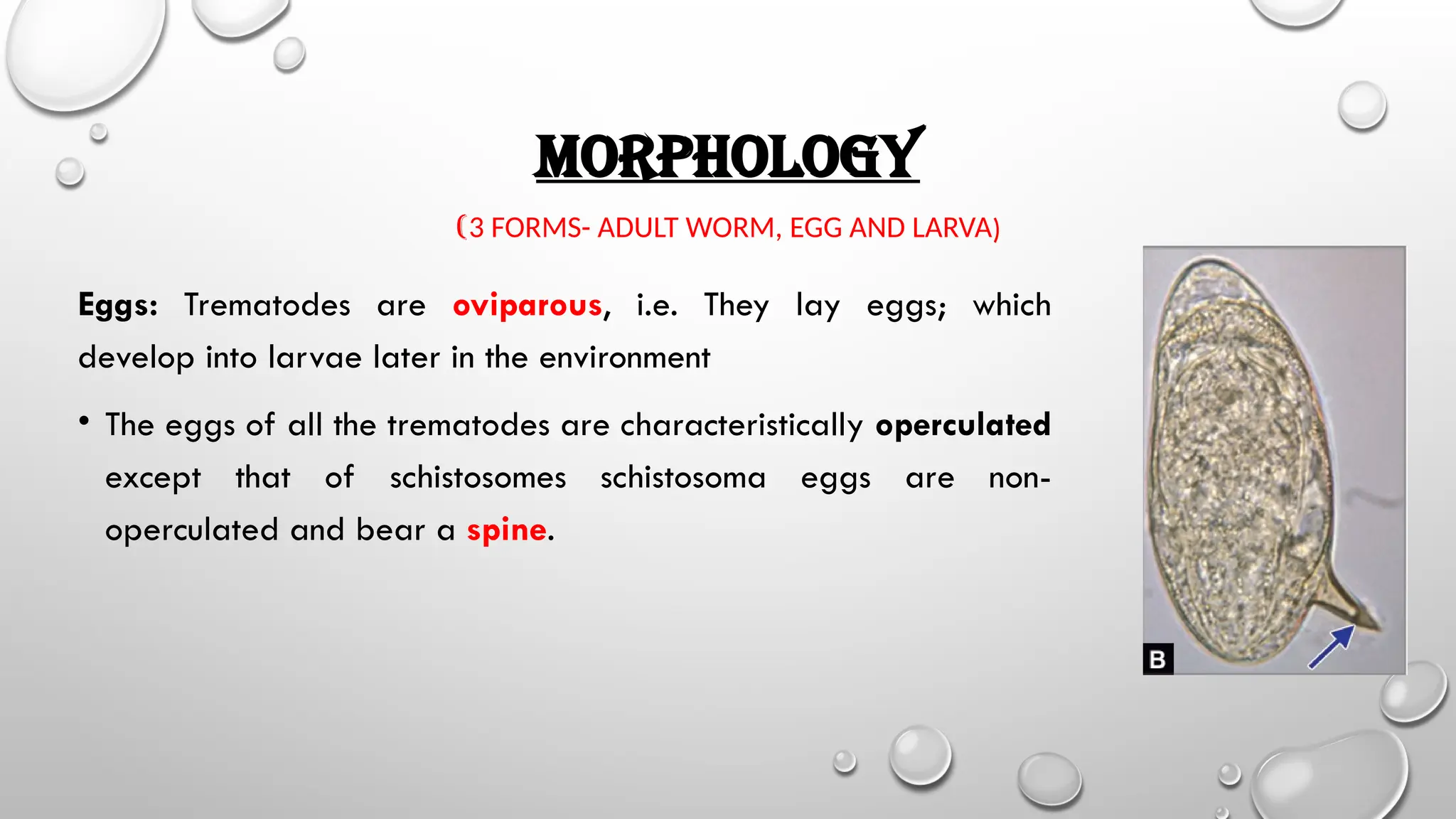
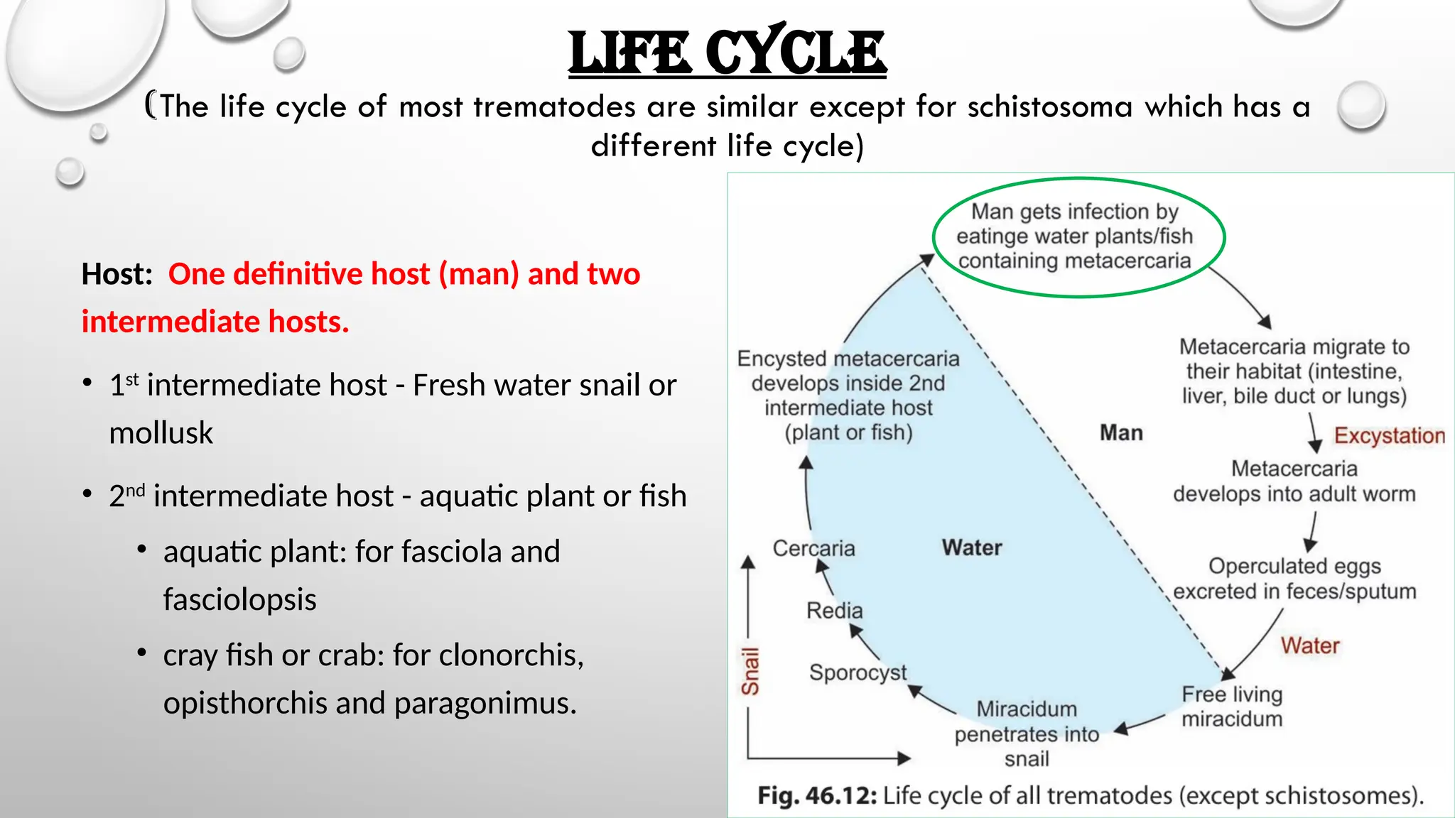
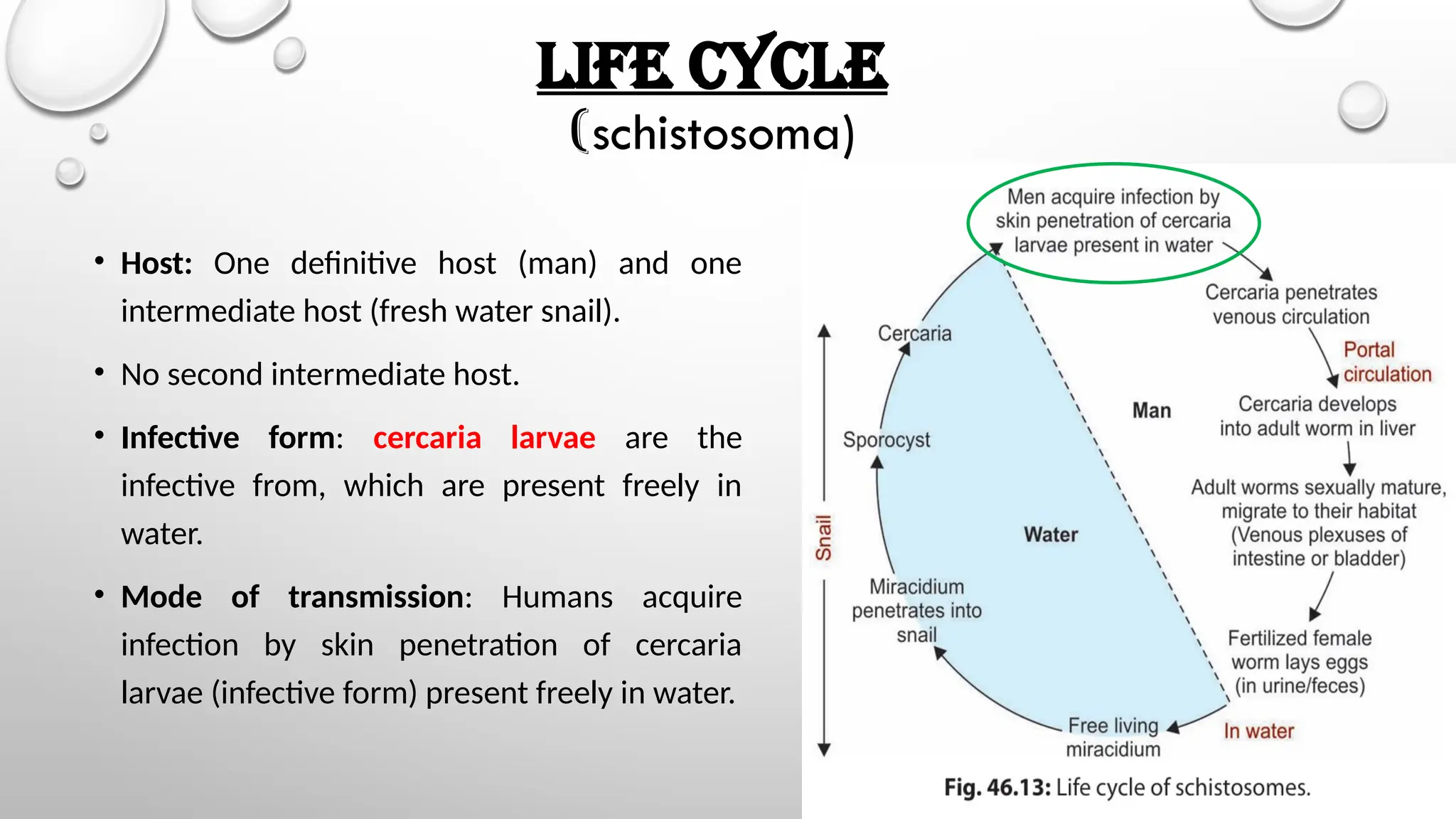


The document provides an overview of platyhelminths, including flat worms and their classifications, characteristics, and life cycles. It highlights the morphology of adult worms, eggs, and larvae, detailing specific examples such as tapeworms and flukes, and their respective hosts and reproductive structures. Additionally, it discusses the clinical manifestations and pathogenesis associated with certain infections caused by these parasites.


