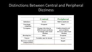
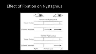
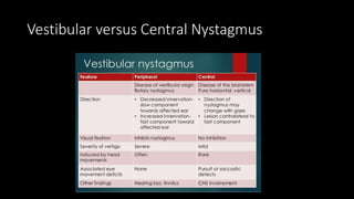
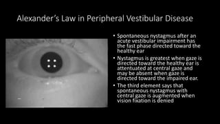
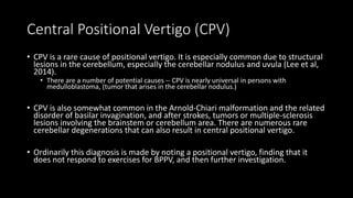
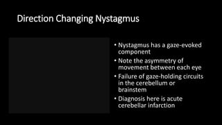
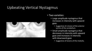
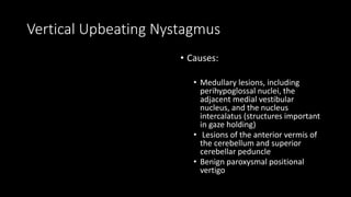
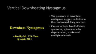
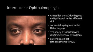
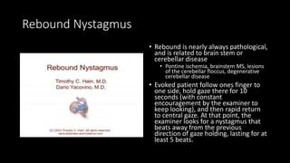
Central Vertigo and
Nystagmus
Distinctions Between Central and Peripheral
Dizziness
Effect of Fixation on Nystagmus
Vestibular versus Central Nystagmus
Alexander’s Law in Peripheral Vestibular Disease
• Spontaneous nystagmus after an
acute vestibular impairment has
the fast phase directed toward the
healthy ear
• Nystagmus is greatest when gaze is
directed toward the healthy ear is
attentuated at central gaze and
may be absent when gaze is
directed toward the impaired ear.
• The third element says that
spontaneous nystagmus with
central gaze is augmented when
vision fixation is denied
Central Positional Vertigo (CPV)
• CPV is a rare cause of positional vertigo. It is especially common due to structural
lesions in the cerebellum, especially the cerebellar nodulus and uvula (Lee et al,
2014).
• There are a number of potential causes -- CPV is nearly universal in persons with
medulloblastoma, (tumor that arises in the cerebellar nodulus.)
• CPV is also somewhat common in the Arnold-Chiari malformation and the related
disorder of basilar invagination, and after strokes, tumors or multiple-sclerosis
lesions involving the brainstem or cerebellum area. There are numerous rare
cerebellar degenerations that can also result in central positional vertigo.
• Ordinarily this diagnosis is made by noting a positional vertigo, finding that it
does not respond to exercises for BPPV, and then further investigation.
Direction Changing Nystagmus
• Nystagmus has a gaze-evoked
component
• Note the asymmetry of
movement between each eye
• Failure of gaze-holding circuits
in the cerebellum or
brainstem
• Diagnosis here is acute
cerebellar infarction
Upbeating Vertical Nystagmus
• Two varieties:
• Large amplitude nystagmus that
increases in intensity with upward
gaze.
• Suggestive of a lesion of the anterior
vermis of the cerebellum.
• Small amplitude nystagmus that
decreases in intensity with upward
gaze and increases in intensity
with downward gaze
• Suggestive of lesions of the medulla.
Vertical Upbeating Nystagmus
• Causes:
• Medullary lesions, including
perihypoglossal nuclei, the
adjacent medial vestibular
nucleus, and the nucleus
intercalatus (structures important
in gaze holding)
• Lesions of the anterior vermis of
the cerebellum and superior
cerebellar peduncle
• Benign paroxysmal positional
vertigo
Vertical Downbeating Nystagmus
• The presence of downbeat
nystagmus suggests a lesion in
the cervicomedullary junction.
• Causes include Arnold-Chiari's
syndrome, spinocerebellar
degeneration, stroke and
multiple sclerosis.
Pendular Nystagmus
• Multivectorial nystagmus (ie,
horizontal, vertical, circular, elliptical)
with an equal velocity in each
direction
• Often there is marked asymmetry and
dissociation between the eyes
• Amplitude of the nystagmus may vary
in different positions of gaze
• Demyelinating disease, monocular or
binocular visual deprivation,
oculopalatal myoclonus, internuclear
ophthalmoplegia, brainstem or
cerebellar dysfunction
Internuclear Ophthalmoplegia
• Named for the ADducting eye
and ipsilateral to the affected
MLF
• Horizontal nystagmus in the
ABducting eye
• Frequently associated with
upbeating vertical nystagmus
• Bilateral is almost
pathognomonic for MS
Convergence-retraction nystagmus (induced
convergence-retraction)
• Part of the dorsal midbrain
syndrome (Piranaud’s Syndrome)
associated with:
• paralysis of upward gaze
• defective convergence
• eyelid retraction
• pupillary light–near dissociation
• Best elicited on attempted
upgaze saccades (eg, by asking
the patient to track a
downwardly rotating OKN drum)
Light Near Dissociation
See Saw Nystagmus
• Localizes to lesions
supra/parasellar region (Large
sellar and hypothalamic lesions)
• In one half cycle, the eye will rise
and intort and the other eye will
fall and extort; then, in the next
half cycle
• The interstitial nucleus of Cajal
(INC), adjacent to the medial
longitudinal fasciculus in the
midbrain tegmentum, has been
frequently implicated in the
pathogenesis of SSN
Rebound Nystagmus
• Rebound is nearly always pathological,
and is related to brain stem or
cerebellar disease
• Pontine ischemia, brainstem MS, lesions
of the cerebellar floccus, degenerative
cerebellar disease
• Evoked patient follow ones finger to
one side, hold gaze there for 10
seconds (with constant
encouragement by the examiner to
keep looking), and then rapid return
to central gaze. At that point, the
examiner looks for a nystagmus that
beats away from the previous
direction of gaze holding, lasting for at
least 5 beats.

Central vertigo and nystagmus

  • 1.
  • 2.
    Distinctions Between Centraland Peripheral Dizziness
  • 3.
    Effect of Fixationon Nystagmus
  • 4.
  • 6.
    Alexander’s Law inPeripheral Vestibular Disease • Spontaneous nystagmus after an acute vestibular impairment has the fast phase directed toward the healthy ear • Nystagmus is greatest when gaze is directed toward the healthy ear is attentuated at central gaze and may be absent when gaze is directed toward the impaired ear. • The third element says that spontaneous nystagmus with central gaze is augmented when vision fixation is denied
  • 7.
    Central Positional Vertigo(CPV) • CPV is a rare cause of positional vertigo. It is especially common due to structural lesions in the cerebellum, especially the cerebellar nodulus and uvula (Lee et al, 2014). • There are a number of potential causes -- CPV is nearly universal in persons with medulloblastoma, (tumor that arises in the cerebellar nodulus.) • CPV is also somewhat common in the Arnold-Chiari malformation and the related disorder of basilar invagination, and after strokes, tumors or multiple-sclerosis lesions involving the brainstem or cerebellum area. There are numerous rare cerebellar degenerations that can also result in central positional vertigo. • Ordinarily this diagnosis is made by noting a positional vertigo, finding that it does not respond to exercises for BPPV, and then further investigation.
  • 8.
    Direction Changing Nystagmus •Nystagmus has a gaze-evoked component • Note the asymmetry of movement between each eye • Failure of gaze-holding circuits in the cerebellum or brainstem • Diagnosis here is acute cerebellar infarction
  • 9.
    Upbeating Vertical Nystagmus •Two varieties: • Large amplitude nystagmus that increases in intensity with upward gaze. • Suggestive of a lesion of the anterior vermis of the cerebellum. • Small amplitude nystagmus that decreases in intensity with upward gaze and increases in intensity with downward gaze • Suggestive of lesions of the medulla.
  • 10.
    Vertical Upbeating Nystagmus •Causes: • Medullary lesions, including perihypoglossal nuclei, the adjacent medial vestibular nucleus, and the nucleus intercalatus (structures important in gaze holding) • Lesions of the anterior vermis of the cerebellum and superior cerebellar peduncle • Benign paroxysmal positional vertigo
  • 11.
    Vertical Downbeating Nystagmus •The presence of downbeat nystagmus suggests a lesion in the cervicomedullary junction. • Causes include Arnold-Chiari's syndrome, spinocerebellar degeneration, stroke and multiple sclerosis.
  • 12.
    Pendular Nystagmus • Multivectorialnystagmus (ie, horizontal, vertical, circular, elliptical) with an equal velocity in each direction • Often there is marked asymmetry and dissociation between the eyes • Amplitude of the nystagmus may vary in different positions of gaze • Demyelinating disease, monocular or binocular visual deprivation, oculopalatal myoclonus, internuclear ophthalmoplegia, brainstem or cerebellar dysfunction
  • 13.
    Internuclear Ophthalmoplegia • Namedfor the ADducting eye and ipsilateral to the affected MLF • Horizontal nystagmus in the ABducting eye • Frequently associated with upbeating vertical nystagmus • Bilateral is almost pathognomonic for MS
  • 14.
    Convergence-retraction nystagmus (induced convergence-retraction) •Part of the dorsal midbrain syndrome (Piranaud’s Syndrome) associated with: • paralysis of upward gaze • defective convergence • eyelid retraction • pupillary light–near dissociation • Best elicited on attempted upgaze saccades (eg, by asking the patient to track a downwardly rotating OKN drum)
  • 15.
  • 16.
    See Saw Nystagmus •Localizes to lesions supra/parasellar region (Large sellar and hypothalamic lesions) • In one half cycle, the eye will rise and intort and the other eye will fall and extort; then, in the next half cycle • The interstitial nucleus of Cajal (INC), adjacent to the medial longitudinal fasciculus in the midbrain tegmentum, has been frequently implicated in the pathogenesis of SSN
  • 17.
    Rebound Nystagmus • Reboundis nearly always pathological, and is related to brain stem or cerebellar disease • Pontine ischemia, brainstem MS, lesions of the cerebellar floccus, degenerative cerebellar disease • Evoked patient follow ones finger to one side, hold gaze there for 10 seconds (with constant encouragement by the examiner to keep looking), and then rapid return to central gaze. At that point, the examiner looks for a nystagmus that beats away from the previous direction of gaze holding, lasting for at least 5 beats.