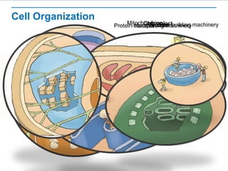
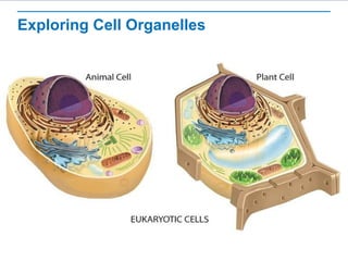
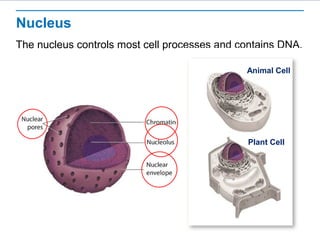
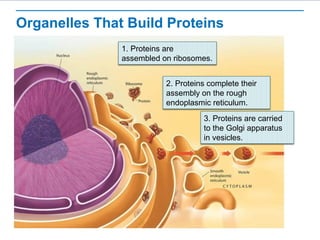
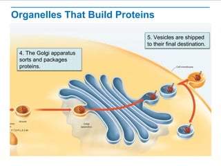
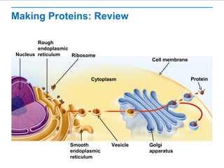
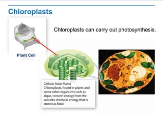
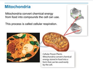
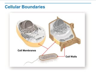
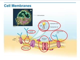
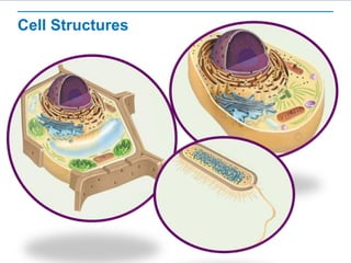
Cell Structure
Learning Objectives
 Describe the structure and function of the cell nucleus.
 Describe the role of vacuoles, lysosomes, and the
cytoskeleton.
 Identify the role of ribosomes, endoplasmic reticulum, and
Golgi apparatus in making proteins.
 Describe the function of the chloroplasts and mitochondria
in the cell.
 Describe the function of the cell membrane.
Cell Organization
NucleusProtein-building machineryProtein-transporting machineryMitochondrionChloroplastVesiclesVacuole
Exploring Cell Organelles
Nucleus
The nucleus controls most cell processes and contains DNA.
Animal Cell
Plant Cell
Vacuoles, Vesicles, and Lysosomes
Animal Cell
Plant Cell
Cytoskeleton
Microtubules
Organelles That Build Proteins
1. Proteins are
assembled on ribosomes.
2. Proteins complete their
assembly on the rough
endoplasmic reticulum.
3. Proteins are carried
to the Golgi apparatus
in vesicles.
Organelles That Build Proteins
4. The Golgi apparatus
sorts and packages
proteins.
5. Vesicles are shipped
to their final destination.
Making Proteins: Review
Nucleus
Rough
endoplasmic
reticulum Ribosome
Protein
Smooth
endoplasmic
reticulum
Cytoplasm
Cell membrane
Vesicle Golgi
apparatus
Chloroplasts
Chloroplasts can carry out photosynthesis.
Plant Cell
Mitochondria
Mitochondria convert chemical energy
from food into compounds the cell can use.
This process is called cellular respiration.
Cellular Boundaries
Cell Membranes
Cell Walls
Cell Membranes
Cell Structures

Cell Organelles

Editor's Notes

  • #2 Read the lesson title aloud to students.
  • #3 Click to show each of the learning objectives. To assess prior knowledge, pair students and have them work together to draw a model of a cell. Encourage students to be as detailed as possible. Ask: What are some of the structures you included in your cell model? Write student answers on the board and ask them to describe each structure’s function if possible. Ask: Are all cells the same? If not, how are they different? Accept all reasonable answers. Hand out the Student Worksheet.
  • #4 Tell students: Cells can be thought of as a complex factory. Point out that the cell can be divided into two major parts: the nucleus and cytoplasm. The cytoplasm has specialized organs called organelles. Click to zoom in on the following parts of the image. Ask students to describe what the analogy is showing. Then identify and describe the cellular component. Nucleus Protein building machinery Protein transport machinery Mitochondrion Chloroplast Vesicles Vacuole Click again to zoom back out. Ask: If cells are like a factory, what can be inferred about the nature of cells? Answer: Cells are complex and dynamic, with various parts that work together.
  • #5 Tell students: These are representations of plant and animal cells and will be used throughout the lesson. As each cellular component is discussed, students will see an image highlighting the component
  • #6 The nucleus contains nearly all the cell’s DNA and, with it, the coded instructions for making proteins and other important molecules. By contrast, prokaryotic cells lack a nucleus. Their DNA, containing the same kinds of instructions, is found in the cytoplasm. Click to highlight each component of the nucleus. Ask students to describe the structure of each component and hypothesize about its function. Describe the function of each: Nuclear envelope: a double-layered membrane barrier that protects the DNA Nuclear pores: tiny holes in the nuclear envelope that allow certain components to enter and leave the nucleus Chromatin: uncoiled DNA + proteins (the form DNA is in when it is not condensed into a chromosome) Nucleolus: a specific area in the nucleus where ribosome assembly initiates
  • #7 Tell students: Different types of vacuoles exist. Click to highlight the plant vacuole. Explain that plant cells generally have large central vacuoles whose sizes change depending on water availability. Ask: What is likely true about the vacuoles of a wilting plant? Answer: The vacuoles are low on water. Explain that once the plant is watered, the vacuoles refill and the plant stands upright again. Tell students: Look at the paramecium picture. Click to highlight the paramecium vacuole. Explain that a paramecium regulates water balance with its contractile vacuole. The contractile vacuole continually fills with excess water from cytoplasm and then expels it. Click to remove the circle. Tell students: The smaller vesicles function to both store and transport materials within the cell and to and from the external cellular environment. The vesicles transport materials to the cell membrane. Activity: Ask for two volunteers to come to the front of the class. Give one student a bag or box of small items. Give the other student a bunch of loose items into his or her hand that are not easy to hold, like a large quantity of paperclips or bouncy balls. Have the students do a simple task, like take the items to another location in the room. Ask: Why is it better for a cell to transport materials using vesicles than without? Answer: Vesicles keep materials segregated from one another, contained and tidy, and prevents things from interacting that should not. Other answers may be reasonable. Tell students: Lysosomes break down lipids, carbohydrates, and proteins into small molecules that can be used by the rest of the cell. They are also involved in breaking down organelles that have outlived their usefulness.
  • #8 Tell students: Look at the figure. Explain that cells have an extensive network of filaments in the cytoplasm called the cytoskeleton. Microfilaments (pale purple) and microtubules (yellow) are two of the principal protein filaments that make up the cytoskeleton. Microfilaments are threadlike structures made up of a protein called actin. They form extensive networks in some cells and produce a tough, flexible framework that supports the cell. Microfilaments also help cells move. Tell students: The cytoskeleton not only forms cell shape and framework but also acts like a highway in the cell, moving materials and organelles to the appropriate locations. In other words, the cellular components are not a random combination of things floating around in the cell, but are rather placed deliberately and moved along specific pathways.
  • #9 Tell students: Microtubules are hollow structures made up of proteins known as tubulins. In many cells, they play critical roles in maintaining cell shape. Microtubules form the mitotic spindle during cell division. In animal cells, centrioles are also formed from tubulins. Centrioles are not found in plant cells. Microtubules also help build projections from the cell surface, known as cilia and flagella. This is a cross-section of a flagellum showing the 9+2 arrangement of microtubules. Click to highlight the cross-section.
  • #10 Tell students that proteins carry out so many of the essential functions of living things, including the synthesis of other biomolecules such as lipids and carbohydrates, that a big part of the cell is devoted to their production and distribution. This is a large picture, and will be seen over two slides (there will also be a third slide showing the entire picture). Help students interpret this picture by asking the following questions. Encourage students to describe what they see, then fill in the details for them. Ask: What happens first? Students will likely notice that a protein is coming off the ribosome. If students are having a difficult time, help them follow the arrows backwards. Click to reveal the answer. Explain that ribosomes are small particles of RNA and protein found throughout the cytoplasm in all cells. Ribosomes assemble proteins by following coded instructions that come from DNA. Ask: What happens next? Students will likely notice that the protein gets fed into the rough ER. Click to reveal the answer. Explain that proteins targeted for export to the cell membrane or to specialized locations within the cell complete their assembly on ribosomes bound to the rough ER. Introduce the difference between the smooth and rough ER. Ask: What happens next? Students should notice that the protein ends up in a vesicle. Click to reveal the answer. Explain that newly assembled proteins are carried from the rough endoplasmic reticulum to the Golgi apparatus in vesicles.
  • #11 Orient students by pointing out that the vesicle is the same one from the previous image. Ask: What does the vesicle do? Answer: The vesicle binds to the Golgi apparatus. Click to reveal the answer. Explain that the vesicle fuses with the Golgi apparatus. The Golgi apparatus modifies, sorts, and packages proteins and other materials from the endoplasmic reticulum. Ask: What happens next? Answer: Vesicles are sent to different places. Click to reveal the answer. Explain that vesicles from the Golgi apparatus are shipped to their final destination within or out of the cell.
  • #12 Orient students by pointing out that this is the complete process shown in the last two slides. Ask the following questions to assess comprehension. Ask: Where are proteins assembled? Answer: ribosomes Ask: What can you infer about a cell that is packed with more than the typical number of ribosomes? Answer: When a cell has more than the typical number of ribosomes, you might infer that it produces more proteins than other cells. Ask: Where is the synthesis of membrane proteins completed? Answer: in the ER Ask: How are proteins transported to the Golgi apparatus? Answer: in vesicles Ask: What happens to proteins leaving the Golgi apparatus? Answer: They are sent out of the cell or back into the cytoplasm to be used. Divide students into groups and have them create flowcharts summarizing the role of each of the following cellular structures in protein synthesis: nucleus, nuclear envelope, ribosomes, endoplasmic reticulum, Golgi apparatus, vesicles, vacuole, and cell membrane. Remind students that proteins can follow multiple processing paths depending on their destination, so the flowcharts may branch at some points.
  • #13 Tell students: Chloroplasts are only present in plant cells and some other organisms such as algae and some bacteria. Animals do not have chloroplasts. Ask: Does any student recall what photosynthesis is? Help students understand the concept by saying chloroplasts might be referred to as “solar collectors.” Answer: In photosynthesis, plants convert solar energy into food molecules (chemical energy). Both plants and animals then break down this food to access energy for cellular activities. (See next slide.)
  • #14 Tell students: Cells need energy to carry out many processes, but can only use it in a certain form. Tell students: Cellular respiration converts energy from food into a form cells can use. Mitochondria carry out cellular respiration. Ask: Why are mitochondria called “powerhouses” of the cell? Answer: They make energy available from food to fuel cell activities. Some students may think that mitochondria are found only in animal cells and chloroplasts are found only in plant cells. Clarify that mitochondria are found in nearly all eukaryotes, including plants. Have students trace a meal of their own back to photosynthesis and cellular respiration (chloroplasts and mitochondria).
  • #15 Ask: Consider what happens when a property owner puts up a fence. What purpose does the fence serve? How do people get in and out? Are there different kinds of fences? Sample answer: The fence keeps people and animals out of the yard and can also keep people and animals from leaving the yard. People enter and exit through a gate. There are many types of fences. Explain that a cell’s contents are also confined within a barrier. Ask: Which cells have cell walls, and where are cell walls located in cells? Answer: Plant cells and prokaryotes have cell walls. Ask: True or false: All cells have a cell membrane. Answer: true Click to reveal the label for cell membranes. Ask: True or false: All cells have a cell wall. Answer: false Click to reveal the label for cell walls.
  • #16 Tell students: The cell membrane regulates what enters and leaves the cell and also protects and supports the cell. The membrane only allows certain things to cross it. Scientists call this semipermeable. Tell students: Look carefully at the picture of the membrane. Point out the inside and outside of the cell labels. Click to highlight. Tell students: The picture shows only a tiny part of a cell membrane, similar to the membrane that surrounds the cell in the micrograph. Explain that the illustration is a cross section. Model a cross section by cutting a lemon or orange in half and showing how the cut reveals a cross section of the rind. Point out that, in the “whoosh” in the illustration, the phospholipid molecule is an enlargement of one tiny part of the cross section of the membrane. Tell students: The phospholipid molecule in the cell membrane has two distinct ends. Click to highlight. Tell students: One end, the hydrophilic end is electrically attracted to water molecules. Click to highlight the ends. On the other end, the hydrophobic end, the phospholipid molecule repels water. Explain how phospholipids move and slide past one another in the membrane. Tell students: Scientists call this the fluid mosaic model. Click to highlight and discuss each of the following components of the membrane: Proteins: A large variety of proteins are located within the phospholipid layer. Functions include assisting materials into and out of the cell and receiving and transmitting messages. Carbohydrates: are attached to the proteins, often act as “flags” identifying the cell in some specific manner Have several students stand and form a “membrane.” Shuffle them into different places to show how the membrane is fluid. Then add other students to act as proteins and carbohydrates. Proteins can be asked to hold up a hand. Carbohydrates can be asked to hold the hand of a protein. The entire class can participate in making a model. Be sure most of the students are phospholipids.
  • #17 Using the Student Worksheet, have students create a compare and contrast table that includes the structures present in plant, animal, and bacterial cells. You may also want to ask students to list the functions of each structure.
  • #18 Divide students into small groups and instruct them to compare their tables and add any important information that they were missing. Also have each group submit a question about a cell function to be answered by the class. Worksheet Answers: Student tables should have three columns: plant, animal, prokaryotic. Each column should list all the structures present in the cell type. Structures that are the same for multiple cells should be in the same rows.