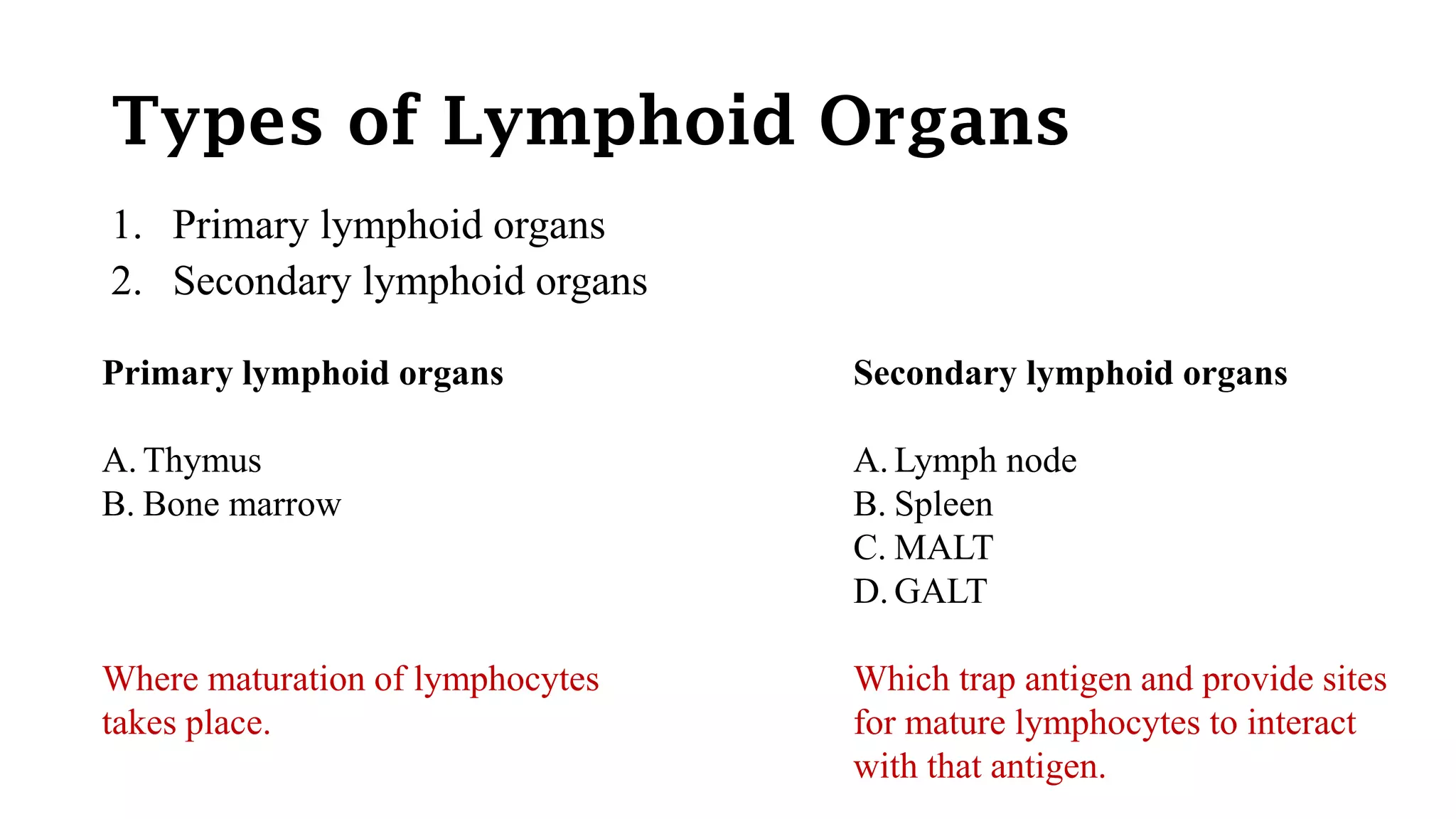
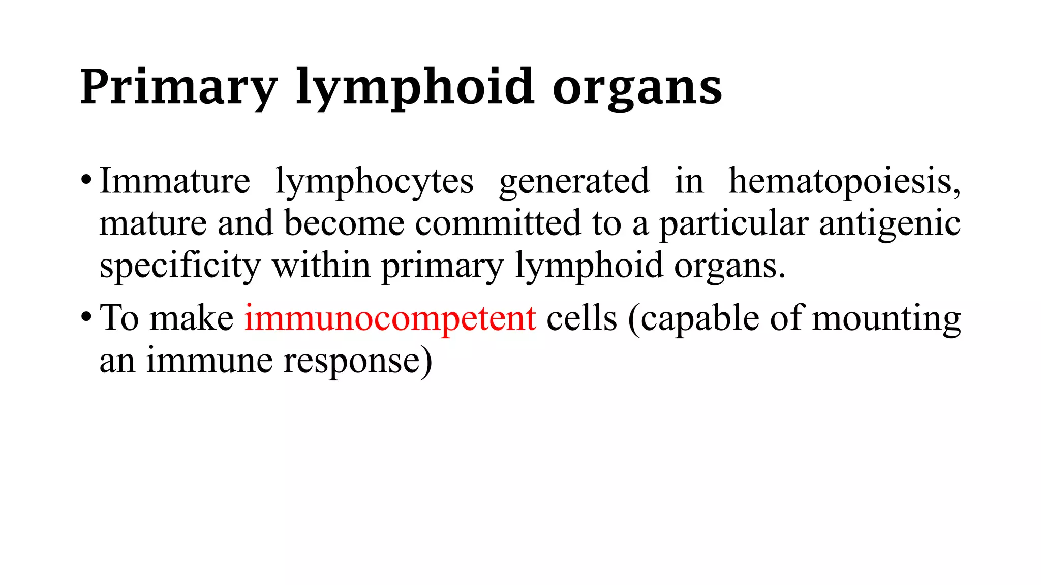
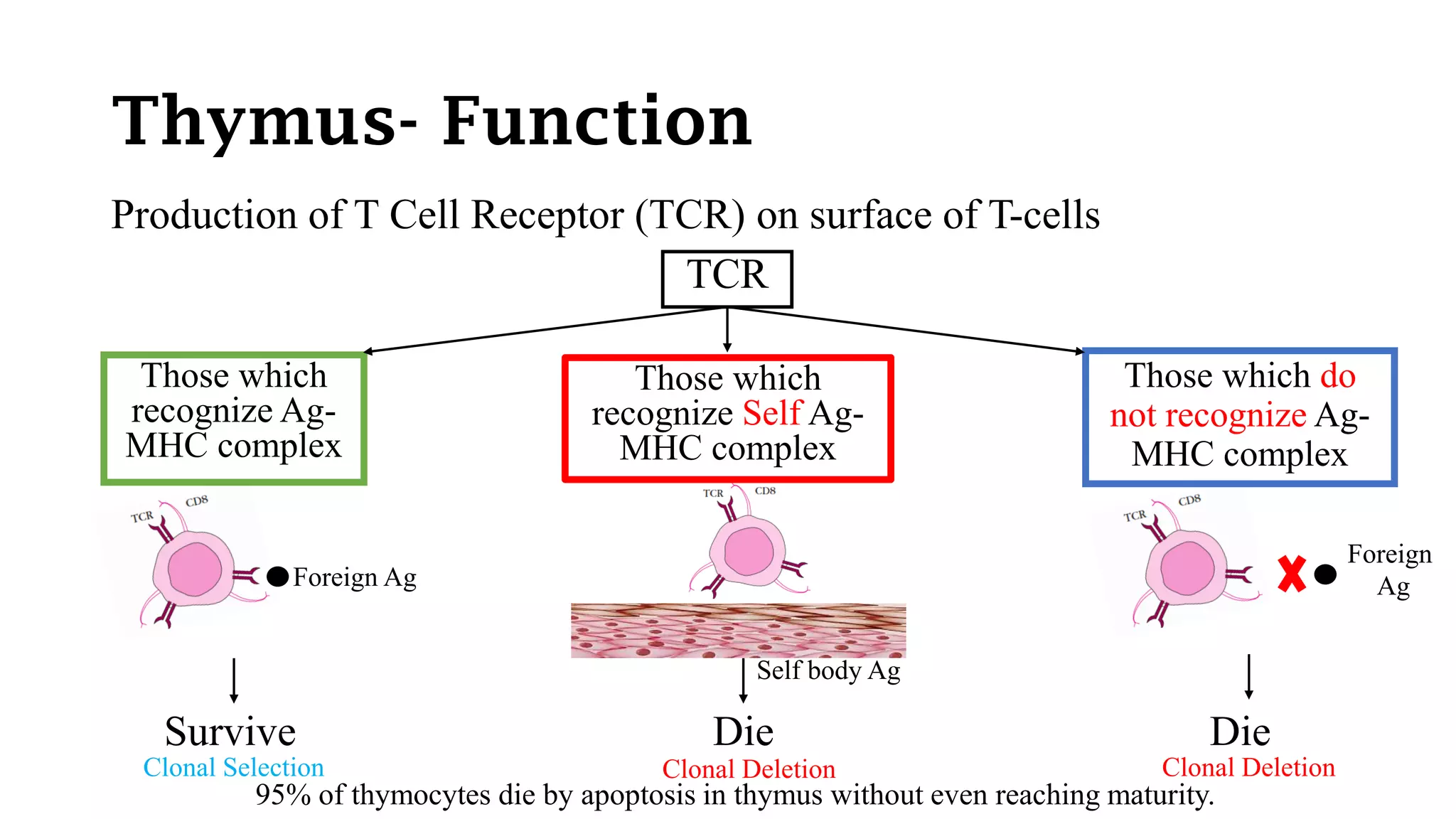
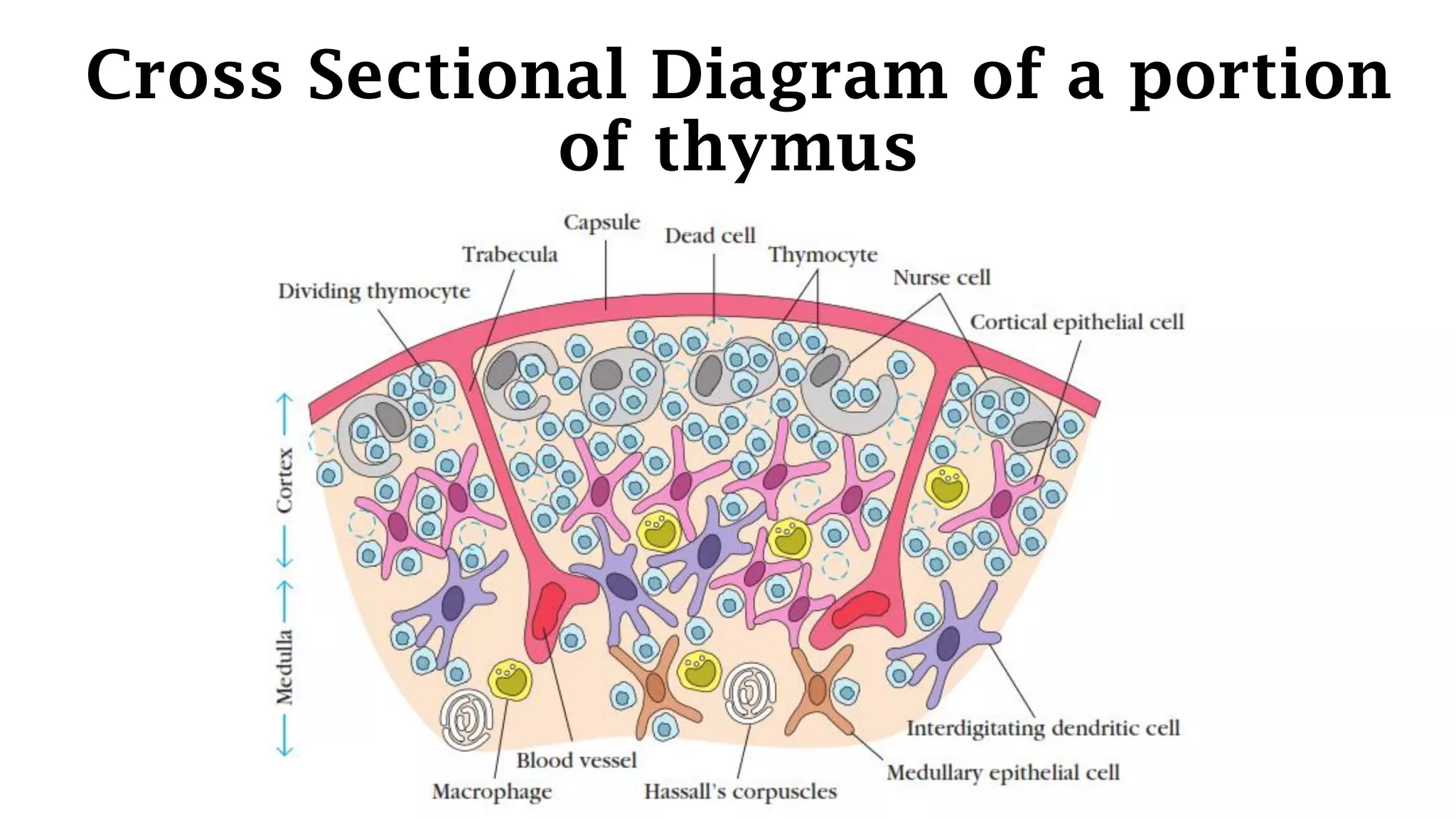
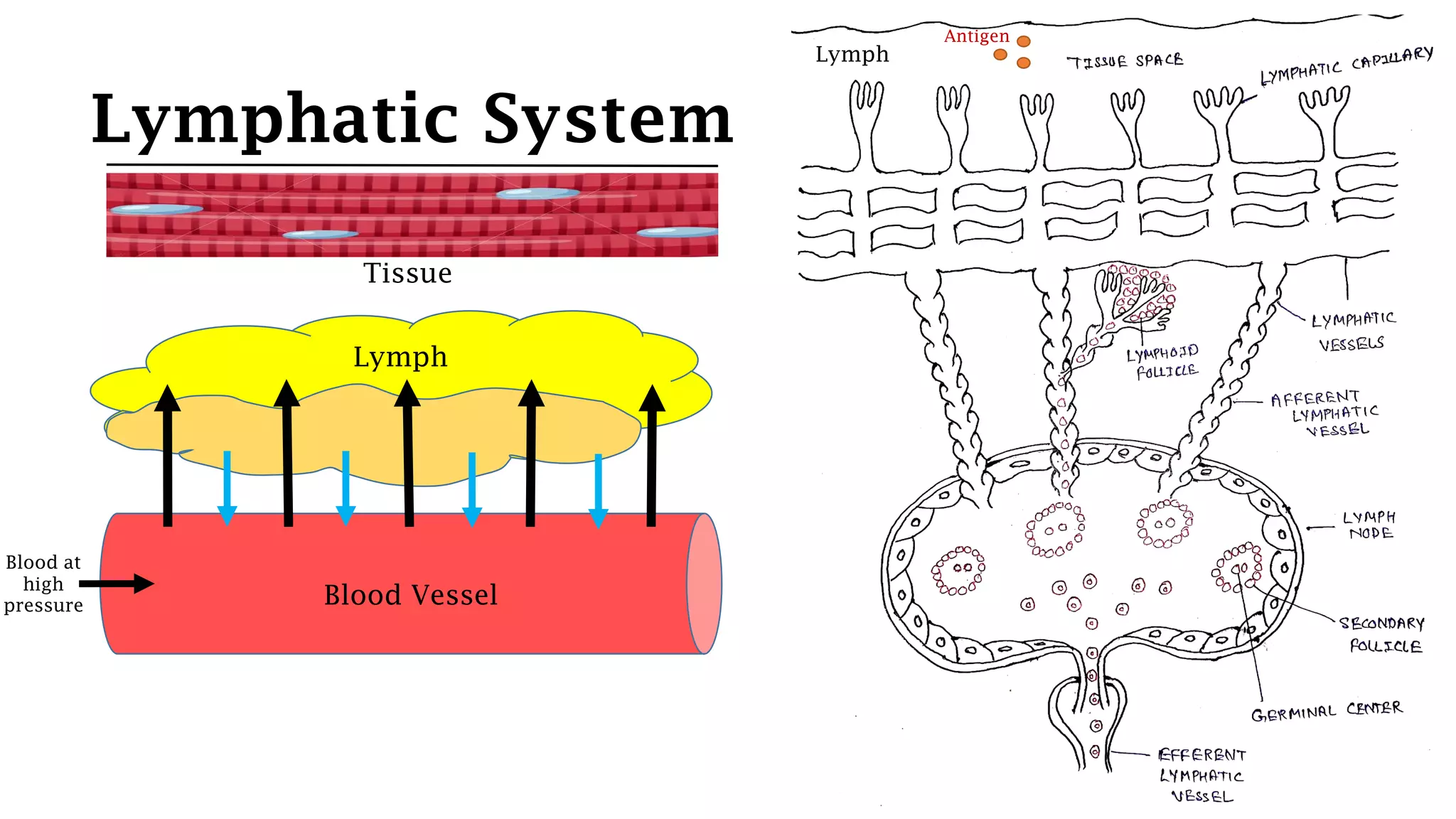
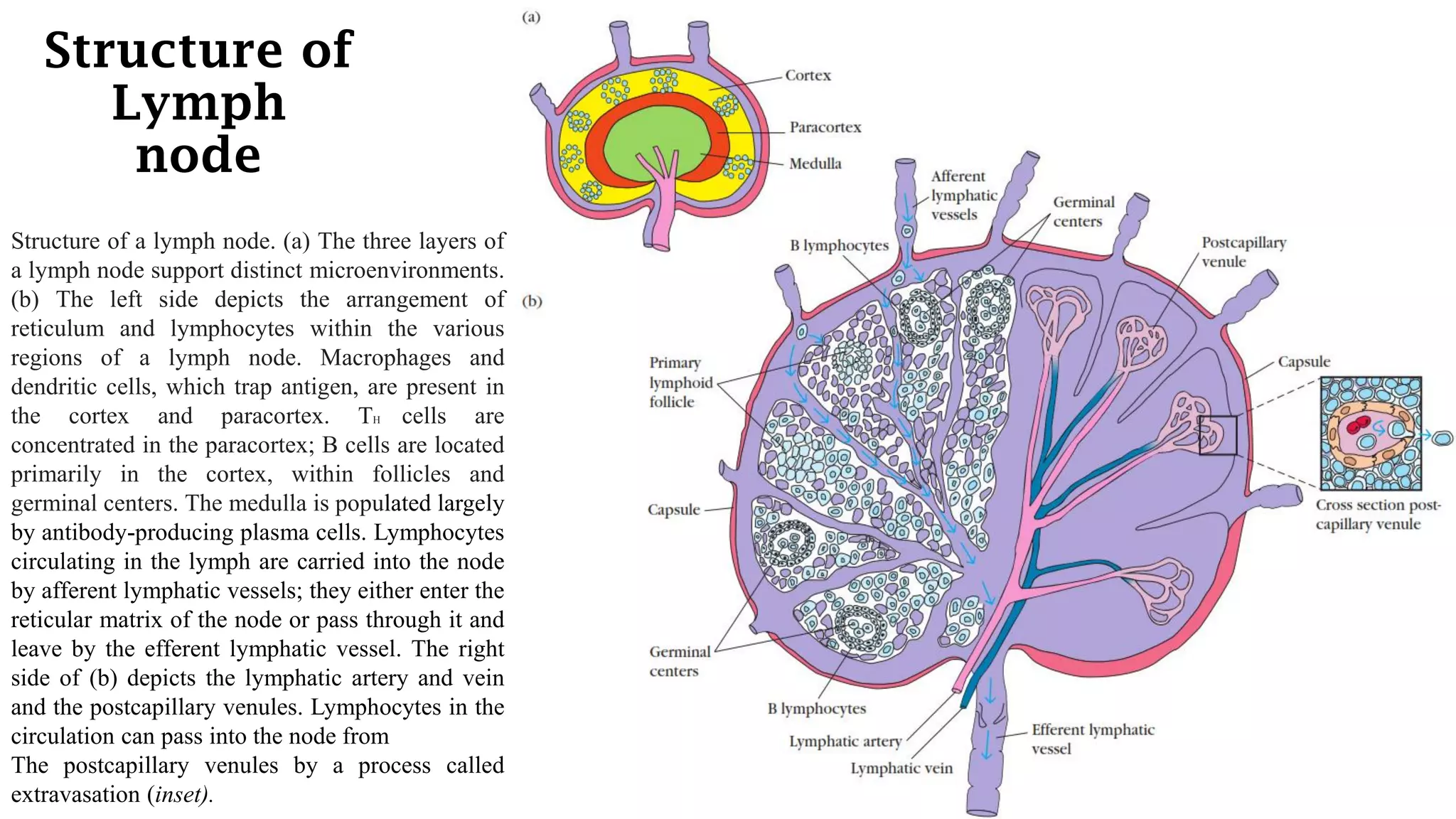
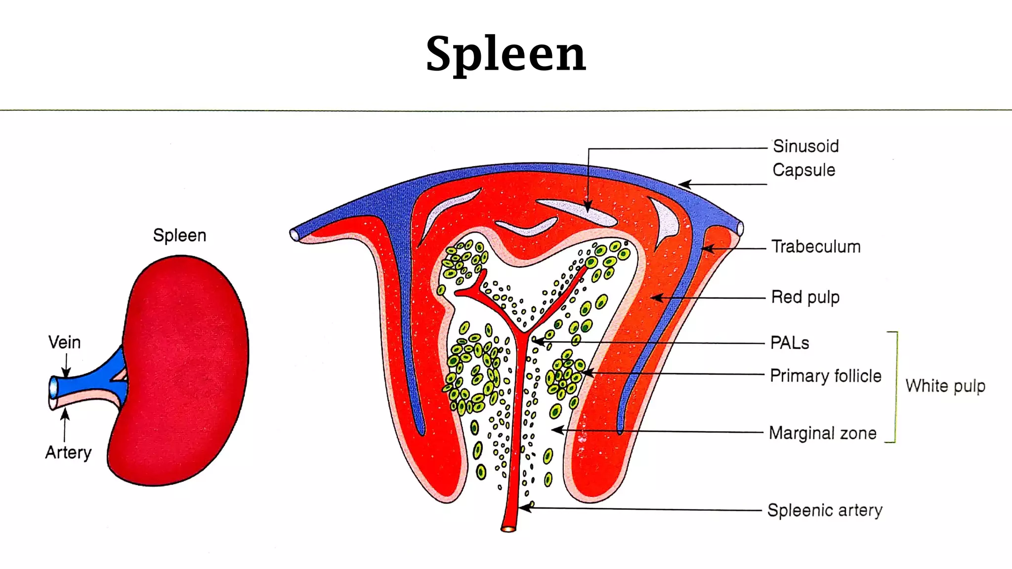
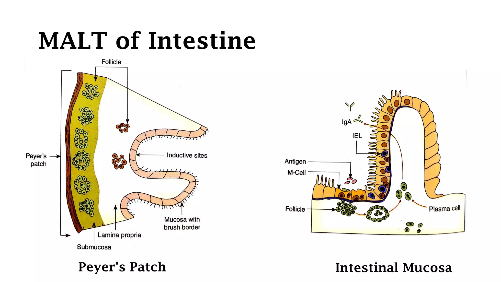
Organs of the
Immune System
Amandeep Singh
Assistant Professor
Department of Biotechnology
GSSDGS Khalsa College Patiala
Types of Lymphoid Organs
1. Primary lymphoid organs
2. Secondary lymphoid organs
Primary lymphoid organs
A. Thymus
B. Bone marrow
Where maturation of lymphocytes
takes place.
Secondary lymphoid organs
A. Lymph node
B. Spleen
C. MALT
D. GALT
Which trap antigen and provide sites
for mature lymphocytes to interact
with that antigen.
Primary lymphoid organs
• Immature lymphocytes generated in hematopoiesis,
mature and become committed to a particular antigenic
specificity within primary lymphoid organs.
• To make immunocompetent cells (capable of mounting
an immune response)
Thymus- Structure
• Site of T-cell maturation.
• Flat, bi-lobed organ situated above the heart, behind the sternum.
• Each lobe is surrounded by a capsule & is divided into lobules, which
are separated from each other by strands of connective tissues called
trabeculae.
• Each lobule is organized into 2 compartments:
1. Cortex: Densely packed with immature T cells or thymocytes
2. Medulla: Lightly packed with thymocytes
Thymus- Structure
• Both Cortex and medulla of thymus are criss-crossed by a
3-D stromal cell network composed of epithelial cells,
dendritic cells and macrophages which lead to the maturation
of thymocytes.
• Some thymic epithelial cells in the outer cortex called nurse
cells, have long membrane extensions that surround as many
as 50 thymocytes, forming large multicellular complex.
Thymus- Function
Production of T Cell Receptor (TCR) on surface of T-cells
TCR
Those which
recognize Ag-
MHC complex
Those which
recognize Self Ag-
MHC complex
Those which do
not recognize Ag-
MHC complex
DieSurvive Die
95% of thymocytes die by apoptosis in thymus without even reaching maturity.
Foreign Ag
Foreign
Ag
Self body Ag
Clonal Selection Clonal Deletion Clonal Deletion
Deficiency of Thymus development
DiGeorge Syndrome Nude Mice
Humans Mice
Thymus fails to develop
• Aging decline thymus function
Avg. wt. = 70 gm in infants
3 gm in elders
Cross Sectional Diagram of a portion
of thymus
Secondary Lymphoid Organs
1. Lymph node
2. Spleen
3. MALT (Mucosal Associated Lymphoid Tissue)
4. GALT (Gut Associated Lymphoid Tissue)
Lymphatic System
Tissue
Lymph
Blood at
high
pressure Blood Vessel
Lymph
Antigen
Lymph node
• Encapsulated bean shaped structures containing a reticular
network packed with lymphocytes, macrophages, dendritic cells.
• Lymph node is divided into 3 regions:
1. Cortex
2. Paracortex
3. Medulla
Cortex Paracortex Medulla
B-cells, macrophages,
follicular dendritic cells.
T-cells, Interdigitating
dendritic cells.
Antibody secreting
plasma cells.
These cells lead to the
production of primary
follicles.
Have MHC-II on the surface
and present Ag to Helper-T
cells.
Primary follicle + Antigen =
Secondary follicles
Thymus independent zone. Thymus dependent zone.
Structure of
Lymph
node
Structure of a lymph node. (a) The three layers of
a lymph node support distinct microenvironments.
(b) The left side depicts the arrangement of
reticulum and lymphocytes within the various
regions of a lymph node. Macrophages and
dendritic cells, which trap antigen, are present in
the cortex and paracortex. TH cells are
concentrated in the paracortex; B cells are located
primarily in the cortex, within follicles and
germinal centers. The medulla is populated largely
by antibody-producing plasma cells. Lymphocytes
circulating in the lymph are carried into the node
by afferent lymphatic vessels; they either enter the
reticular matrix of the node or pass through it and
leave by the efferent lymphatic vessel. The right
side of (b) depicts the lymphatic artery and vein
and the postcapillary venules. Lymphocytes in the
circulation can pass into the node from
The postcapillary venules by a process called
extravasation (inset).
Spleen
• Graveyard of RBCs
• Destroy blood born antigens
• Antigens are supplied by blood to the spleen
Structure:
Surrounded by a capsule that extend projections (trabeculae) into the interior
to form a compartmentalized structures.
2 compartments:
1. Red Pulp
2. White Pulp
Both are separated by marginal zone in between.
Structure of Spleen
Red Pulp
• Network of sinusoids populated by macrophages and RBCs
• Site where old and defective RBCs are destroyed and removed.
• Macrophages contain engulfed RBCs or iron pigments from degraded Hb.
White Pulp
• It surrounds the branch of splenic artery, forming a Peri-Arteriolar Lymphoid
Sheath (PALS).
• Populated mainly by T-lymphocytes
• Primary lymphoid follicles (B-cells) are attached to PALS.
Marginal Zone
• Peripheral to PALS
• Lymphocytes and macrophages
Mechanism of action of Spleen
• Blood born antigens and lymphocytes enter the spleen through splenic
artery.
• This empty into marginal zone.
• In marginal zone, Ag trapped by dendritic cells, carry it to PALS.
• In PALS, Initial activation of B and T cells occur.
• Which move to Primary follicle in marginal zone to give rise to
Secondary follicle.
Spleen
MALT (Mucosal Associated
Lymphoid Tissue)
• Mucus lining of respiratory, digestive and genito-
urinary tract are protected by group of lymphoid
tissues called MALT.
Example:
• MALT of Intestine, which consist of M-cells, IEL,
Lamina Propia, Peyer’s Patches in Submucosa.
MALT of Intestine
IEL
Majority of these ells are T-cells which express TCR (αδ TCR) on the surface
and suitable for antigens in the GIT.
Lamina Propia Lymphocytes
Contain loose cluster of cells resembling follicles.
Contain large number of B-cells, activated Helper T cells and macrophages.
Submucosal Peyer’s Patches
30-40 lymphoid follicles present in submucosa below lamina propia.
Follicles undergo proliferation following antigen encounter into secondary
follicles with germinal centers.
MALT of Intestine
M Cells
• Flat surfaced epithelial cells (without microvilli)
• Active pinocytic cells
• Capable of endocytosing antigen from lumen.
• Basolateral deep pocket , that contain B cells, T cells and Macrophages
• Site bearing M-cell is called Inductive site.
• M-cells are utilized by Salmonella typhi, Vibrio cholera and Polio virus to infect
lumen.
MALT of Intestine
Peyer’s Patch Intestinal Mucosa
MALT of Intestine
M-Cell at an Inductive site
CALT (Cutaneous Associated
Lymphoid Tissue)
The epidermal cells called keratinocytes
secret cytokines, express MHC-II
molecules and function as antigen
presenting cells.
Dendritic cells also present called as
Langerhan's cells which internalize
antigens and then migrate to lymph node
as veiled cells to activate Helper- T cells.
Dermis contain scattered B and T
lymphocytes and macrophage to further
strengthen the immune system.

Organs of the immune system

  • 1.
    Organs of the ImmuneSystem Amandeep Singh Assistant Professor Department of Biotechnology GSSDGS Khalsa College Patiala
  • 2.
    Types of LymphoidOrgans 1. Primary lymphoid organs 2. Secondary lymphoid organs Primary lymphoid organs A. Thymus B. Bone marrow Where maturation of lymphocytes takes place. Secondary lymphoid organs A. Lymph node B. Spleen C. MALT D. GALT Which trap antigen and provide sites for mature lymphocytes to interact with that antigen.
  • 3.
    Primary lymphoid organs •Immature lymphocytes generated in hematopoiesis, mature and become committed to a particular antigenic specificity within primary lymphoid organs. • To make immunocompetent cells (capable of mounting an immune response)
  • 4.
    Thymus- Structure • Siteof T-cell maturation. • Flat, bi-lobed organ situated above the heart, behind the sternum. • Each lobe is surrounded by a capsule & is divided into lobules, which are separated from each other by strands of connective tissues called trabeculae. • Each lobule is organized into 2 compartments: 1. Cortex: Densely packed with immature T cells or thymocytes 2. Medulla: Lightly packed with thymocytes
  • 5.
    Thymus- Structure • BothCortex and medulla of thymus are criss-crossed by a 3-D stromal cell network composed of epithelial cells, dendritic cells and macrophages which lead to the maturation of thymocytes. • Some thymic epithelial cells in the outer cortex called nurse cells, have long membrane extensions that surround as many as 50 thymocytes, forming large multicellular complex.
  • 6.
    Thymus- Function Production ofT Cell Receptor (TCR) on surface of T-cells TCR Those which recognize Ag- MHC complex Those which recognize Self Ag- MHC complex Those which do not recognize Ag- MHC complex DieSurvive Die 95% of thymocytes die by apoptosis in thymus without even reaching maturity. Foreign Ag Foreign Ag Self body Ag Clonal Selection Clonal Deletion Clonal Deletion
  • 7.
    Deficiency of Thymusdevelopment DiGeorge Syndrome Nude Mice Humans Mice Thymus fails to develop • Aging decline thymus function Avg. wt. = 70 gm in infants 3 gm in elders
  • 8.
    Cross Sectional Diagramof a portion of thymus
  • 9.
    Secondary Lymphoid Organs 1.Lymph node 2. Spleen 3. MALT (Mucosal Associated Lymphoid Tissue) 4. GALT (Gut Associated Lymphoid Tissue)
  • 10.
  • 11.
    Lymph node • Encapsulatedbean shaped structures containing a reticular network packed with lymphocytes, macrophages, dendritic cells. • Lymph node is divided into 3 regions: 1. Cortex 2. Paracortex 3. Medulla
  • 12.
    Cortex Paracortex Medulla B-cells,macrophages, follicular dendritic cells. T-cells, Interdigitating dendritic cells. Antibody secreting plasma cells. These cells lead to the production of primary follicles. Have MHC-II on the surface and present Ag to Helper-T cells. Primary follicle + Antigen = Secondary follicles Thymus independent zone. Thymus dependent zone.
  • 13.
    Structure of Lymph node Structure ofa lymph node. (a) The three layers of a lymph node support distinct microenvironments. (b) The left side depicts the arrangement of reticulum and lymphocytes within the various regions of a lymph node. Macrophages and dendritic cells, which trap antigen, are present in the cortex and paracortex. TH cells are concentrated in the paracortex; B cells are located primarily in the cortex, within follicles and germinal centers. The medulla is populated largely by antibody-producing plasma cells. Lymphocytes circulating in the lymph are carried into the node by afferent lymphatic vessels; they either enter the reticular matrix of the node or pass through it and leave by the efferent lymphatic vessel. The right side of (b) depicts the lymphatic artery and vein and the postcapillary venules. Lymphocytes in the circulation can pass into the node from The postcapillary venules by a process called extravasation (inset).
  • 14.
    Spleen • Graveyard ofRBCs • Destroy blood born antigens • Antigens are supplied by blood to the spleen Structure: Surrounded by a capsule that extend projections (trabeculae) into the interior to form a compartmentalized structures. 2 compartments: 1. Red Pulp 2. White Pulp Both are separated by marginal zone in between.
  • 15.
    Structure of Spleen RedPulp • Network of sinusoids populated by macrophages and RBCs • Site where old and defective RBCs are destroyed and removed. • Macrophages contain engulfed RBCs or iron pigments from degraded Hb. White Pulp • It surrounds the branch of splenic artery, forming a Peri-Arteriolar Lymphoid Sheath (PALS). • Populated mainly by T-lymphocytes • Primary lymphoid follicles (B-cells) are attached to PALS. Marginal Zone • Peripheral to PALS • Lymphocytes and macrophages
  • 16.
    Mechanism of actionof Spleen • Blood born antigens and lymphocytes enter the spleen through splenic artery. • This empty into marginal zone. • In marginal zone, Ag trapped by dendritic cells, carry it to PALS. • In PALS, Initial activation of B and T cells occur. • Which move to Primary follicle in marginal zone to give rise to Secondary follicle.
  • 17.
  • 18.
    MALT (Mucosal Associated LymphoidTissue) • Mucus lining of respiratory, digestive and genito- urinary tract are protected by group of lymphoid tissues called MALT. Example: • MALT of Intestine, which consist of M-cells, IEL, Lamina Propia, Peyer’s Patches in Submucosa.
  • 19.
    MALT of Intestine IEL Majorityof these ells are T-cells which express TCR (αδ TCR) on the surface and suitable for antigens in the GIT. Lamina Propia Lymphocytes Contain loose cluster of cells resembling follicles. Contain large number of B-cells, activated Helper T cells and macrophages. Submucosal Peyer’s Patches 30-40 lymphoid follicles present in submucosa below lamina propia. Follicles undergo proliferation following antigen encounter into secondary follicles with germinal centers.
  • 20.
    MALT of Intestine MCells • Flat surfaced epithelial cells (without microvilli) • Active pinocytic cells • Capable of endocytosing antigen from lumen. • Basolateral deep pocket , that contain B cells, T cells and Macrophages • Site bearing M-cell is called Inductive site. • M-cells are utilized by Salmonella typhi, Vibrio cholera and Polio virus to infect lumen.
  • 21.
    MALT of Intestine Peyer’sPatch Intestinal Mucosa
  • 22.
    MALT of Intestine M-Cellat an Inductive site
  • 23.
    CALT (Cutaneous Associated LymphoidTissue) The epidermal cells called keratinocytes secret cytokines, express MHC-II molecules and function as antigen presenting cells. Dendritic cells also present called as Langerhan's cells which internalize antigens and then migrate to lymph node as veiled cells to activate Helper- T cells. Dermis contain scattered B and T lymphocytes and macrophage to further strengthen the immune system.