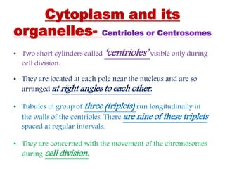
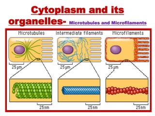
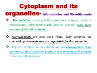
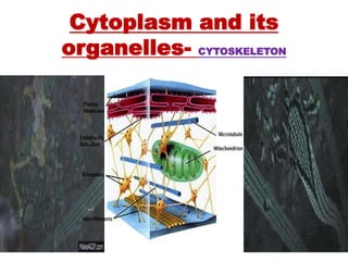
Download as PPSX, PPTX
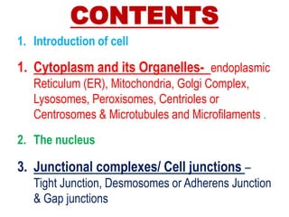
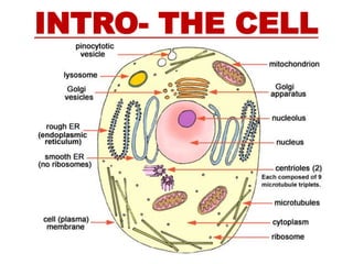
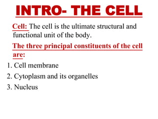
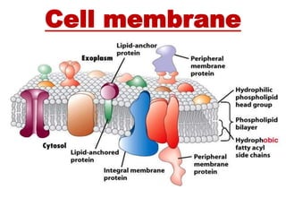
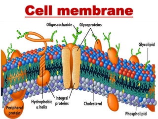
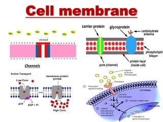
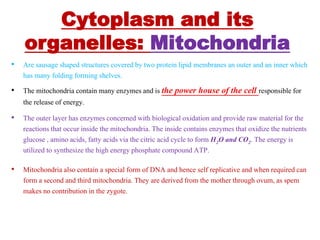
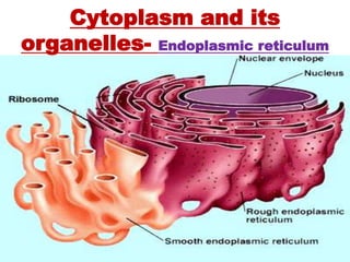
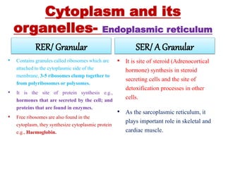
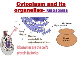
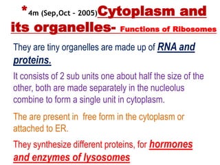
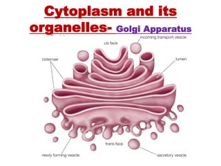
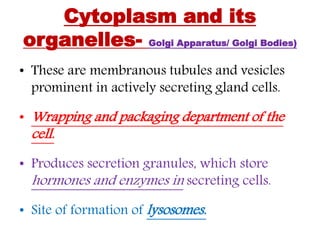
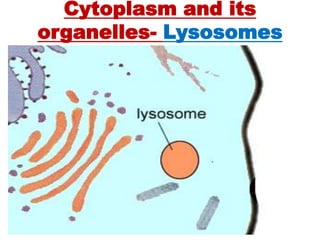

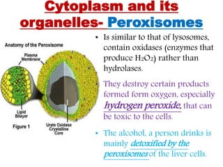
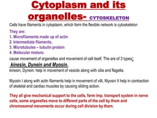
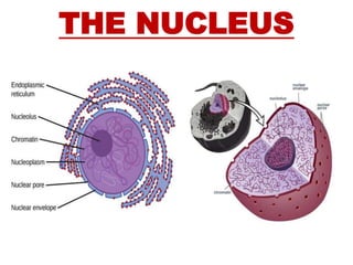
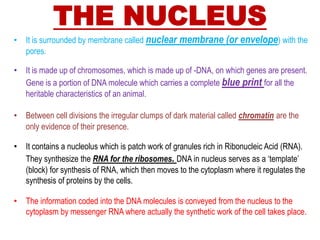
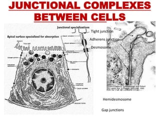
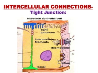
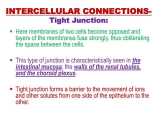
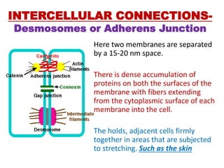
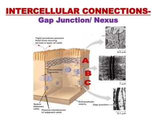
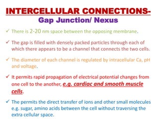


The document provides an overview of cell structure, focusing on the cell membrane, cytoplasm and its organelles, nuclear components, and intercellular connections. It details the functions of organelles such as mitochondria, endoplasmic reticulum, Golgi apparatus, lysosomes, and peroxisomes, as well as types of cell junctions like tight junctions, desmosomes, and gap junctions. Emphasis is placed on how these components contribute to the overall functionality and integrity of cells in the body.


![ONFH[AVN HIP] -TRIPLE REGIME -A NOVAL SURGICAL CONCEPT .pptx](https://cdn.slidesharecdn.com/ss_thumbnails/onfhavnhip2026koaconcalicutdrgokuldevdrmashraf-260210064517-213ec005-thumbnail.jpg?width=640&height=640&fit=bounds)


![PERI-PROSTHETIC FRACTURE NAIL-PLATE CONSTRUCT [NPC].pptx](https://cdn.slidesharecdn.com/ss_thumbnails/drarunkumardrmohamedashrafperiprostheticfrasturenail-plateconstructnpc-260209164459-7e9d15a1-thumbnail.jpg?width=640&height=640&fit=bounds)

