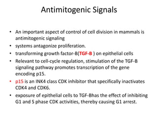
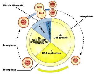
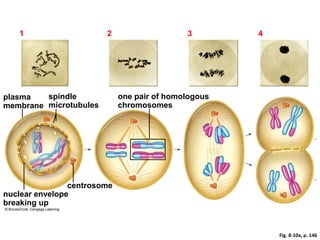
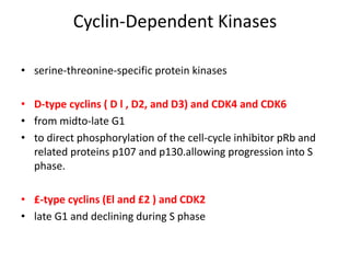
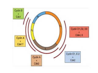
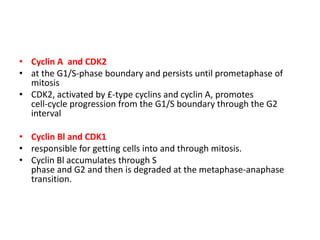
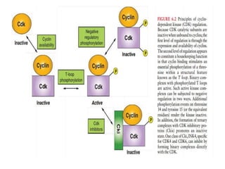
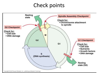
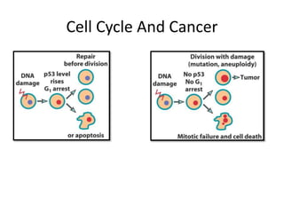
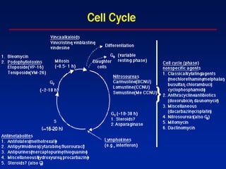
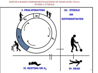
- The cell cycle consists of four main phases - G1, S, G2, and M. The G1, S, and G2 phases make up interphase.
- The cell cycle is tightly regulated by cyclins and cyclin-dependent kinases (CDKs). Different cyclin-CDK complexes control progression through the different cell cycle phases.
- Checkpoints exist to monitor DNA damage before progression into S phase and M phase. These checkpoints are regulated by proteins like ATM, ATR, Chk1, Chk2, and p53.
- Dysregulation of cell cycle control and checkpoint pathways contributes to uncontrolled cell proliferation in cancer. Both oncogenes and tumor
The combined use of radiation therapy and chemotherapy in cancer treatment is a logical and reasonable approach that has already proven beneficial for several malignancies.
It describes relationship between radiation dose and the fraction of cells that “survive” that dose.
This is mainly used to assess biological effectiveness of radiation.
To understand it better, we need to know about a few basic things e.g.
Cell Death
Estimation of Survival / Plating Efficiency
Nature of Cell killing etc.
A cell survival curve is the relationship between the fraction of cells retaining their reproductive integrity and absorbed dose.
Conventionally, surviving fraction on a logarithmic scale is plotted on the Y-axis, the dose is on the X-axis . The shape of the survival curve is important.
The cell-survival curve for densely ionizing radiations (α-particles and low-energy neutrons) is a straight line on a log-linear plot, that is survival is an exponential function of dose.
The cell-survival curve for sparsely ionizing radiations (X-rays, gamma-rays has an initial slope, followed by a shoulder after which it tends to straighten again at higher doses.
The phenomenon of signal transduction, also known as cell signaling, pertains to the intricate mechanisms that facilitate the transfer of biological information between cells. The effective coordination of diverse specialized cell types in various tissues and organs is a prerequisite for the proper functioning of complex multicellular organisms, necessitating intercellular communication. This communication must be continuous and dynamic to maintain coordination. Additionally, cell signaling pathways play a crucial role in the mechanisms of action of numerous drugs, including both local and general anesthetics. Consequently, a fundamental understanding of cell signaling mechanisms is imperative for comprehending various pathophysiologic and pharmacologic mechanisms.
Molecular event during Cell cycle By KK Sahu SirKAUSHAL SAHU
WHAT IS CELL?
WHAT IS CELL DIVISION OR CELL CYCLE?
WHY DO CELL DIVIDE?
HISTORY
CELL CYCLE
INTERPHASE
M-PHASE
MOLECULAR EVENT DURING CELL CYCLE AND CELL REGULATION
TYPES OF CELL DIVISION
IMPORTANCE OF CELL DIVISION
ABNORMALTIES OF CELL CYCLE
REFRENCES
This presentation on "Cell Cycle regulation" takes you to the cell cycle describing the stages and checkpoints involved providing some of the evidences of cell cycle regulation. Then we will move to cyclins and cyclin dependent kinases and the mechanism they follow.
This journey in regulation of cell cycle will take a halt after a general discussion of positive and negative cell cycle regulators.
Thankyou.
263778731218 Abortion Clinic /Pills In Harare ,sisternakatoto
263778731218 Abortion Clinic /Pills In Harare ,ABORTION WOMEN’S CLINIC +27730423979 IN women clinic we believe that every woman should be able to make choices in her pregnancy. Our job is to provide compassionate care, safety,affordable and confidential services. That’s why we have won the trust from all generations of women all over the world. we use non surgical method(Abortion pills) to terminate…Dr.LISA +27730423979women Clinic is committed to providing the highest quality of obstetrical and gynecological care to women of all ages. Our dedicated staff aim to treat each patient and her health concerns with compassion and respect.Our dedicated group ABORTION WOMEN’S CLINIC +27730423979 IN women clinic we believe that every woman should be able to make choices in her pregnancy. Our job is to provide compassionate care, safety,affordable and confidential services. That’s why we have won the trust from all generations of women all over the world. we use non surgical method(Abortion pills) to terminate…Dr.LISA +27730423979women Clinic is committed to providing the highest quality of obstetrical and gynecological care to women of all ages. Our dedicated staff aim to treat each patient and her health concerns with compassion and respect.Our dedicated group of receptionists, nurses, and physicians have worked together as a teamof receptionists, nurses, and physicians have worked together as a team wwww.lisywomensclinic.co.za/
Couples presenting to the infertility clinic- Do they really have infertility...Sujoy Dasgupta
Dr Sujoy Dasgupta presented the study on "Couples presenting to the infertility clinic- Do they really have infertility? – The unexplored stories of non-consummation" in the 13th Congress of the Asia Pacific Initiative on Reproduction (ASPIRE 2024) at Manila on 24 May, 2024.
The prostate is an exocrine gland of the male mammalian reproductive system
It is a walnut-sized gland that forms part of the male reproductive system and is located in front of the rectum and just below the urinary bladder
Function is to store and secrete a clear, slightly alkaline fluid that constitutes 10-30% of the volume of the seminal fluid that along with the spermatozoa, constitutes semen
A healthy human prostate measures (4cm-vertical, by 3cm-horizontal, 2cm ant-post ).
It surrounds the urethra just below the urinary bladder. It has anterior, median, posterior and two lateral lobes
It’s work is regulated by androgens which are responsible for male sex characteristics
Generalised disease of the prostate due to hormonal derangement which leads to non malignant enlargement of the gland (increase in the number of epithelial cells and stromal tissue)to cause compression of the urethra leading to symptoms (LUTS
NVBDCP.pptx Nation vector borne disease control programSapna Thakur
NVBDCP was launched in 2003-2004 . Vector-Borne Disease: Disease that results from an infection transmitted to humans and other animals by blood-feeding arthropods, such as mosquitoes, ticks, and fleas. Examples of vector-borne diseases include Dengue fever, West Nile Virus, Lyme disease, and malaria.
Lung Cancer: Artificial Intelligence, Synergetics, Complex System Analysis, S...Oleg Kshivets
RESULTS: Overall life span (LS) was 2252.1±1742.5 days and cumulative 5-year survival (5YS) reached 73.2%, 10 years – 64.8%, 20 years – 42.5%. 513 LCP lived more than 5 years (LS=3124.6±1525.6 days), 148 LCP – more than 10 years (LS=5054.4±1504.1 days).199 LCP died because of LC (LS=562.7±374.5 days). 5YS of LCP after bi/lobectomies was significantly superior in comparison with LCP after pneumonectomies (78.1% vs.63.7%, P=0.00001 by log-rank test). AT significantly improved 5YS (66.3% vs. 34.8%) (P=0.00000 by log-rank test) only for LCP with N1-2. Cox modeling displayed that 5YS of LCP significantly depended on: phase transition (PT) early-invasive LC in terms of synergetics, PT N0—N12, cell ratio factors (ratio between cancer cells- CC and blood cells subpopulations), G1-3, histology, glucose, AT, blood cell circuit, prothrombin index, heparin tolerance, recalcification time (P=0.000-0.038). Neural networks, genetic algorithm selection and bootstrap simulation revealed relationships between 5YS and PT early-invasive LC (rank=1), PT N0—N12 (rank=2), thrombocytes/CC (3), erythrocytes/CC (4), eosinophils/CC (5), healthy cells/CC (6), lymphocytes/CC (7), segmented neutrophils/CC (8), stick neutrophils/CC (9), monocytes/CC (10); leucocytes/CC (11). Correct prediction of 5YS was 100% by neural networks computing (area under ROC curve=1.0; error=0.0).
CONCLUSIONS: 5YS of LCP after radical procedures significantly depended on: 1) PT early-invasive cancer; 2) PT N0--N12; 3) cell ratio factors; 4) blood cell circuit; 5) biochemical factors; 6) hemostasis system; 7) AT; 8) LC characteristics; 9) LC cell dynamics; 10) surgery type: lobectomy/pneumonectomy; 11) anthropometric data. Optimal diagnosis and treatment strategies for LC are: 1) screening and early detection of LC; 2) availability of experienced thoracic surgeons because of complexity of radical procedures; 3) aggressive en block surgery and adequate lymph node dissection for completeness; 4) precise prediction; 5) adjuvant chemoimmunoradiotherapy for LCP with unfavorable prognosis.
Ethanol (CH3CH2OH), or beverage alcohol, is a two-carbon alcohol
that is rapidly distributed in the body and brain. Ethanol alters many
neurochemical systems and has rewarding and addictive properties. It
is the oldest recreational drug and likely contributes to more morbidity,
mortality, and public health costs than all illicit drugs combined. The
5th edition of the Diagnostic and Statistical Manual of Mental Disorders
(DSM-5) integrates alcohol abuse and alcohol dependence into a single
disorder called alcohol use disorder (AUD), with mild, moderate,
and severe subclassifications (American Psychiatric Association, 2013).
In the DSM-5, all types of substance abuse and dependence have been
combined into a single substance use disorder (SUD) on a continuum
from mild to severe. A diagnosis of AUD requires that at least two of
the 11 DSM-5 behaviors be present within a 12-month period (mild
AUD: 2–3 criteria; moderate AUD: 4–5 criteria; severe AUD: 6–11 criteria).
The four main behavioral effects of AUD are impaired control over
drinking, negative social consequences, risky use, and altered physiological
effects (tolerance, withdrawal). This chapter presents an overview
of the prevalence and harmful consequences of AUD in the U.S.,
the systemic nature of the disease, neurocircuitry and stages of AUD,
comorbidities, fetal alcohol spectrum disorders, genetic risk factors, and
pharmacotherapies for AUD.
Title: Sense of Smell
Presenter: Dr. Faiza, Assistant Professor of Physiology
Qualifications:
MBBS (Best Graduate, AIMC Lahore)
FCPS Physiology
ICMT, CHPE, DHPE (STMU)
MPH (GC University, Faisalabad)
MBA (Virtual University of Pakistan)
Learning Objectives:
Describe the primary categories of smells and the concept of odor blindness.
Explain the structure and location of the olfactory membrane and mucosa, including the types and roles of cells involved in olfaction.
Describe the pathway and mechanisms of olfactory signal transmission from the olfactory receptors to the brain.
Illustrate the biochemical cascade triggered by odorant binding to olfactory receptors, including the role of G-proteins and second messengers in generating an action potential.
Identify different types of olfactory disorders such as anosmia, hyposmia, hyperosmia, and dysosmia, including their potential causes.
Key Topics:
Olfactory Genes:
3% of the human genome accounts for olfactory genes.
400 genes for odorant receptors.
Olfactory Membrane:
Located in the superior part of the nasal cavity.
Medially: Folds downward along the superior septum.
Laterally: Folds over the superior turbinate and upper surface of the middle turbinate.
Total surface area: 5-10 square centimeters.
Olfactory Mucosa:
Olfactory Cells: Bipolar nerve cells derived from the CNS (100 million), with 4-25 olfactory cilia per cell.
Sustentacular Cells: Produce mucus and maintain ionic and molecular environment.
Basal Cells: Replace worn-out olfactory cells with an average lifespan of 1-2 months.
Bowman’s Gland: Secretes mucus.
Stimulation of Olfactory Cells:
Odorant dissolves in mucus and attaches to receptors on olfactory cilia.
Involves a cascade effect through G-proteins and second messengers, leading to depolarization and action potential generation in the olfactory nerve.
Quality of a Good Odorant:
Small (3-20 Carbon atoms), volatile, water-soluble, and lipid-soluble.
Facilitated by odorant-binding proteins in mucus.
Membrane Potential and Action Potential:
Resting membrane potential: -55mV.
Action potential frequency in the olfactory nerve increases with odorant strength.
Adaptation Towards the Sense of Smell:
Rapid adaptation within the first second, with further slow adaptation.
Psychological adaptation greater than receptor adaptation, involving feedback inhibition from the central nervous system.
Primary Sensations of Smell:
Camphoraceous, Musky, Floral, Pepperminty, Ethereal, Pungent, Putrid.
Odor Detection Threshold:
Examples: Hydrogen sulfide (0.0005 ppm), Methyl-mercaptan (0.002 ppm).
Some toxic substances are odorless at lethal concentrations.
Characteristics of Smell:
Odor blindness for single substances due to lack of appropriate receptor protein.
Behavioral and emotional influences of smell.
Transmission of Olfactory Signals:
From olfactory cells to glomeruli in the olfactory bulb, involving lateral inhibition.
Primitive, less old, and new olfactory systems with different path
Pulmonary Thromboembolism - etilogy, types, medical- Surgical and nursing man...VarunMahajani
Disruption of blood supply to lung alveoli due to blockage of one or more pulmonary blood vessels is called as Pulmonary thromboembolism. In this presentation we will discuss its causes, types and its management in depth.
These simplified slides by Dr. Sidra Arshad present an overview of the non-respiratory functions of the respiratory tract.
Learning objectives:
1. Enlist the non-respiratory functions of the respiratory tract
2. Briefly explain how these functions are carried out
3. Discuss the significance of dead space
4. Differentiate between minute ventilation and alveolar ventilation
5. Describe the cough and sneeze reflexes
Study Resources:
1. Chapter 39, Guyton and Hall Textbook of Medical Physiology, 14th edition
2. Chapter 34, Ganong’s Review of Medical Physiology, 26th edition
3. Chapter 17, Human Physiology by Lauralee Sherwood, 9th edition
4. Non-respiratory functions of the lungs https://academic.oup.com/bjaed/article/13/3/98/278874
Report Back from SGO 2024: What’s the Latest in Cervical Cancer?bkling
Are you curious about what’s new in cervical cancer research or unsure what the findings mean? Join Dr. Emily Ko, a gynecologic oncologist at Penn Medicine, to learn about the latest updates from the Society of Gynecologic Oncology (SGO) 2024 Annual Meeting on Women’s Cancer. Dr. Ko will discuss what the research presented at the conference means for you and answer your questions about the new developments.
2. • In 1 9 5 1 ,Howard and Pelc, studying the division o f plant
root cells,separated the process into four phases eventually
referred to as GAPl , synthetic phase, GAP2, and mitosis.
• four successive phases are referred to collectively as the cell
cycle.
• growth and protein synthesis occur constantly for the most
part.
• synthesis of DNA occurs only during a discrete interval.
3. • “Gap " phases-- it is now known that these are not
idle periods in a cell's life but the intervals in which most
regulation of the cell cycle is specifically exerted
• A large amount of information, originating from the external
environment and the cell's internal milieu, is integrated during
the G1 and G2 intervals and used to determine whether and
when to proceed into S phase and M phase, respectively.
4. Cell Cycle
fundamental cellular process
Wonderful convergence of cell biology, biochemistry and genetics
G0 – Resting phase
Interphase: time between divisions
• G1 Phase
– Begins immediately after division
– New organelles formed
– End of G1, cell has doubled in size
• S phase
– Duplicate copy of each chromosome
• G2 phase
– Cell prepares to begin mitosis
7. Phases of the Cell Cycle
• G0 – Resting phase
• G1 – organelles double, accumulates material for DNA synthesis.
• S – DNA synthesis & DNA doubles ( genetic material duplicated)
(10hrs)
• G2 –(4.5hrs) DNA synthesis ↓, protein and RNA synthesis-
continues & micro-tubular precursor are produced, ( proteins
necessary for cell division synthesized).
• M – genetic material is segregated into daughter cells (
components of mother cell are divided in to two daughter cells-
each contains the exact same chromosomes as the original
mother cell) (30 min)
• Cytokinesis – cytoplasm divides & two daughter cells formed.
8. Mitosis subdivided into five phases:
• Prophase: internal membranous compartments of the cell, including
the nucleus, are disassembled and dispersed
• Prometaphase: chromosomes form bivalent attachments to the
spindle, driving them to the cellular equator
• Metaphase: Proper alignment of paired chromatids on the spindle.
• Anaphase: paired sister chromatids lose cohesion and microtubule
forces separate the chromatids and pull them to opposite poles of
the cell
• Telophase: the events of prophase are reversed (nuclei and other
membrane structures reassemble, the chromosomes decondense,
and protein synthesis resumes)
9. Fig. 8-10a, p. 146
1 Prophase I 2 Metaphase I 3 Anaphase I 4 Telophase I
one pair of homologous
chromosomes
plasma
membrane
spindle
microtubules
nuclear envelope
breaking up
centrosome
10. Cyclin-Dependent Kinases
• serine-threonine-specific protein kinases
• D-type cyclins ( D l , D2, and D3) and CDK4 and CDK6
• from midto-late G1
• to direct phosphorylation of the cell-cycle inhibitor pRb and
related proteins p107 and p130.allowing progression into S
phase.
• £-type cyclins (El and £2 ) and CDK2
• late G1 and declining during S phase
11.
12. • Cyclin A and CDK2
• at the G1/S-phase boundary and persists until prometaphase of
mitosis
• CDK2, activated by £-type cyclins and cyclin A, promotes
cell-cycle progression from the G1/S boundary through the G2
interval
• Cyclin Bl and CDK1
• responsible for getting cells into and through mitosis.
• Cyclin Bl accumulates through S
phase and G2 and then is degraded at the metaphase-anaphase
transition.
13.
14.
15. INDUCTION OF CELL-CYCLE
PHASE TRANSITIONS
• The cell cycle is composed of two action phases, S phase and
M phase, in which the genetic material is duplicated and the
components of a mother cell are divided into two daughter
cells, respectively
• The intervening phases, G1 and G2, are
thought to exist primarily to allow time for cell growth
• cell proliferation is controlled operationally
at two key transitions: that between G1 and S phase and that
between G2 and M phase
16. • cyclins E and A--for the G1 -S phase transition
• These kinases are kept in check by the action of Cip/Kip family
inhibitors.
• If the internal and external environments are permissive for
proliferation, the continued accumulation of
cyclins will eventually titrate the inhibitors, allowing the latter
to be phosphorylated by free cyclin-CDK complexes.
• Phosphorylation then marks these inhibitors as targets of
ubiquitin-mediated proteolysis
• The concerted destruction of CDK inhibitors and concomitant
activation of the entire pool of CDK complexes assure that the
transition into S phase is rapid and irreversible.
17. • Cyclin B-CDK1 complexes accumulate starting near
the end of S phase but are held in check not by CDK inhibitors
but by negative regulatory phosphorylation of CDK l .
• This phosphorylation on threonine 14 and tyrosine 15 is
carried out by kinases Weel and Mytl.
• Entry into M phase is signaled
by the rapid dephosphorylation of T14 and Y15, resulting in
activation of CDKl.
• These positive feedback
dynamics leading to the simultaneous activation of a large
accumulated pool of cyclin B-CDK1 assures that entry into
mitosis is decisive.
18. UBIQUITIN-MEDIATED
PROTEOLYSIS
• Ubiquitin is a 76-amino acid polypeptide
• The enzymes that transfer ubiquitin to target proteins are
known as protein-ubiquitin ligases.
• From the perspective of cell-cycle control, two families of
protein-ubiquitin ligases have predominant roles.
• The first family, SCF
(Skp1 -Cullin-F-box protein), specifically targets proteins that
are marked for destruction by phosphorylation.
• The second family of protein-ubiquitin ligases that is critical
for cell-cycle control is known collectively as the APC/C
19.
20. Quiescence and Differentiation
• The most fundamental aspect of cell-cycle control is the
regulation of entry and exit.
• For mammalian cells, the decision to
enter or exit the proliferative mode is based on environmental
signals such as mitogens, growth factors, hormones, and
cellcell contact, as well as on internal differentiation programs
• If the state of cell-cycle exit is reversible, it is referred to as
quiescence
• Entry into and exit from quiescence are mediated largely by
growth factors and mitogens that interact with cell surface
receptors
21. • These in turn are linked to intracellular signaling cascades that
up-regulate the rate of protein synthesis as well as the
transcription of genes that promote proliferation, such as
those encoding CDKs and cyclins.
22.
23. Antimitogenic Signals
• An important aspect of control of cell division in mammals is
antimitogenic signaling
• systems antagonize proliferation.
• transforming growth factor-B(TGF-B ) on epithelial cells
• Relevant to cell-cycle regulation, stimulation of the TGF-B
signaling pathway promotes transcription of the gene
encoding p15.
• p15 is an INK4 class CDK inhibitor that specifically inactivates
CDK4 and CDK6.
• exposure of epithelial cells to TGF-Bhas the effect of inhibiting
G1 and S phase CDK activities, thereby causing G1 arrest.
24.
25. Checkpoints
• Cells are constantly faced with insults, resulting in damage
that can threaten their survival
• These insults can be generated internally as chemically active
by-products of metabolism or can originate in the external
environment; for example, chemical agents or radiation.
• As a result, mechanisms have evolved to remove damaged
molecules and make necessary repairs.
• cell-cycle progression would be harmful or catastrophic before
repair of damage, further mechanisms have evolved to delay
progression pending repair.
• These are called cell-cycle checkpoints
26. • Cells are particularly susceptible to the harmful effects
of DNA damage at two points in the cell cycle: S phase and M
phase.
• Replication that does traverse regions of unrepaired DNA
damage is likely to be error-prone, resulting in accumulation
of mutations
• segregation of severely damaged chromosomes at
mitosis might lead to loss of genetic information, seriously
threatening the survival or integrity of daughter cells.
•
28. DNA Damage Checkpoints
• cell-cycle progression is blocked at three points
before S phase entry (the G1 DNA damage checkpoint)
during S phase (the intra-S phase DNA damage checkpoint)
before M phase entry (the G2 DNA damage checkpoint)
• DNA damage of various forms is first detected by DNA-bound
protein complexes that serve as sensors.
29. • ATM and ATR are activated by DNA damage at all points in the
cell cycle
• The principal transcriptional target of p53 in the context of
the G1 checkpoint is the Cip/Kip inhibitor p2 1 Cipl
• int is the Cip/Kip inhibitor p2 1 Cipl . The resulting high levels
of p2 1 block CDK2 activity and possibly CDK4 and CDK6
activity, leading to G1 arrest
30. • G2 DNA damage checkpoint is p53- independent
• It involves one of two effector protein kinases
known as chk l and chk2 --inhibits cyclin B-CDK1 complexes
• The intra-S phase DNAdamage checkpoint response appears
to be p53-independent but requires the chk1 or chk2 kinases,
• It affects CDK2
• Because ongoing DNA replication requires the
activity of CDK2, DNA synthesis ceases until damage is
repaired.
31. Replication Checkpoint
• Although the signaling pathways are somewhat
different, the replication checkpoint ultimately functions like
the G2 DNA damage checkpoint in that mitotic entry is
blocked by inhibiting CDC25C via the action of chk1 , thus
preventing activation of CDKl.
32. Spindle Integrity Checkpoint
• assembling a mitotic spindle and attaching chromosomes to it
are extensively monitored processes.
• The mechanism of delay at prometaphase or metaphase in
response to spindle defects or improper chromosome
attachment is referred to as the spindle integrity checkpoint
• The target is the essential APC/C cofactor, CDC20
• Unattached or improperly attached kinetochores not
experiencing an appropriate level of tension indicative of
bipolar attachment inhibit CDC20 function.
• As a result, cells are prevented from initiating anaphase until
all kinetochores are properly attached to
a bipolar spindle
33. Restriction Point
• Cells deprived of an essential nutrient or growth factor are
blocked from cell-cycle progression at a point in mid-G1–
known as Restriction point.
• most malignant cells do not have a functional restriction
point, which presumably helps them evade normal growth
control signals.
34. Senescence
• All normal mammalian cells have a finite proliferative lifespan.
As cells approach the end of their proliferative capacity, they
enter a state referred to as replicative senescence.
• it has been speculated that restricting cells to a finite number
of divisions may be a protective mechanism against malignant
growth
• Senescence is characterized by the accumulation of high
levels of CDK inhibitors and ultimately permanent G1 arrest
• It is one of the requirements of malignant transformation of
cells is to overcome the senescence barrier so as to provide
tumor cells with unlimited proliferative capacity.
35. CELL CYCLE AND CANCER
• Cancer is partly a disease of uncontrolled proliferation.
• cell-cycle and checkpoint genes are often found misregulated
or mutated in cancer.
Protooncogenes--Genes in which mutations give rise to a gain
of function or an enhanced level of function, leading to
malignancy.
• usually encode growth- or division-promoting proteins
• Dominant
Tumor suppressor genes--Genes that give rise to loss of
function mutations that lead to malignancy
• Usually encode negative regulators of growth and
proliferation that protect cells from malignancy
• Recessive
37. • to achieve uncontrolled cell division, two basic requirements
must be met.
• First, cells need a strong constitutive proliferation signal
capable of overriding the environmental and internal
restraints on division that normal cells
experience.
• E.g.-Mutation in Rb, p107, p130 gene…
38. • Second, the barrier of senescence needs to be dismantled to
render tumor cells immortal.
• E.g.—p53
39. Cell cycle & cancer therapy
Cell cycle phase specific agents:
• Drugs that inhibit or kill during a specific phase of the
cell cycle
• Generally produces the greatest cell kill if the amount
of drug is divided and given in repeated fractions
instead of a single large dose
• More effective against tumors with high growth
fraction
40. • Cell cycle phase non specific agents:
• Work by DNA cross linking, strand breakage or interfering with
DNA/RNA synthesis
• Exert a cytotoxic effect throughout the cell cycle
• Some of them are effective against cells in resting phase
• Commonly see a dose-response relationship
• Concentration dependent – more effective when given in larger
intermittent doses
-
41. • cell-cycle proteins have been suggested as targets for
therapeutic exploitation.
• Notably, CDKs have been extensively screened for small-
molecule inhibitors, some of which are in clinical trials.
• An alternative approach being explored is to develop agents
that undermine checkpoint responses.
• However, it is noteworthy that many therapeutic approaches
currently use compounds that normally trigger checkpoint
responses, such as genotoxic agents or spindle poisons.
42. • Oncolytic adenoviruses have therefore been engineered to
not express E1B p55K.66
• These adenoviruses are harmless to normal cells but can
productively infect and lyse p53-defective tumor cells in tissue
culture and mouse xenograft models. However, technical
issues such as low tumor infectivity, rapid viral clearance and
neutralizing immune responses in clinical trials have limited
the efficacy of this approach.66
• On the other hand, if new generations of oncolytic viruses
that circumvent these problems can be developed, this may
constitute one of the more promising new therapeutic
approaches.
45. 45
Cellular Kinetics
• Human body contains 5x10¹³ cells
• Cells can either
-non dividing and terminally differentiated
- continually proliferating
- rest but may be recruited into cell cycle
• Tumors becomes clinically detectable when there is a
mass of 109 cells (1g)
46. 46
• number of
• cancer cells
diagnostic
threshold
(1cm)
time
undetectable
cancer
detectable
cancer
limit of
clinical
detection
host
death
10 12
10 9
Tumor Growth
47. 47
Tumour kinetic
Growth rate and Volume doubling time (Td)
dependson:
growth fraction (GF)
cell cycle time (TC)
rate of cell loss
Tumors grow faster if :
TC decreases ,GF increases and cell loss
decreases
48. 48
Factors Affecting Tumor Growth
• Growth fraction (fraction of cells in population which are
actually cycling)
– Even in tumors most cells are not cycling
– Cycling cells are well oxygenated and fed
– Large growth fraction will usually result in rapid tumor
growth.
• Cell cycle time
– Cell cycle times vary widely within a given tumor.
– Tumors of the same type may have different average
cell cycle times
– Slow is generally equated with benign tumors
– Fast is generally equated with malignancy
– The difference in total cell cycle time bet the two cells is
due to diff in the length of the G1- phase.
49. 49
• Cell loss fraction
– Cells are lost from the tumor population in
several ways.
– Nonviable replication of deranged cells will
result in loss of those cells
– DNA is too altered for a functional cell to
exist
– Anoxia, cell death from poor blood supply
– Attack of antigenetic cells by immune
system
51. 51
THE VARIOUS TUMOUR COMPARTMENTS
Tumour cells are distributed in the following compartments :
Growth Fraction
• Contributes to tumor volume by prodn of all new
tumor.. cells
– All cells actively go through the ‘Cell Cycle’
– These cells are also designated ‘P’ (proliferating) Cells
II. Resting (G0)Compartment –
– Cells capable of entering the ‘P’ compartment (i.e. re-
entering Cell Cycle)
– Some G0 cells may be clonogenic (capacity to
repopulate the tumor), hence risk to host & need to be
eliminated with appropriate Rx
52. III. Sterile (Differentiated) Compartment –
– No longer capable of cell division
– Hence, also called ‘Q’ (quiescent) Cells
– At times difficult to differentiate bet. G0 from Q cells.
IV. Dead & Dying Cell Compartment –
– Present in most tumors, due to deficient blood supply
– Comprises of pyknotic cells and varying areas of
necrosis also
53. 53
Gompertzian Growth
• Growth rates are exponential at early stages of
development and slower at later stages of development.
• Growth fraction of the tumor
is not constant but decreases
exponentially with time
- Biological growth follows this characteristic curve.
54. 54
Gompertzian Kinetics and tumour response
Although murine leukemias follow exponential cell kinetics, most solid
tumours show Gompertzian model of growth & regression since the GF is not
constant but decreases exponentially with time while exponential growth is
always matched by exponential retardation.
A typical tumour grows rapidly with a fixed GF depending on cell type and
shows peak activity when it reaches 37% of its max. size, after which there is
retardation in its growth.
Under the Gompertzian model, when a patient with advanced cancer is
treated, tumor mass is LARGE, it’s GF is LOW and the FRACTION OF CELL KILL
is therefore SMALL.
Tumour Negligible - GF
Tumour enlarges but GF regresses, thus causing effect of CT
GF
Tumour Mass
56. 56
Cell Cycle.
• The point that a cell is in the cell cycle has a
marked influence on its response and survival
of irradiation.
• G1 & G0 are relatively insensitive to radiation
injury.
• S phase is generally considered to be the most
resistant to radiation injury.
• G2M is the most sensitive phase .
57. 57
Repair Of Radiation Damage
• Repair of sublethal damage
• Reassortment
• Repopulation
• Reoxygenation
Repair of SLD–
• Following a D0 level dose there is repair of radiation
injury in surviving cells
– Cells with long cell cycle times generally have a
wider repair shoulder on the survival curve
– Cells with short cell cycle time generally have a
narrow repair shoulder.
58. 58
Repair (Cont)
– Tumor cells are considered to have short cell
cycle times
• Fractionation will broaden the survival shoulder
more for late responding tissue than early responding
tissues.
– At high doses the cell survival curve actually
indicates lower survival for late responding cells
59. 59
Reassortment (Redistribution)
• Cells in G2 and M are most sensitive and more
likely to be killed.
• Cells in S are more resistant and likely to survive
• A radiation induce mitotic arrest is likely present
60. 60
– Reirrradition will then again selectively kill
cells in the radiation sensitive portions of the
cell cycle
– It improves chances of cells being irradiated in
a sensitive part of the cycle
– Tumor cells on average have shorter cell cycle
times than normal tissues
– It favors survival of normal late responding
tissues
61. 61
Regeneration (Repopulation)
– Following irradiation some cell populations
will exhibit increased cell division.
• Usually follows a period of mitotic arrest
– It tends to begin more quickly in normal
early responding tissues than in tumors.
– It favors survival of normal early responding
tissues over tumors.
62. 62
Reoxygenation
– Hypoxia in many tumors blunts radiation injury
• 2-3 times as much dose required to kill hypoxic
cells
– Normal tissues are not hypoxic as a rule
– However, of the well oxygenated cells in a tumor
there is usually a high percentage of cycling cells.
Large numbers of cycling tumor cells are killed
– Cells previously of marginal oxygenation survive and
move into the oxygenated zone
– These newly oxygenated cells then start to cycle and
are then susceptible to the next dose due to being
oxygenated and cycling
– All tumor cells can be reoxygenated this way if
enough fractions used
64. • The goal of radiation therapy is to maximize the radiation
injury to tumor cells while minimizing the injury to normal
cells
• Greater cell killing effect for rapidly cycling cell than for slowly
cycling cells
• The repair shoulder is broader for late responding tissue than
for acute ones in dose range.
• Fractionation promotes reoxygenation
• Fractionation promotes repeated reassortment
65. • Normal early responding tissues and tumor tissues respond
similarly
• Possible slight advantage for normal cells for repopulation