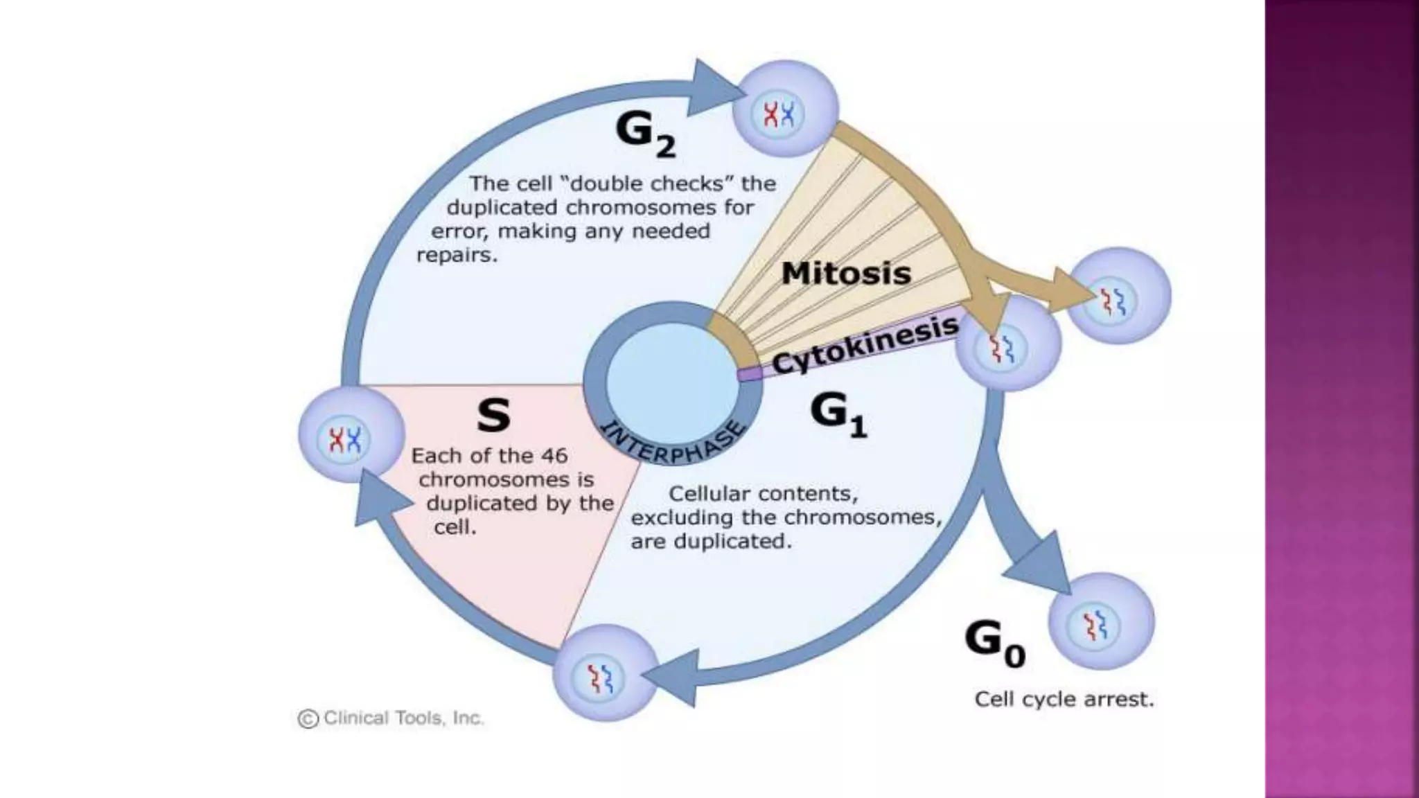
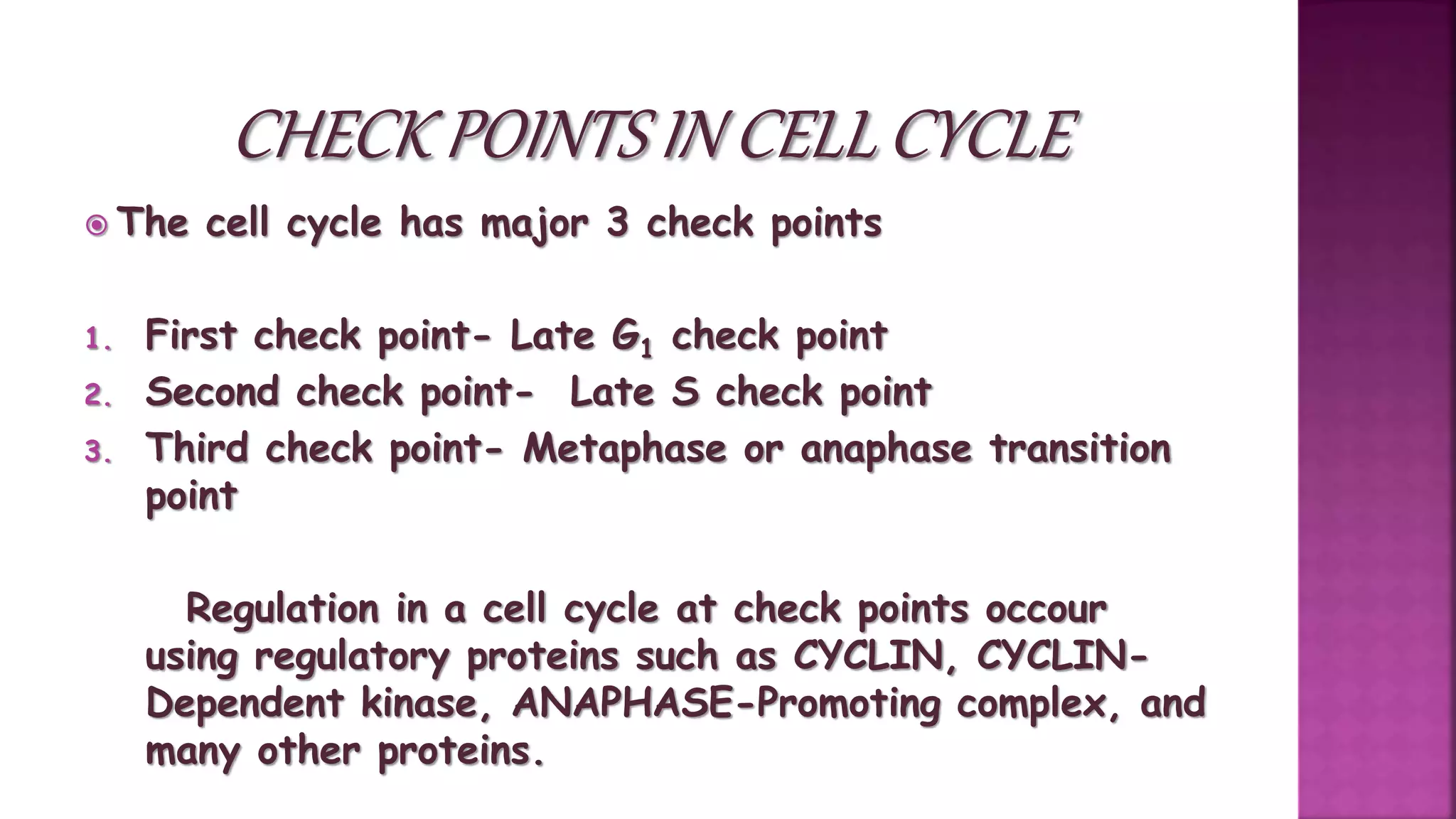
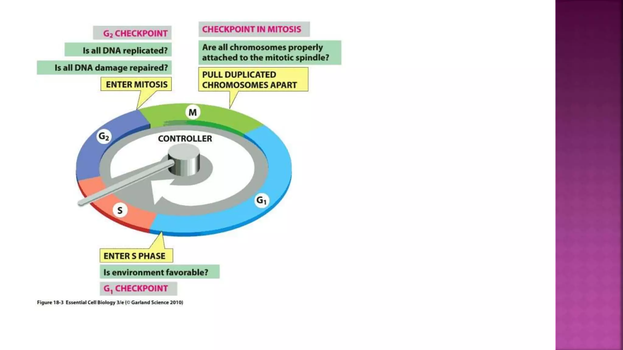
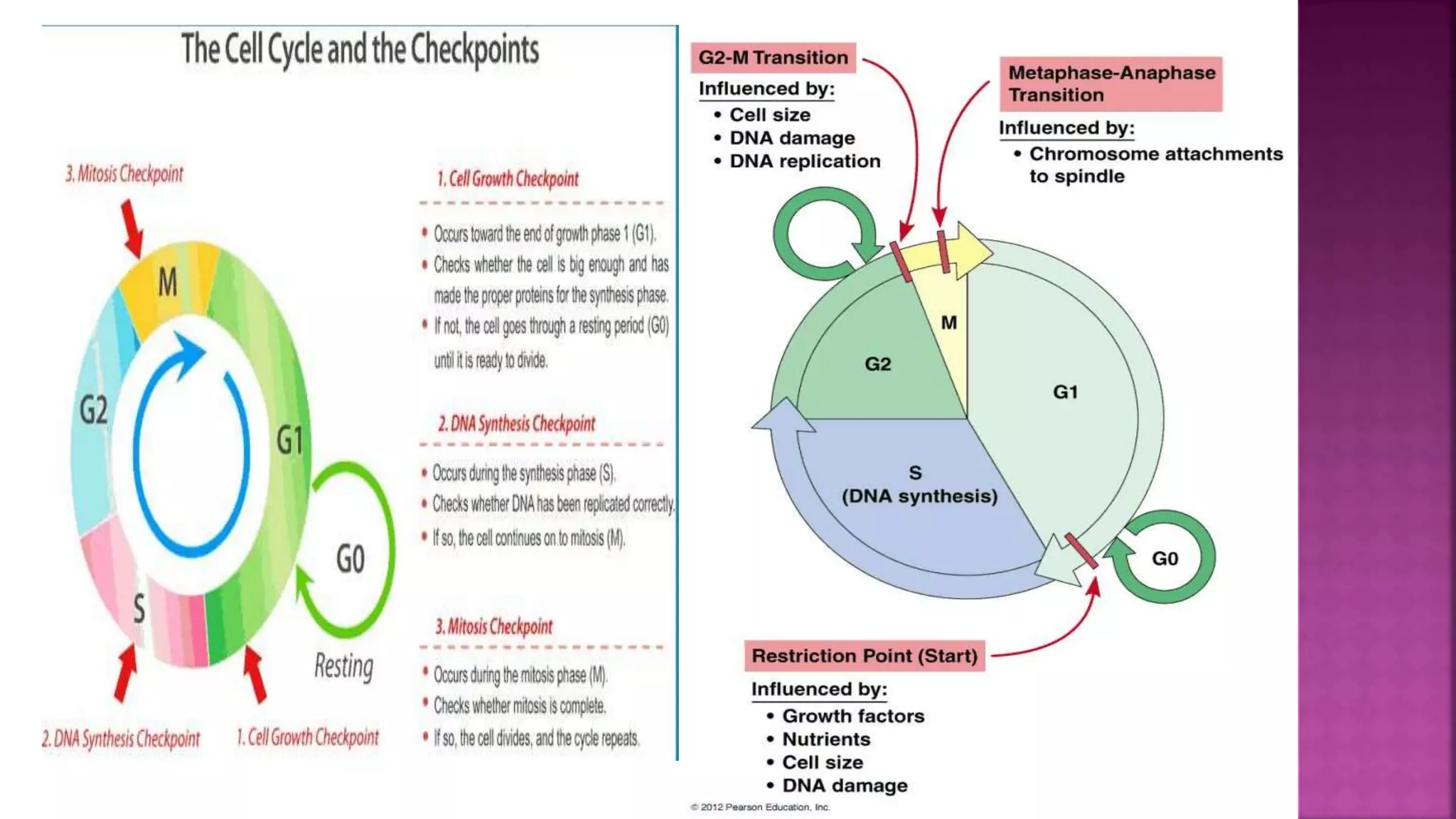
This document summarizes the cell cycle and its regulation. It describes that the cell cycle consists of interphase and M-phase. Interphase includes G1, S, and G2 phases where the cell grows and duplicates its DNA. M-phase is where the cell divides. Regulation occurs through cyclins, cyclin-dependent kinases (CDKs), and checkpoint proteins that control phase transitions. CDK activity is regulated by binding with cyclins to form active complexes, or being inactivated through phosphorylation. Precise coordination of these elements ensures orderly cell division and replication.
Basic Cell cycle regulation suitable for undergraduate students.
This presentation has been started from the basics to enable easy understanding. It covers all the details of cell cycle regulation in yeast as well as higher eukaryotes.
Basic Cell cycle regulation suitable for undergraduate students.
This presentation has been started from the basics to enable easy understanding. It covers all the details of cell cycle regulation in yeast as well as higher eukaryotes.
This presentation on "Cell Cycle regulation" takes you to the cell cycle describing the stages and checkpoints involved providing some of the evidences of cell cycle regulation. Then we will move to cyclins and cyclin dependent kinases and the mechanism they follow.
This journey in regulation of cell cycle will take a halt after a general discussion of positive and negative cell cycle regulators.
Thankyou.
A detailed description of molecular level of cell cycle. Its regulation by different checkpoints. The Structure and Function of MPF. Description of MPF discovery.
Apoptosis is an orderly process in which the cell's contents are packaged into small packets of membrane for “garbage collection” by immune cells. Apoptosis removes cells during development, eliminates potentially cancerous and virus-infected cells, and maintains balance in the body.
Cell cycle regulation presentation by me and my colleagues. Not the Best work but still it will give a general idea about DNA damage checkpoints, roles of Cdk-Cyclin complexes, Rb proteins, ATM&ATR kinases, p51, etc.
Reference : Nature reviews & The Cell a molecular approach. (cooper)
A detailed description of programmed cell death mechanism also called Apoptosis.
It explains about the factors, mechanism and pathways involved in the apoptosis.
Cell cycle and Regulation
* cell Division is occur in every human but these have certaint check point to preventing from the forming the defective cell or cancerious cell.
ONCOGENE AND PROTOONCOGENE
P53 GENE AND ITS APPLICATION IN CANCER ETIOLOGY
TUMOUR SUPPRESSOR GENE AND BCA AND BAC GENE AND ITS APPLICATION ON THE APOPTOSIS AND DEATH RECEPTORS
Why do different cell types' rates of the cell cycle differ?
The cell cycle is swiftly completed by injured or lost cell types to produce replacements.
Adult skin and digestive tract cells go through the cell cycle quite fast, whereas nervous system cells divide very seldom.
Cells divide regularly during embryonic development, perhaps as frequently as once or twice an hour, moving through the cell cycle very quickly.
What is the cell cycle?
The regular sequence of activities that cells go through as they develop and divide is known as the cell cycle. Prokaryotic cells go through a rapid cycle of cell division, DNA replication, and expansion. In prokaryotes, cell division occurs in a single stage known as binary fission (shown right).Compared to prokaryotic cells, eukaryotic cells have a more complicated cell cycle.
How is the eukaryotic cell cycle divided?
Interphase is the period between cell divisions. Depending on the kind of cell, the interphase might be shorter or longer.
The three stages or phases of the eukaryotic interphase are G1, S, and G2.
The M phase of the cell cycle is when eukaryotic cells divide. Mitosis and cytokinesis are the two stages that make up the M phase.
What happens during each phase of eukaryotic interphase?
G1: Cells do most of their growing during this phase. It begins when mitosis is complete and ends when DNA replication begins.
S: DNA is synthesized as chromosomes are replicated.
G2: Many of the molecules and cell structures required for cell division are produced; usually the shortest phase of the cell cycle.
What happens during the M phase of the eukaryotic cell cycle?
The M phase is usually much shorter than interphase and results in two daughter cells.
The first step of the M phase is mitosis. The cell’s nucleus divides during mitosis.
The second step of the M phase is cytokinesis, during which the cell’s cytoplasm is divided.
What are the steps of mitosis?
Mitosis consists of four steps: prophase, metaphase, anaphase, and telophase.
Prophase: nuclear envelope breaks down, DNA condenses, spindle begins to form.
Metaphase: replicated chromosomes, which appear as paired sister chromatids, line up across the center of the cell and attach to spindle.
Anaphase: sister chromatids separate and move toward ends of the cell.
Telophase: chromosomes disperse, nuclear envelope reforms.
What completes the M phase of the cell cycle?
Cytokinesis completes the M phase of the cell cycle. It may begin while telophase is still taking place.
During cytokinesis, the cytoplasm (which includes all of the contents of a eukaryotic cell outside the nucleus) draws inward, eventually pinching off into two nearly equal parts. Each part contains a nucleus.
In plant cells and other eukaryotic cells that have a cell wall, a cell plate forms halfway between the divided nuclei. It gradually develops into cell membranes and forms a complete cell wall surrounding each daughter cell.
Upon the completion of cytokinesis and the M phase, a
The cell cycle, or cell-division cycle, is the series of events that take place in a cell leading to duplication of its DNA (DNA replication) and division of cytoplasm and organelles to produce two daughter cells.
Seminar of U.V. Spectroscopy by SAMIR PANDASAMIR PANDA
Spectroscopy is a branch of science dealing the study of interaction of electromagnetic radiation with matter.
Ultraviolet-visible spectroscopy refers to absorption spectroscopy or reflect spectroscopy in the UV-VIS spectral region.
Ultraviolet-visible spectroscopy is an analytical method that can measure the amount of light received by the analyte.
(May 29th, 2024) Advancements in Intravital Microscopy- Insights for Preclini...Scintica Instrumentation
Intravital microscopy (IVM) is a powerful tool utilized to study cellular behavior over time and space in vivo. Much of our understanding of cell biology has been accomplished using various in vitro and ex vivo methods; however, these studies do not necessarily reflect the natural dynamics of biological processes. Unlike traditional cell culture or fixed tissue imaging, IVM allows for the ultra-fast high-resolution imaging of cellular processes over time and space and were studied in its natural environment. Real-time visualization of biological processes in the context of an intact organism helps maintain physiological relevance and provide insights into the progression of disease, response to treatments or developmental processes.
In this webinar we give an overview of advanced applications of the IVM system in preclinical research. IVIM technology is a provider of all-in-one intravital microscopy systems and solutions optimized for in vivo imaging of live animal models at sub-micron resolution. The system’s unique features and user-friendly software enables researchers to probe fast dynamic biological processes such as immune cell tracking, cell-cell interaction as well as vascularization and tumor metastasis with exceptional detail. This webinar will also give an overview of IVM being utilized in drug development, offering a view into the intricate interaction between drugs/nanoparticles and tissues in vivo and allows for the evaluation of therapeutic intervention in a variety of tissues and organs. This interdisciplinary collaboration continues to drive the advancements of novel therapeutic strategies.
The ability to recreate computational results with minimal effort and actionable metrics provides a solid foundation for scientific research and software development. When people can replicate an analysis at the touch of a button using open-source software, open data, and methods to assess and compare proposals, it significantly eases verification of results, engagement with a diverse range of contributors, and progress. However, we have yet to fully achieve this; there are still many sociotechnical frictions.
Inspired by David Donoho's vision, this talk aims to revisit the three crucial pillars of frictionless reproducibility (data sharing, code sharing, and competitive challenges) with the perspective of deep software variability.
Our observation is that multiple layers — hardware, operating systems, third-party libraries, software versions, input data, compile-time options, and parameters — are subject to variability that exacerbates frictions but is also essential for achieving robust, generalizable results and fostering innovation. I will first review the literature, providing evidence of how the complex variability interactions across these layers affect qualitative and quantitative software properties, thereby complicating the reproduction and replication of scientific studies in various fields.
I will then present some software engineering and AI techniques that can support the strategic exploration of variability spaces. These include the use of abstractions and models (e.g., feature models), sampling strategies (e.g., uniform, random), cost-effective measurements (e.g., incremental build of software configurations), and dimensionality reduction methods (e.g., transfer learning, feature selection, software debloating).
I will finally argue that deep variability is both the problem and solution of frictionless reproducibility, calling the software science community to develop new methods and tools to manage variability and foster reproducibility in software systems.
Exposé invité Journées Nationales du GDR GPL 2024
Toxic effects of heavy metals : Lead and Arsenicsanjana502982
Heavy metals are naturally occuring metallic chemical elements that have relatively high density, and are toxic at even low concentrations. All toxic metals are termed as heavy metals irrespective of their atomic mass and density, eg. arsenic, lead, mercury, cadmium, thallium, chromium, etc.
Comparing Evolved Extractive Text Summary Scores of Bidirectional Encoder Rep...University of Maribor
Slides from:
11th International Conference on Electrical, Electronics and Computer Engineering (IcETRAN), Niš, 3-6 June 2024
Track: Artificial Intelligence
https://www.etran.rs/2024/en/home-english/
Observation of Io’s Resurfacing via Plume Deposition Using Ground-based Adapt...Sérgio Sacani
Since volcanic activity was first discovered on Io from Voyager images in 1979, changes
on Io’s surface have been monitored from both spacecraft and ground-based telescopes.
Here, we present the highest spatial resolution images of Io ever obtained from a groundbased telescope. These images, acquired by the SHARK-VIS instrument on the Large
Binocular Telescope, show evidence of a major resurfacing event on Io’s trailing hemisphere. When compared to the most recent spacecraft images, the SHARK-VIS images
show that a plume deposit from a powerful eruption at Pillan Patera has covered part
of the long-lived Pele plume deposit. Although this type of resurfacing event may be common on Io, few have been detected due to the rarity of spacecraft visits and the previously low spatial resolution available from Earth-based telescopes. The SHARK-VIS instrument ushers in a new era of high resolution imaging of Io’s surface using adaptive
optics at visible wavelengths.
Deep Behavioral Phenotyping in Systems Neuroscience for Functional Atlasing a...Ana Luísa Pinho
Functional Magnetic Resonance Imaging (fMRI) provides means to characterize brain activations in response to behavior. However, cognitive neuroscience has been limited to group-level effects referring to the performance of specific tasks. To obtain the functional profile of elementary cognitive mechanisms, the combination of brain responses to many tasks is required. Yet, to date, both structural atlases and parcellation-based activations do not fully account for cognitive function and still present several limitations. Further, they do not adapt overall to individual characteristics. In this talk, I will give an account of deep-behavioral phenotyping strategies, namely data-driven methods in large task-fMRI datasets, to optimize functional brain-data collection and improve inference of effects-of-interest related to mental processes. Key to this approach is the employment of fast multi-functional paradigms rich on features that can be well parametrized and, consequently, facilitate the creation of psycho-physiological constructs to be modelled with imaging data. Particular emphasis will be given to music stimuli when studying high-order cognitive mechanisms, due to their ecological nature and quality to enable complex behavior compounded by discrete entities. I will also discuss how deep-behavioral phenotyping and individualized models applied to neuroimaging data can better account for the subject-specific organization of domain-general cognitive systems in the human brain. Finally, the accumulation of functional brain signatures brings the possibility to clarify relationships among tasks and create a univocal link between brain systems and mental functions through: (1) the development of ontologies proposing an organization of cognitive processes; and (2) brain-network taxonomies describing functional specialization. To this end, tools to improve commensurability in cognitive science are necessary, such as public repositories, ontology-based platforms and automated meta-analysis tools. I will thus discuss some brain-atlasing resources currently under development, and their applicability in cognitive as well as clinical neuroscience.
2. • Cell division is very important process in a living organisms. During the division of a cell, DNA replication and cell growth also
tales place.
• All the processes, i.e., cell division, DNA replication, and cell growth, hence have to take place in a coordinated way to ensure
correct division and formation of progeny cells containing intact genomes.
• Although growth (in terms of cytoplasmic increase) is a continuous process, DNA synthesis occurs only during one specific stage in
the cell cycle.
• The replicated chromosomes (DNA) are then distributed to the daughter nuclei by a complex series of events during cell division.
These events are themselves under genetic control. Complete life cycle is called as cell cycle.
3. There are 2 phases of cell cycle :-
1. Interphase
2. M-phase
4.
5. The most active phase of the whole cell cycle.
Constitutes 95% of whole cell cycle.
It is the preparatory phase for cell division.
In interphase metabolism of cell increases. A series of metabolic changes
occurs during interphase in cell. These changes are not visible under
microscope, so some scientists termed interphase as resting phase.
6. Howard and Pelc classifieds interphase into 3 subjects categories.
1. G1 phase or Pre DNA synthesis phase (1st gap phase)
2. S phase (DNA Synthesis phase)
3. G2 phase (2nd Gap phase) or Post DNA synthesis phase (Pre mitosis
phase).
7. G1 phase also known as pre synthetic phase as it occurs between mitosis and initiation between
DNA replication,
During this stage cell is metabolically active and continuously grows.
Cell organelle increase and rapidly synthesizes different types of RNA and proteins.
As now proteins are available, so synthesis of new protoplasm takes place and start growing in
size.
Cell grows maximum in this phase.
8. Replication of nuclear DNA and synthesis of histone protein.
The DNA amount becomes double.
Note: Amount of DNA becomes double but Chromosome
duplication do not takes place.
9. Occurs during this phase.
Proteins are synthesized in Final preparation of M- Phase
preparation for mitosis Example- formation of Tubulin Protein
which is required for spindle fibres formation while cell growth
continues.
12. •Centrioles which had
undergone duplication
during S-Phase start
moving towards the
opposite pole.
•End of prophase do not
show golgi apparatus,
Endoplasmic nucleus and
nuclear envelope.
13. •Best stage to
study morphology
of chromosome.
•Disintegration of
nuclear envelope
14.
15. •Also known as
reverse prophase.
•Cytokinesis takes
place which starts
in late anaphase.
16.
17. HOW THE CELL IS
CONTROLLED? IS IT ONLY THIS
MUCH? WITHOUT ANY
PREPARATION HOW CAN IT BE
DONE?
PREPARATION FOR THIS IS
ALSO NEDED. THIS ALL WILL
BE TAUGHT TO YOU IN THE
TOPIC CELL REGULATION ,
CHECK POINTS AND
PROTIENS REQUIRED FOR
THIS.
18. Leland and H. Hartwell, R. Timothy Hunt and Paul M. Nurse won the
2001 Nobel Prize in Physiology or Medicine for the discovery of the
central molecules like cyclins and cyclin dependent kinase (CDKs).
Nurse, T. Hunt and Hartwell in 2001 studied cell cycle in
sachcharomyces (Baker yeast).
19. The cell cycle has major 3 check points
1. First check point- Late G1 check point
2. Second check point- Late S check point
3. Third check point- Metaphase or anaphase transition
point
Regulation in a cell cycle at check points occour
using regulatory proteins such as CYCLIN, CYCLIN-
Dependent kinase, ANAPHASE-Promoting complex, and
many other proteins.
20.
21.
22. Phosphorylates the Threonine or Serine
Residues of its target protein.
Phosphorylated proteins in turn result in the
initiation or regulation of major cell cycle
events such as replication (S phase), mitosis,
and cytokinesis (M phase).
But the phosphrylation role of CdK is
mediated by Cyclin. cyclin combines with Cdk
forming an active cyclin- Cdk
complex(CCC).CCC is functional only in
23. There are 3 types of Cyclin , depending on
the phases of cell cycle at which they
function:
a) G1/S cyclin, which binds to the CdK at the
end of the G1 and function in the S phase
by promoting DNA Synthesis.
b) S cyclin, which binds to the CdK in the
Sphase and stimulates DNA replication
c) M cyclin, which binds to CdK in the M phase
and promotes mitosis.
25. •The activity of CDK is regulated
by the formation or dissociation
of CCC.
•The active site of CdK is a cleft
blocked by a T-loop.
•Binding of cyclin with CdK
dislocates the loop and exposes
the active site, forming CCC.
•Phosphorylation of threonine
residue induces changes in the
loop and forms completely active
CdK, capable of phosphorylating
the proteins involved in the cell
regulation. .
26. •CdK contains tyrosine
residue at the roof of
active site which is
the site of inhibition .
•An enzyme called wee
1 kinase
phosphorylates the
tyrosine, leading to
the inactivation of
27. Done with the help
of CAK CdK
activating kinase .
CAK
dephosphorylates
the roof of active
site and reactivates
the CCC.
28.
29. In cancer cells check points do not work
properly leading to uncontrolled division.
The total heredity material which occur
outside the chromosome is called
plasmogene.
Some cells in adult animals do not
appear to exhibit division are heart cells
The cells which do not divide in Go phase
are neurons.
30. Thank you
For any query or doubts mail me at-
ritisha.gupta9719@gmail.com
Ritisha
Gupta