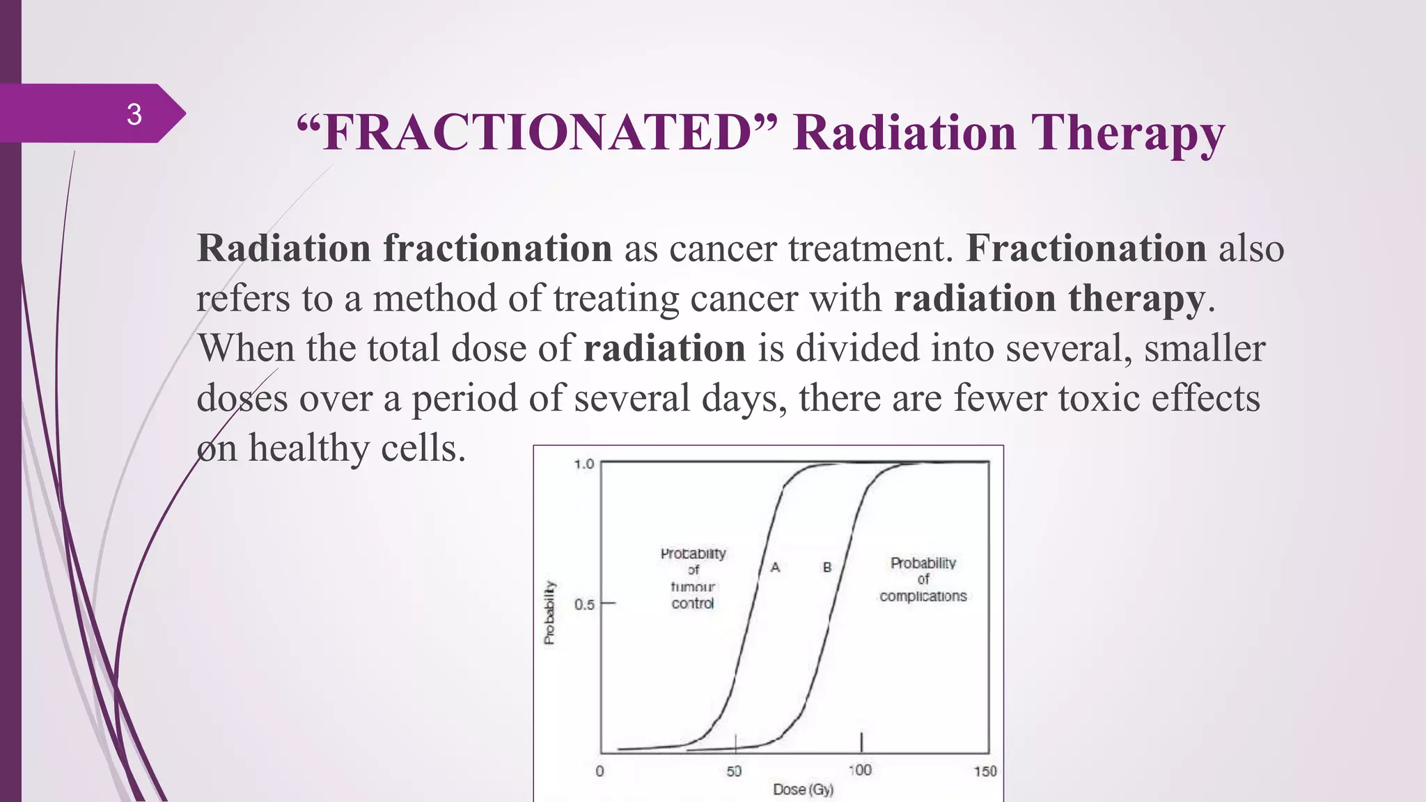
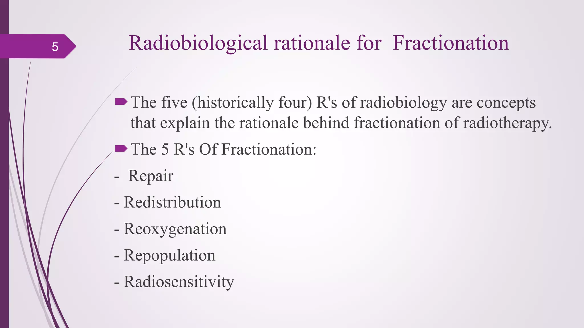
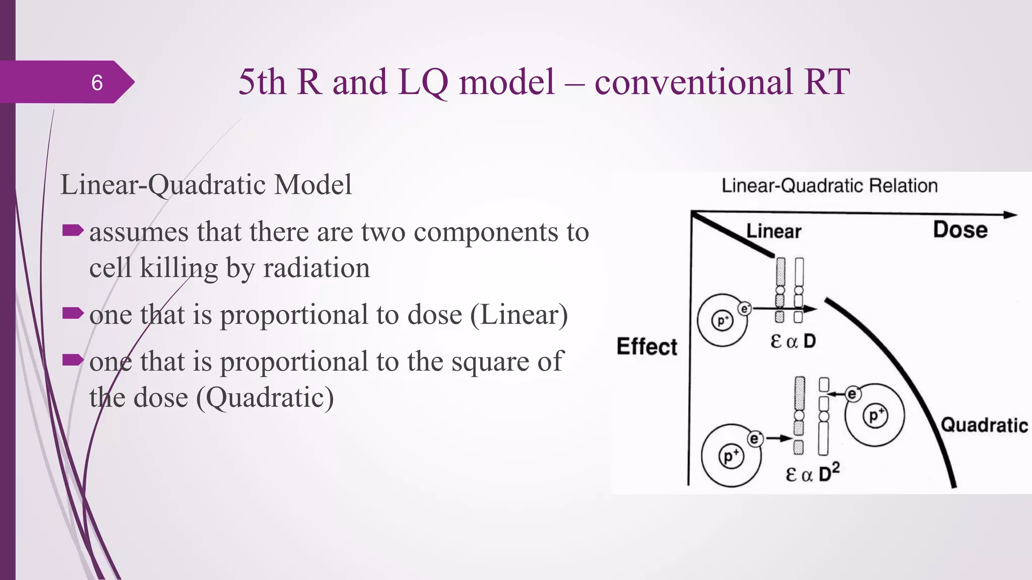
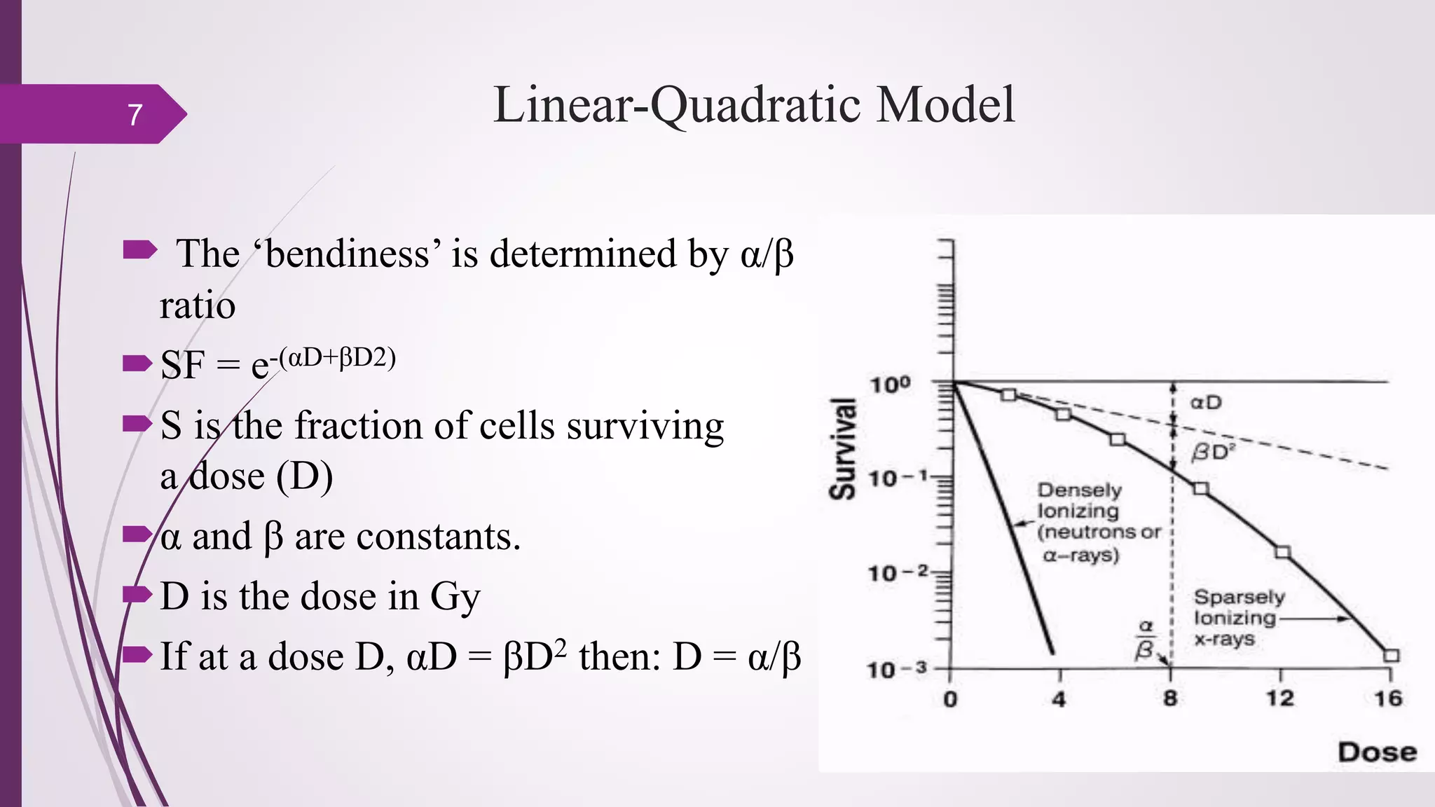
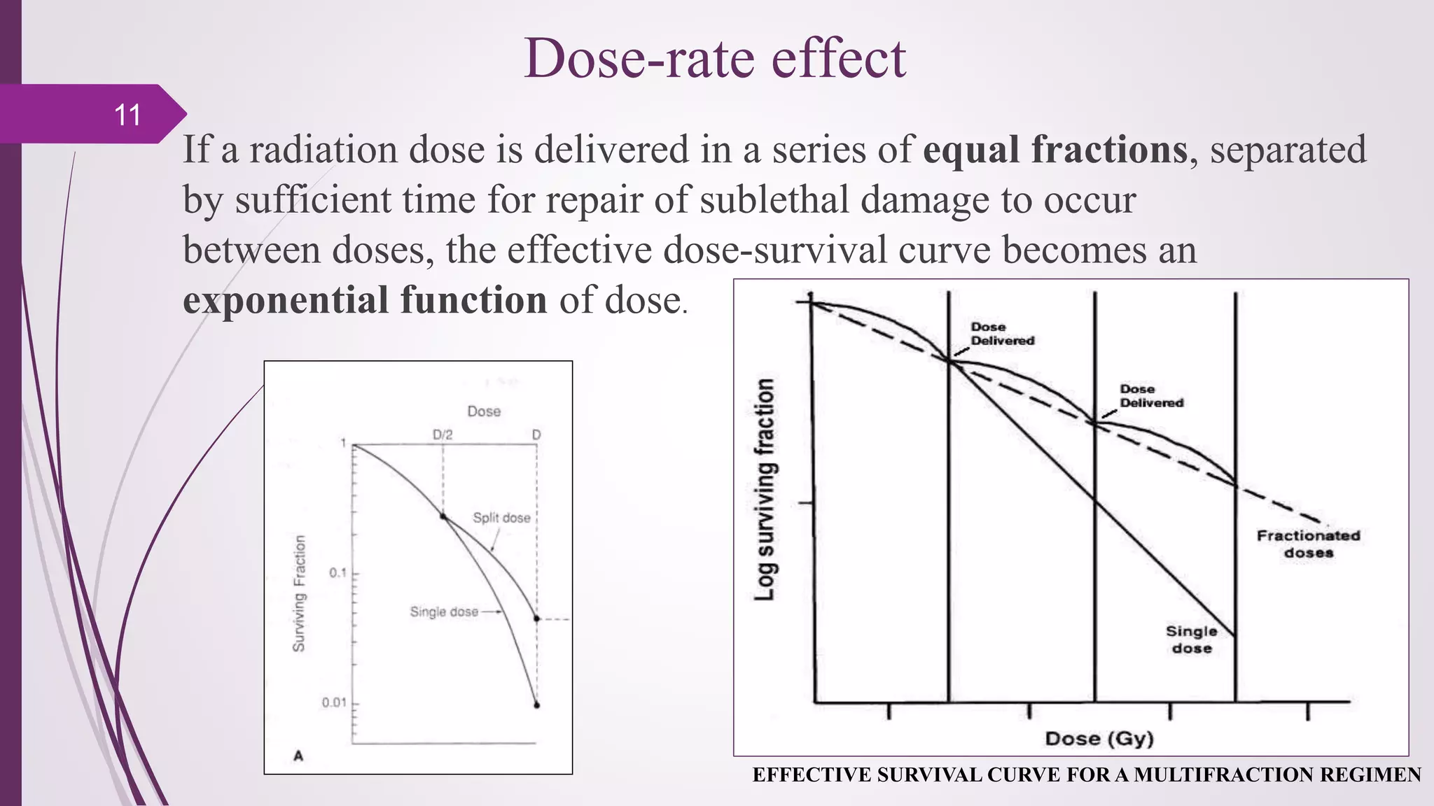
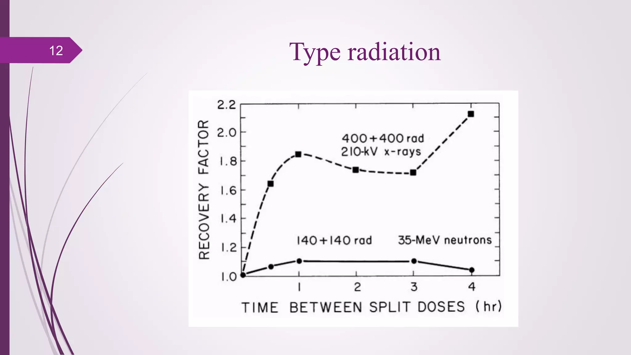
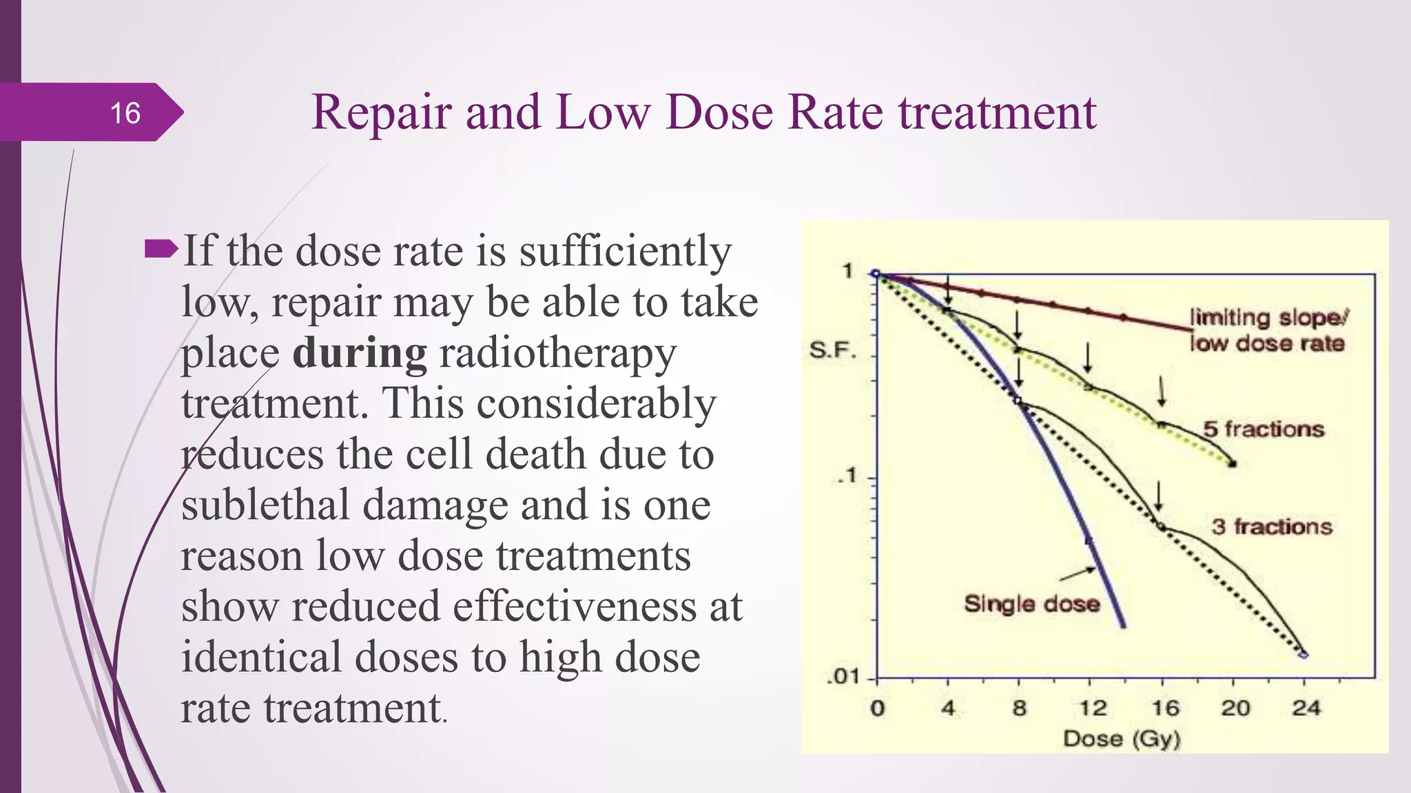
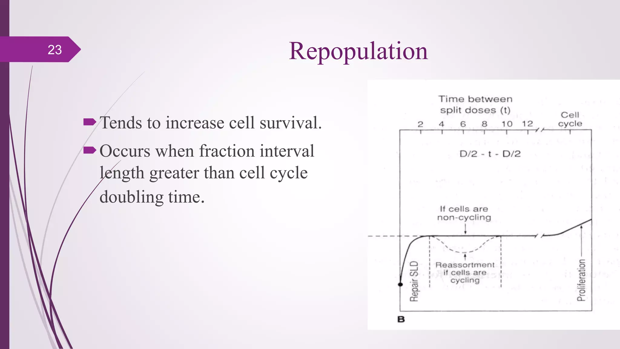
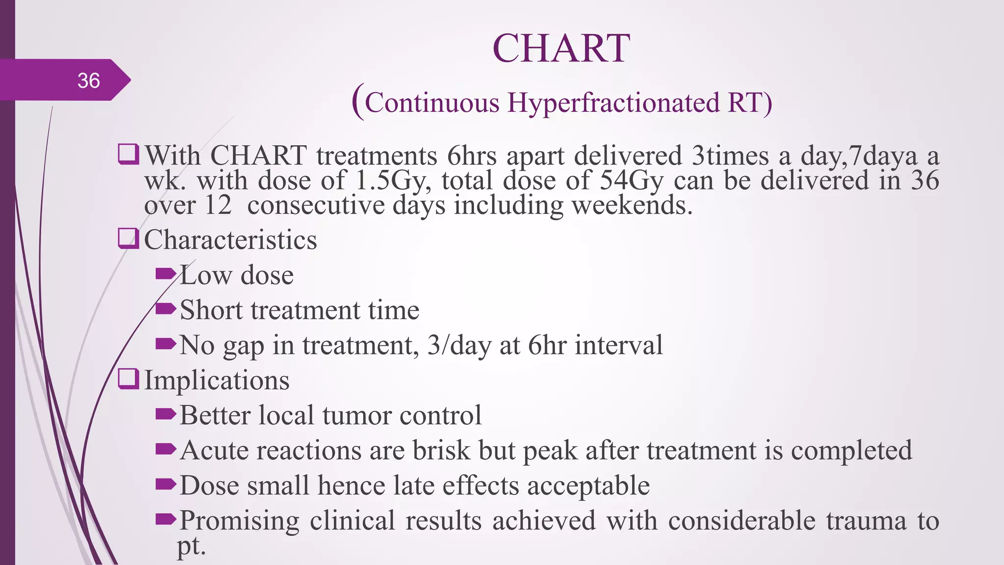
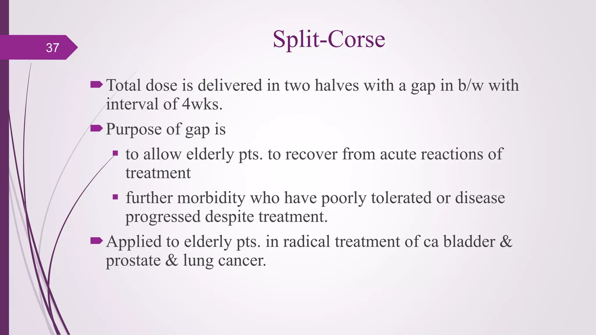
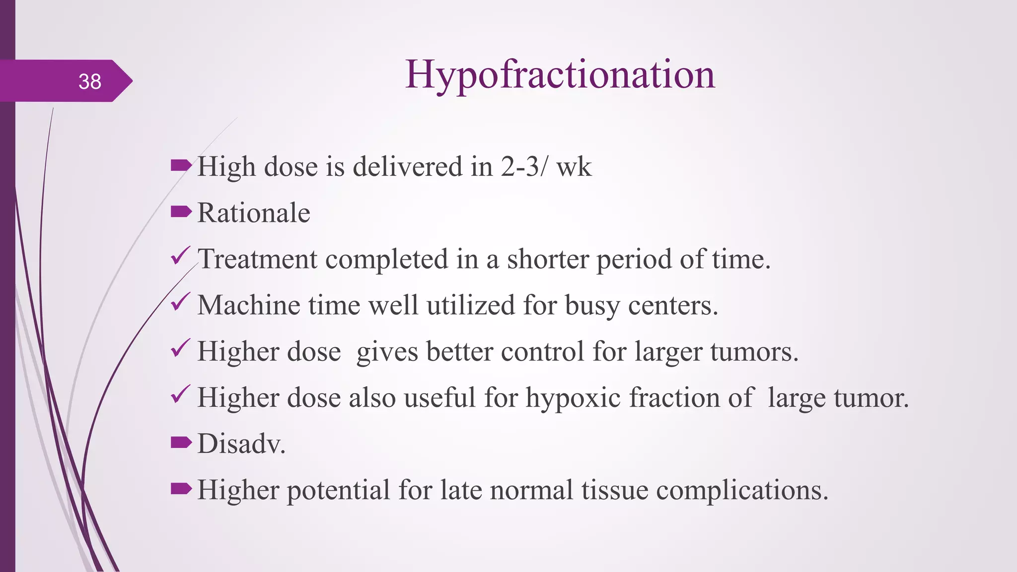
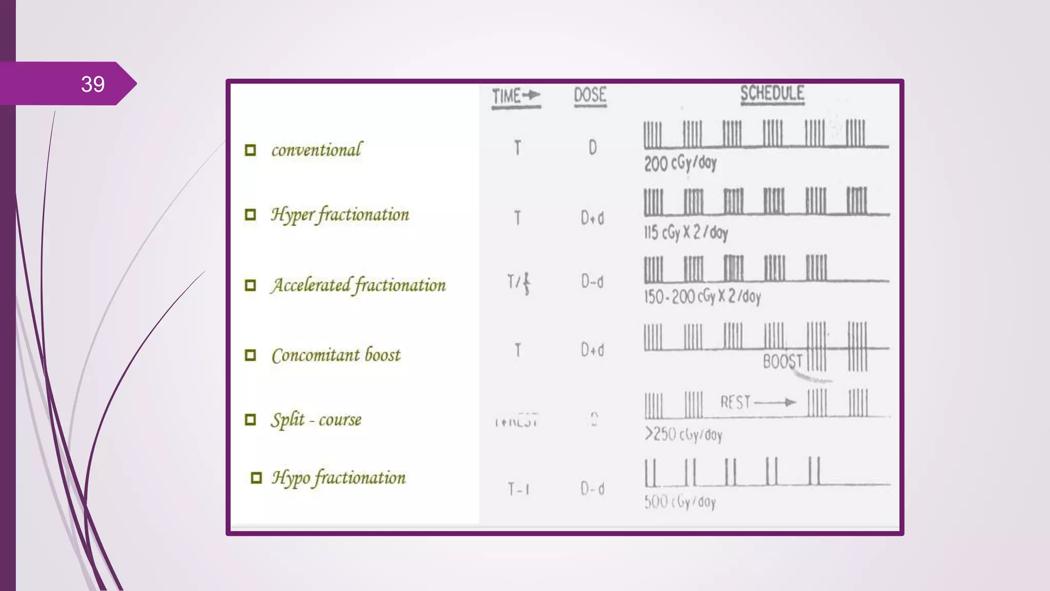
Fractionation in radiotherapy refers to dividing the total radiation dose into smaller doses given over multiple treatment sessions. This allows healthy cells to repair sublethal damage between fractions while maximizing cancer cell kill through mechanisms like redistribution and reoxygenation. The "5 R's" of radiobiology explain fractionation: repair of sublethal damage in normal cells; redistribution of tumor cells to sensitive phases; reoxygenation of hypoxic tumor cells; repopulation of tumor cells during prolonged treatment; and intrinsic radiosensitivity differences between cell types. Fractionation schedules are tailored based on these factors to improve the therapeutic ratio for different cancers and patients.
3. Radiation fractionation as cancer treatment. Fractionation also
refers to a method of treating cancer with radiation therapy.
When the total dose of radiation is divided into several, smaller
doses over a period of several days, there are fewer toxic effects
on healthy cells.
“FRACTIONATED” Radiation Therapy3
4. Historical Review
Earlier some radiotherapists believed that fractionated
treatment was inferior & single dose was necessary to cure
cancer.
While radiobiological experiments conducted in France
favored fractionated regimen for radiotherapy which
allows cancerocidal dose to be delivered without
exceeding normal tissue tolerance.
4
5. Radiobiological rationale for Fractionation
The five (historically four) R's of radiobiology are concepts
that explain the rationale behind fractionation of radiotherapy.
The 5 R's Of Fractionation:
- Repair
- Redistribution
- Reoxygenation
- Repopulation
- Radiosensitivity
5
6. 5th R and LQ model – conventional RT
Linear-Quadratic Model
assumes that there are two components to
cell killing by radiation
one that is proportional to dose (Linear)
one that is proportional to the square of
the dose (Quadratic)
6
7. Linear-Quadratic Model
The ‘bendiness’ is determined by α/β
ratio
SF = e-(αD+βD2)
S is the fraction of cells surviving
a dose (D)
α and β are constants.
D is the dose in Gy
If at a dose D, αD = βD2 then: D = α/β
7
8. 5th R and LQ model – conventional RT
The LQ model is simple and convenient
– better fit in the low dose–high survival region
– α (lethal/non-repairable) & β (sub-lethal/reparable)
– α/β ratio for early and late reactions in human normal
tissues consistent with results from experimental models
8
9. Repair
Repair is the one of the primary reasons to fractionate
radiotherapy. As discussed in DNA Damage and Repair
There are three types of damage that ionising radiation can
cause to cells:
-Lethal Damage :which is irreversible and irreparable and leads
irrevocably to cell death
-Potentially Lethal Damage(PLD):The component of radiation
damage that can be modified by postirradiation environmental
conditions
-Sublethal Damage(SLD):which under normal circumstances can
be repaired in hours unless additional sublethal damage is added
9
10. Repair of Sublethal Damage
Dose rate effect
Type radiation
Cell in different cell cycle phase
Type cell
10
11. Dose-rate effect
If a radiation dose is delivered in a series of equal fractions, separated
by sufficient time for repair of sublethal damage to occur
between doses, the effective dose-survival curve becomes an
exponential function of dose.
EFFECTIVE SURVIVAL CURVE FOR A MULTIFRACTION REGIMEN
11
15. Type cell
Cell survival is a function of cell type and radiation type
Non- or slowly proliferating cells (nerve, muscle,
secretory) are less susceptible to radiation damage.
Highly-proliferating cells (epithelial, stem cells) are
more susceptible.
15
16. Repair and Low Dose Rate treatment
If the dose rate is sufficiently
low, repair may be able to take
place during radiotherapy
treatment. This considerably
reduces the cell death due to
sublethal damage and is one
reason low dose treatments
show reduced effectiveness at
identical doses to high dose
rate treatment.
16
17. Repair Half Life
Intefraction interval and the repair half life is an important
consideration when fractionating radiotherapy. Some
tissues, notably the spinal cord, appear to have a slow
repair mechanism with a half life of about 4 hours. It is
important to separate dose by at least 6 hours and
preferably 8 hours if two fractions are given on the same
day.
17
18. Redistribution
Cells move to more radiosensitive phase in the cell cycle
between fractions.
M and G2 most sensitive phases.
Late S most resistant phase
18
19. Redistribution
Redistribution of proliferating cell
populations throughout the cell cycle
increases cell kill in fractionated
treatment relative to a single session
treatment.
Cells are most sensitive during M & G2
phase & are resistant during S phase of
cell cycle .
Redistribution can be a benefit in
fractionated course of RT if cells are
caught in sensitive phase after each
fraction
19
20. Reoygenation
Cells at the center of tumor are hypoxic
& are resistant to low LET radiation.
Hypoxic cells get reoxygenated occurs
during a fractionated course of treatment,
making them more radiosensitive to
subsequent doses of radiation.
Tumours may be acutely or
chronically hypoxic. This oxygenation
status may change during treatment.
20
25. Radiosensitivity
Radiosensitivity is a newer member of the R's
It reminds us, that apart from repair pathways, redistribution of
cells, reoxygenation of malignant cells and repopulation there is
an intrinsic radiosensitivity or radioresistance in different cell
types.
Radio sensitivity expresses the response of the tumor to
irradiation.
Malignant cells have greater reproductive capacity hence are
more radiosensitive.
25
26. Factors affecting the radiosensitivity
Physical
LET (linear energy transfer): RS
Dose rate: RS
Chemical
Increase RS: OXYGEN, cytotoxic drugs.
Decrease RS: SULFHYDRL compounds (cys,
cysteamine…)
Biological
Cycle status:
RS: G2, M
RS: S
26
28. Radiation Response
Response of all normal tissues to radn is
not same
Depending on their response tissues are
either
Early responding – constitute fast
proliferating cells such as skin, mucosa,
intestinal epithelium, colon, testis etc.
Late responding – have large no. of cells
in the resting phase e.g. spinal cord,
bladder, lung, kidneys etc.
28
29. Various Fractionation Schedule
Fractionated radiation exploits difference in 4R’s between tumors
and normal tissue thereby improving therapeutic index
Types
Conventional
Altered
• Hyper fractionation
• Accelerated fractionation
• Split course
• Hypofractionation
29
30. Conventional fractionation
Division of dose into multiple spares normal tissue through repair
of SLD & repopulation of cells.
Concurrently , fractionation increases tumor damage through
reoxygenation & redistribution of tumor cells.
Hence a balance is achieved the response of tumor & early & late
reacting normal tissue.
Most common fractionation for curative radiotherapy is 1.8 to
2.2Gy
30
31. Conventional fractionation
Evolved as conventional regimen because it is
Convenient (no weekend treatment)
Efficient (treatment every weekday)
Effective (high doses can be delivered without exceeding
either acute or chronic normal tissue tolerance)
Allows upkeep of machines.
Rationale for using conventional fractionation
Most tried & trusted method
Both tumorocidal & tolerance doses are well documented
31
32. Hyperfractionation
the delivery of radiation in small-dose fractions( 2-3 times per day)
aims to improve the therapeutic ratio, reducing the dose given in
each fraction, so as to reduce the late side effects while also
permitting an increased total dose to the tumor
hyperfractionation provided the greatest benefit to patients with
head and neck cancer
32
33. Hyperfractionation
A hyper fractionated schedule of 80.5Gy/70(1.15Gy
twice/day)/7wks compared with 70Gy/35/7wks in head &
neck cancer.
Implications
• Increased local tumor control at 5yr from 40 to59%
• Reflected in improved survival
• No increase in side effects
33
34. Accelerated Treatment
Alternative to hyper fractionation
Rationale – To reduce repopulation in rapidly proliferating
tumors by reducing overall treatment time.
Pure accelerated treatment – same total dose delivered in half
the overall time by giving 2or more s/day. but it is not possible
to achieve as acute effects become limiting factor.
Impure accelerated treatment – dose is reduced or rest period is
interposed in the middle of treatment.
34
35. Types of accelerated fraction
Comparison of head & neck cases accelerated regimen
72Gy/45 (1.6Gy,3/day)/5wks with 70Gy/35/7wks
Implications –
15% increase in loco regional control
No survival adv.
Increased acute effects
Unexpected increase in late complications
35
36. CHART
(Continuous Hyperfractionated RT)
With CHART treatments 6hrs apart delivered 3times a day,7daya a
wk. with dose of 1.5Gy, total dose of 54Gy can be delivered in 36
over 12 consecutive days including weekends.
Characteristics
Low dose
Short treatment time
No gap in treatment, 3/day at 6hr interval
Implications
Better local tumor control
Acute reactions are brisk but peak after treatment is completed
Dose small hence late effects acceptable
Promising clinical results achieved with considerable trauma to
pt.
36
37. Split-Corse
Total dose is delivered in two halves with a gap in b/w with
interval of 4wks.
Purpose of gap is
to allow elderly pts. to recover from acute reactions of
treatment
further morbidity who have poorly tolerated or disease
progressed despite treatment.
Applied to elderly pts. in radical treatment of ca bladder &
prostate & lung cancer.
37
38. Hypofractionation
High dose is delivered in 2-3/ wk
Rationale
Treatment completed in a shorter period of time.
Machine time well utilized for busy centers.
Higher dose gives better control for larger tumors.
Higher dose also useful for hypoxic fraction of large tumor.
Disadv.
Higher potential for late normal tissue complications.
38