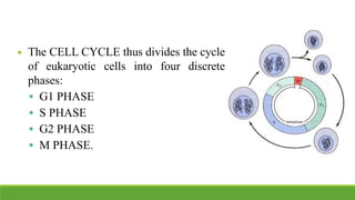
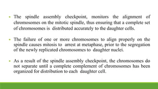
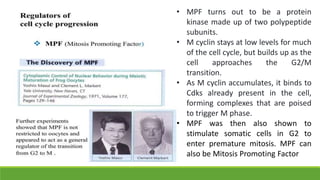
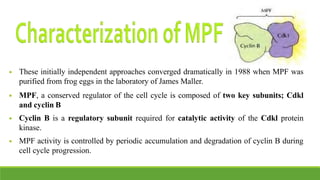
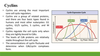
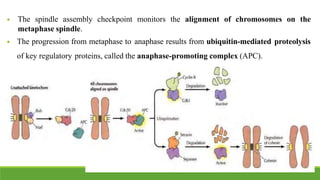
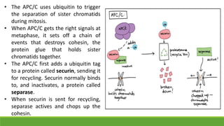
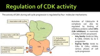
Cell cycle and its regulation
◾ A typical eukaryotic cell cycle is illustrated by human cells in
culture, which divide approximately every 24 hours.
◾ It was studied by Howard and Pelc 1953.
◾ The cell cycle is divided into two basic parts:
 Interphase
 Mitosis (or meiosis)
◾ Mitosis and cytokinesis last only about an hour, so approximately
95% of the cell
cycle is spent in interphase-the period between mitoses.
◾ However, interphase is the time during which both cell growth and
DNA replication occur in an orderly manner in preparation for cell
division.
◾ The CELL CYCLE thus divides the cycle
of eukaryotic cells into four discrete
phases:
 G1 PHASE
 S PHASE
 G2 PHASE
 M PHASE.
◾ Cells at different stages of cell cycle can be
differentiated by their DNA content.
◾ Animal cells in G1 are diploid (2n). During S
phase, replication increase the DNA content from
2n to 4n. It remains in the 4n stage in G2 phase as
well and is then decreased to 2n stage after M
phase.
◾ Some cells in adult animals cease division
altogether (e.g., nerve cells) and many other cells
divide only occasionally (e.g. skin fibroblasts).
◾ The progression of cells through the division cycle is regulated by
extracellular signals from the environment, as well as by internal
signals that monitor and coordinate the various processes.
◾ This is accomplished by some control points that regulate progression
through the cell cycle:
 START
 Restriction point
 Control of G2 to M transition
REGULATION OF CELL CYCLE BY EXTERNAL SIGNALS
◾ Occurs during late G1 and controls
progression from G1 to S.
◾ First defined by studies of budding yeast
(Saccharomyces cerevisiae), where it is
known as START.
◾ The importance of this regulation is
particularly evident in budding yeasts in
which cell division produces progeny cells
of very different sizes: a large mother cell
and a small daughter cell.
START checkpoint
◾ Some of the activities monitored at this
check point are:
 Optimum cell size
 Nutrient status
 Polypeptide factors that signal yeast
mating also arrest the cell cycle at
START, allowing haploid yeast cells
to fuse with one another instead of
progressing to S phase.
RESTRICTION POINT
Skin fibroblasts are arrested
in G0 until they are
stimulated to divide as
required to repair damage
resulting from a wound.
◾ In fission yeast Saccharomyces pombe, the cellcycle is regulated
primarily by control of the transition from G2 to M, at which cell size
and nutrient availability are monitored.
◾ Vertebrate oocytes can remain arrested in G2 for long periods of time
(several decades in humans) until their progression to M phase is
triggered by hormonal stimulation.
◾ In addition, some haploid mosses use G2 control point.
CONTROL OF G2 – M TRANSITION
◾ This coordination between
different phases of the cell
cycle is dependent on a
series of cell cycle
checkpoints that prevent
entry into the next phase of
the cell cycle until the
events of the preceding
phase have been
completed.
◾ Several cell cycle checkpoints function to ensure that incomplete or
damaged chromosomes are not replicated and passed on to daughter
cells.
◾ Cell cycle arrest at the G1, S, and G2 checkpoints is mediated by two
related protein kinases, designated ATM and ATR, that recognize
damaged or unreplicated DNA and are activated in response to DNA
damage.
◾ ATM and ATR then activate a signaling pathway that leads not only to
cell cycle arrest, but also to the activation of DNA repair and, in some
cases, programmed cell death.
◾ The spindle assembly checkpoint, monitors the alignment of
chromosomes on the mitotic spindle, thus ensuring that a complete set
of chromosomes is distributed accurately to the daughter cells.
◾ The failure of one or more chromosomes to align properly on the
spindle causes mitosis to arrest at metaphase, prior to the segregation
of the newly replicated chromosomes to daughter nuclei.
◾ As a result of the spindle assembly checkpoint, the chromosomes do
not separate until a complete complement of chromosomes has been
organized for distribution to each daughter cell.
◾ DNA replication is restricted to once per
cell cycle by the MCM helicase proteins
that bind to origins of replication together
with ORC (origin recognition complex)
proteins and are required for the initiation
of DNA replication.
◾ MCM proteins are only able to bind to
DNA in G1, allowing DNA replication to
initiate in S phase.
◾ Once initiation has occurred, the MCM
proteins are displaced so that replication
cannot initiate again until after mitosis.
• MPF turns out to be a protein
kinase made up of two polypeptide
subunits.
• M cyclin stays at low levels for much
of the cell cycle, but builds up as the
cell approaches the G2/M
transition.
• As M cyclin accumulates, it binds to
Cdks already present in the cell,
forming complexes that are poised
to trigger M phase.
• MPF was then also shown to
stimulate somatic cells in G2 to
enter premature mitosis. MPF can
also be Mitosis Promoting Factor
◾ These initially independent approaches converged dramatically in 1988 when MPF was
purified from frog eggs in the laboratory of James Maller.
◾ MPF, a conserved regulator of the cell cycle is composed of two key subunits; Cdkl
and cyclin B
◾ Cyclin B is a regulatory subunit required for catalytic activity of the Cdkl protein
kinase.
◾ MPF activity is controlled by periodic accumulation and degradation of cyclin B during
cell cycle progression.
Cyclins
• Cyclins are among the most important
core cell cycle regulators.
• Cyclins are a group of related proteins,
and there are four basic types found in
humans and most other eukaryotes: G1
cyclins, G1/S cyclins, S cyclins, and M
cyclins.
• Cyclins regulate the cell cycle only when
they are tightly bound to Cdks.
• The levels of Cdk proteins are relatively
stable throughout the cell cycle.
• The concentrations of cyclin fluctuate and
determine when Cdk/cyclin complexes
form.
Cyclin-dependent kinases
• Cyclin activate or inactivate many target
proteins inside of the cell.
• Cyclins drive the events of the cell cycle by
partnering with a family of enzymes called the
cyclin-dependent kinases (Cdks).
• Cdks are kinases, enzymes that phosphorylate
(attach phosphate groups to) specific target
proteins.
• The attached phosphate group acts like a
switch, making the target protein more or less
active.
• When a cyclin attaches to a Cdk, it has two important effects: it activates the Cdk as a
kinase, but it also directs the Cdk to a specific set of target proteins, ones appropriate to
the cell cycle period controlled by the cyclin.
The anaphase-promoting complex/cyclosome (APC/C)
• MPF also triggers its own destruction by activating the anaphase-promoting
complex/cyclosome (APC/C), a protein complex that causes M cyclins to be destroyed
starting in anaphase.
• The destruction of M cyclins pushes the cell out of mitosis, allowing the new daughter
cells to enter G1
• The APC/C also causes destruction of the proteins that hold the sister chromatids
together, allowing them to separate in anaphase and move to opposite poles of the cell.
• It adds a small protein tag called ubiquitin (Ub). When a target is tagged with ubiquitin, it
is sent to the proteasome, which can be thought of as the recycle bin of the cell, and
destroyed.
◾ The spindle assembly checkpoint monitors the alignment of chromosomes on the
metaphase spindle.
◾ The progression from metaphase to anaphase results from ubiquitin-mediated proteolysis
of key regulatory proteins, called the anaphase-promoting complex (APC).
• The APC/C uses ubiquitin to trigger
the separation of sister chromatids
during mitosis.
• When APC/C gets the right signals at
metaphase, it sets off a chain of
events that destroys cohesin, the
protein glue that holds sister
chromatids together.
• The APC/C first adds a ubiquitin tag
to a protein called securin, sending it
for recycling. Securin normally binds
to, and inactivates, a protein called
separase.
• When securin is sent for recycling,
separase actives and chops up the
cohesin.
Mechanism of cell cycle regulation
◾ Cdkl forms complexes with cyclin B during G2.
◾ CdkI is then phosphorylated on threonine-161 by MO15, which is required for Cdkl
activity.
◾ It is then phosphorylated on tyrosine-15/ 14 (vertebrate cells) by a protein kinase called
Wee1, which inhibits Cdk l activity and leads to the accumulation of inactive
Cdkl/cyclin B complexes throughout G2 phase.
◾ Dephosphorylation of Thr14 and Tyr15 by a protein phosphatase called Cdc25C
activates MPF at the G2 to M transition.
◾ Activated Cdkl protein phosphorylates a variety kinase proteins.
◾ MPF activity is terminated at the end of mitosis by degradation of cyclin B.
The activity ofCdk's during cell cycle progression is regulated by four molecular mechanisms:
Activities
complexes
regulated
can also
by binding
of CdkI/cyclin B
be
of
inhibitory proteins called CKI’s
(Cdk inhibitors). In mammals
2 families ofCKI’s are present:
◾ Ink4 family: binds to Cdk4
& Cdk6; inhibits G1 to S
progression
◾ Kip/Cip family: binds to
Cdk1 & Cdk2; inhibits
various phases of cell
cycle progression
◾ At fertilization, the sperm binds to a receptor on the surface of the egg and fuses with the egg
plasma membrane, initiating the development of a new diploid organism containing genetic
information derived from both parents.
◾ A sperm binds to a plasma membrane receptor of the egg and this induces an increase in Ca2+
level in egg cytoplasm,via hydrolysis of PIP2 (phosphatidylinositol4,5-isphosphate).
◾ The Ca2+ induces exocytosis of secretory vesicles that are present in large numbers beneath the egg
plasma membrane. This, in turn, induces surface alterations that prevent additional sperm from
entering the egg.
◾ The increase in cytosolic Ca2+ following fertilization also signals the completion of meiosis.
◾ Following completion of oocyte meiosis, the fertilized egg (zygote) contains two haploid nuclei
(called pronuclei), one derived from each parent.
◾ The two pronuclei then enter S phase and replicate their DNA as they migrate toward each other.
◾ As they meet, the zygote enters M phase of its first mitotic division.

cellcycle_regulation.pptx

  • 1.
    Cell cycle andits regulation
  • 2.
    ◾ A typicaleukaryotic cell cycle is illustrated by human cells in culture, which divide approximately every 24 hours. ◾ It was studied by Howard and Pelc 1953. ◾ The cell cycle is divided into two basic parts:  Interphase  Mitosis (or meiosis) ◾ Mitosis and cytokinesis last only about an hour, so approximately 95% of the cell cycle is spent in interphase-the period between mitoses. ◾ However, interphase is the time during which both cell growth and DNA replication occur in an orderly manner in preparation for cell division.
  • 3.
    ◾ The CELLCYCLE thus divides the cycle of eukaryotic cells into four discrete phases:  G1 PHASE  S PHASE  G2 PHASE  M PHASE.
  • 4.
    ◾ Cells atdifferent stages of cell cycle can be differentiated by their DNA content. ◾ Animal cells in G1 are diploid (2n). During S phase, replication increase the DNA content from 2n to 4n. It remains in the 4n stage in G2 phase as well and is then decreased to 2n stage after M phase. ◾ Some cells in adult animals cease division altogether (e.g., nerve cells) and many other cells divide only occasionally (e.g. skin fibroblasts).
  • 5.
    ◾ The progressionof cells through the division cycle is regulated by extracellular signals from the environment, as well as by internal signals that monitor and coordinate the various processes. ◾ This is accomplished by some control points that regulate progression through the cell cycle:  START  Restriction point  Control of G2 to M transition REGULATION OF CELL CYCLE BY EXTERNAL SIGNALS
  • 6.
    ◾ Occurs duringlate G1 and controls progression from G1 to S. ◾ First defined by studies of budding yeast (Saccharomyces cerevisiae), where it is known as START. ◾ The importance of this regulation is particularly evident in budding yeasts in which cell division produces progeny cells of very different sizes: a large mother cell and a small daughter cell. START checkpoint
  • 7.
    ◾ Some ofthe activities monitored at this check point are:  Optimum cell size  Nutrient status  Polypeptide factors that signal yeast mating also arrest the cell cycle at START, allowing haploid yeast cells to fuse with one another instead of progressing to S phase.
  • 8.
    RESTRICTION POINT Skin fibroblastsare arrested in G0 until they are stimulated to divide as required to repair damage resulting from a wound.
  • 9.
    ◾ In fissionyeast Saccharomyces pombe, the cellcycle is regulated primarily by control of the transition from G2 to M, at which cell size and nutrient availability are monitored. ◾ Vertebrate oocytes can remain arrested in G2 for long periods of time (several decades in humans) until their progression to M phase is triggered by hormonal stimulation. ◾ In addition, some haploid mosses use G2 control point. CONTROL OF G2 – M TRANSITION
  • 11.
    ◾ This coordinationbetween different phases of the cell cycle is dependent on a series of cell cycle checkpoints that prevent entry into the next phase of the cell cycle until the events of the preceding phase have been completed.
  • 12.
    ◾ Several cellcycle checkpoints function to ensure that incomplete or damaged chromosomes are not replicated and passed on to daughter cells. ◾ Cell cycle arrest at the G1, S, and G2 checkpoints is mediated by two related protein kinases, designated ATM and ATR, that recognize damaged or unreplicated DNA and are activated in response to DNA damage. ◾ ATM and ATR then activate a signaling pathway that leads not only to cell cycle arrest, but also to the activation of DNA repair and, in some cases, programmed cell death.
  • 13.
    ◾ The spindleassembly checkpoint, monitors the alignment of chromosomes on the mitotic spindle, thus ensuring that a complete set of chromosomes is distributed accurately to the daughter cells. ◾ The failure of one or more chromosomes to align properly on the spindle causes mitosis to arrest at metaphase, prior to the segregation of the newly replicated chromosomes to daughter nuclei. ◾ As a result of the spindle assembly checkpoint, the chromosomes do not separate until a complete complement of chromosomes has been organized for distribution to each daughter cell.
  • 14.
    ◾ DNA replicationis restricted to once per cell cycle by the MCM helicase proteins that bind to origins of replication together with ORC (origin recognition complex) proteins and are required for the initiation of DNA replication. ◾ MCM proteins are only able to bind to DNA in G1, allowing DNA replication to initiate in S phase. ◾ Once initiation has occurred, the MCM proteins are displaced so that replication cannot initiate again until after mitosis.
  • 15.
    • MPF turnsout to be a protein kinase made up of two polypeptide subunits. • M cyclin stays at low levels for much of the cell cycle, but builds up as the cell approaches the G2/M transition. • As M cyclin accumulates, it binds to Cdks already present in the cell, forming complexes that are poised to trigger M phase. • MPF was then also shown to stimulate somatic cells in G2 to enter premature mitosis. MPF can also be Mitosis Promoting Factor
  • 17.
    ◾ These initiallyindependent approaches converged dramatically in 1988 when MPF was purified from frog eggs in the laboratory of James Maller. ◾ MPF, a conserved regulator of the cell cycle is composed of two key subunits; Cdkl and cyclin B ◾ Cyclin B is a regulatory subunit required for catalytic activity of the Cdkl protein kinase. ◾ MPF activity is controlled by periodic accumulation and degradation of cyclin B during cell cycle progression.
  • 18.
    Cyclins • Cyclins areamong the most important core cell cycle regulators. • Cyclins are a group of related proteins, and there are four basic types found in humans and most other eukaryotes: G1 cyclins, G1/S cyclins, S cyclins, and M cyclins. • Cyclins regulate the cell cycle only when they are tightly bound to Cdks. • The levels of Cdk proteins are relatively stable throughout the cell cycle. • The concentrations of cyclin fluctuate and determine when Cdk/cyclin complexes form.
  • 19.
    Cyclin-dependent kinases • Cyclinactivate or inactivate many target proteins inside of the cell. • Cyclins drive the events of the cell cycle by partnering with a family of enzymes called the cyclin-dependent kinases (Cdks). • Cdks are kinases, enzymes that phosphorylate (attach phosphate groups to) specific target proteins. • The attached phosphate group acts like a switch, making the target protein more or less active.
  • 20.
    • When acyclin attaches to a Cdk, it has two important effects: it activates the Cdk as a kinase, but it also directs the Cdk to a specific set of target proteins, ones appropriate to the cell cycle period controlled by the cyclin. The anaphase-promoting complex/cyclosome (APC/C) • MPF also triggers its own destruction by activating the anaphase-promoting complex/cyclosome (APC/C), a protein complex that causes M cyclins to be destroyed starting in anaphase. • The destruction of M cyclins pushes the cell out of mitosis, allowing the new daughter cells to enter G1 • The APC/C also causes destruction of the proteins that hold the sister chromatids together, allowing them to separate in anaphase and move to opposite poles of the cell. • It adds a small protein tag called ubiquitin (Ub). When a target is tagged with ubiquitin, it is sent to the proteasome, which can be thought of as the recycle bin of the cell, and destroyed.
  • 21.
    ◾ The spindleassembly checkpoint monitors the alignment of chromosomes on the metaphase spindle. ◾ The progression from metaphase to anaphase results from ubiquitin-mediated proteolysis of key regulatory proteins, called the anaphase-promoting complex (APC).
  • 22.
    • The APC/Cuses ubiquitin to trigger the separation of sister chromatids during mitosis. • When APC/C gets the right signals at metaphase, it sets off a chain of events that destroys cohesin, the protein glue that holds sister chromatids together. • The APC/C first adds a ubiquitin tag to a protein called securin, sending it for recycling. Securin normally binds to, and inactivates, a protein called separase. • When securin is sent for recycling, separase actives and chops up the cohesin.
  • 24.
    Mechanism of cellcycle regulation ◾ Cdkl forms complexes with cyclin B during G2. ◾ CdkI is then phosphorylated on threonine-161 by MO15, which is required for Cdkl activity. ◾ It is then phosphorylated on tyrosine-15/ 14 (vertebrate cells) by a protein kinase called Wee1, which inhibits Cdk l activity and leads to the accumulation of inactive Cdkl/cyclin B complexes throughout G2 phase. ◾ Dephosphorylation of Thr14 and Tyr15 by a protein phosphatase called Cdc25C activates MPF at the G2 to M transition. ◾ Activated Cdkl protein phosphorylates a variety kinase proteins. ◾ MPF activity is terminated at the end of mitosis by degradation of cyclin B.
  • 26.
    The activity ofCdk'sduring cell cycle progression is regulated by four molecular mechanisms: Activities complexes regulated can also by binding of CdkI/cyclin B be of inhibitory proteins called CKI’s (Cdk inhibitors). In mammals 2 families ofCKI’s are present: ◾ Ink4 family: binds to Cdk4 & Cdk6; inhibits G1 to S progression ◾ Kip/Cip family: binds to Cdk1 & Cdk2; inhibits various phases of cell cycle progression
  • 27.
    ◾ At fertilization,the sperm binds to a receptor on the surface of the egg and fuses with the egg plasma membrane, initiating the development of a new diploid organism containing genetic information derived from both parents. ◾ A sperm binds to a plasma membrane receptor of the egg and this induces an increase in Ca2+ level in egg cytoplasm,via hydrolysis of PIP2 (phosphatidylinositol4,5-isphosphate). ◾ The Ca2+ induces exocytosis of secretory vesicles that are present in large numbers beneath the egg plasma membrane. This, in turn, induces surface alterations that prevent additional sperm from entering the egg. ◾ The increase in cytosolic Ca2+ following fertilization also signals the completion of meiosis. ◾ Following completion of oocyte meiosis, the fertilized egg (zygote) contains two haploid nuclei (called pronuclei), one derived from each parent. ◾ The two pronuclei then enter S phase and replicate their DNA as they migrate toward each other. ◾ As they meet, the zygote enters M phase of its first mitotic division.