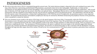
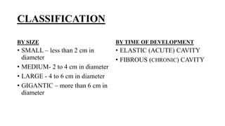
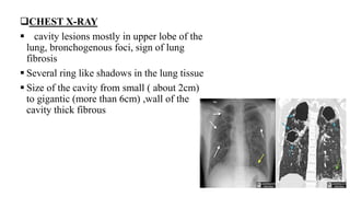
Cavitary TB involves the upper lobes of the lung. The bacteria cause progressive lung destruction by forming cavities, or enlarged air spaces.
is a characteristic feature of "secondary" tuberculosis.
This type of TB occurs in reactivation disease.
The upper lobes of the lung are affected because they are highly oxygenated (an environment in which M. tuberculosis thrives). Cavitary TB can, rarely, occur soon after primary infection.
♛VVIP Hyderabad Call Girls Chintalkunta🖕7001035870🖕Riya Kappor Top Call Girl ...
Cavitary tuberculosis
1. CAVITARY FORMS OF TUBERCULOSIS
BY,
ROUF ABDUR
GROUP NO 7
8TH SEMESTER
2. INTRODUCTION
Cavitary TB involves the upper lobes of the lung. The bacteria cause progressive
lung destruction by forming cavities, or enlarged air spaces.
is a characteristic feature of "secondary" tuberculosis.
This type of TB occurs in reactivation disease.
The upper lobes of the lung are affected because they are highly oxygenated (an
environment in which M. tuberculosis thrives). Cavitary TB can, rarely, occur
soon after primary infection.
3. PATHOGENESIS
Mycobacterium tuberculosis (M.tb) is transmitted through the aerosol route. The initial infectious droplet is deposited in the well-ventilated lower parts of the
lung, where Ghon foci, representing calcified healed TB granulomas, are primarily observed on chest radiographs. Traditionally, it is believed that
mycobacteria are first phagocytosed by alveolar macrophages, where M.tb persists in immature phagosomes by interfering with the acidification . After several
days of infection, M.tb may escape into the cytosol, where they can replicate freely . Macrophage infection drives a localized inflammatory response recruiting
mononuclear cells that contribute to host defense but may also provide new uninfected cells for mycobacterial growth . The inflammatory cells that die then
form the caseous center of the granuloma, a feature that is pathognomonic of TB. With the arrival of lymphocytes, the granuloma acquires a more organized
structure. The granuloma then becomes surrounded by lymphocytes that may be enclosed by fibroblasts demarcating the peripheral structure of the granuloma,
which is proposed to contain the infection .
TB cavity formation occurs usually in the apices of the lungs or in the apical segment of the lower lobes. Consequently, within the TB life cycle, a
dissemination event to the apices must occur from the initial deposition event in the lung bases. Once TB cavities form in the lung apex and connect with an
airway, there is exponential growth of the M.tb, which ultimately results in the release of large quantities of viable infectious bacilli, which can spread to a new
human host, completing the cycle . The mechanism of TB dissemination to the apices is unknown, but recent studies in the zebrafish suggest that infected
monocytes may transport mycobacteria through the blood circulation. The presence of a blood-borne phase in TB is consistent with the distribution of lesions
throughout the lungs and other organs observed in miliary TB and by the very occasional positive M.tb blood cultures in patients, particularly those coinfected
with HIV.
4. CLASSIFICATION
BY SIZE
• SMALL – less than 2 cm in
diameter
• MEDIUM- 2 to 4 cm in diameter
• LARGE - 4 to 6 cm in diameter
• GIGANTIC – more than 6 cm in
diameter
BY TIME OF DEVELOPMENT
• ELASTIC (ACUTE) CAVITY
• FIBROUS (CHRONIC) CAVITY
5. SYMPTOMS
• Productive cough
• Night sweats,
• Fever,
• Weight loss, loss of appetite and weakness.
• There may be hemoptysis (coughing up blood)
• Patients with cavitary TB are highly contagious. Occasionally, disease spreads into
the pleural space and causes TB empyema (pus in the pleural fluid).
6. DIAGNOSIS
PHYSICAL EXAMINATION
oINSPECTION- Deformation of the affected side of chest
oPERCUSSION- Dullness of the sound , consolidated areas of lungs are
detected
oAUSCULTATION- Bronchial Breathing , crepitation, wheezes, crackle
SPUTUM MICROSCOPY
SPUTUM CULTURE TEST
COMPLETE BLOOD COUNT- increase ESR, Leucocytosis,
lymphocytosis
7. CHEST X-RAY
cavity lesions mostly in upper lobe of the
lung, bronchogenous foci, sign of lung
fibrosis
Several ring like shadows in the lung tissue
Size of the cavity from small ( about 2cm)
to gigantic (more than 6cm) ,wall of the
cavity thick fibrous