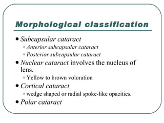
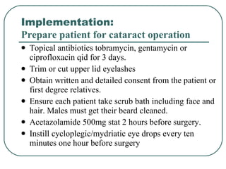
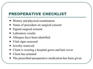
Cataract Gauri S. Shrestha, M.Optom, FIACLE
What is a cataract? Opacification of human crystalline lens  Functionally, it includes the cases which interferes with vision
Morphological classification Subcapsular cataract Anterior subcapsular cataract  Posterior subcapsular cataract  Nuclear cataract  involves the nucleus of lens. Yellow to brown voloration  Cortical cataract  wedge shaped or radial spoke-like opacities. Polar cataract
Classification according to maturity An immature cataract  A mature cataract  A hypermature cataract  A morgagnian cataract
Etiological classification Congenital and developmental cataract Acquired cataract Senile cataract Traumatic cataract Complicated (secondary) cataract .  eg, uveitis, pathological myopia, glaucoma, retinal detachment, retinitis pigmentosa etc, Metabolic cataract . Eg, Diabetes, hypocalcaemia, Wilson’s disease, Lowe’s syndrome
Etiological classification Acquired cataract Electric cataract   Radiational cataract Drug induced cataract  eg, corticosteroid, copper, iron, Chlorpromazine, Busulphan, Allopurinol, Amiodarone, etc, Cataract associated with skin diseases . Eg, atopic dermatitis, scleroderma, etc, Cataract with miscellaneous syndromes . Eg, Dystrophica myotonica, Down’s syndrome etc,
CONGENITAL AND DEVELOPMENTAL CATARACT Congenital cataract develops from some disturbance to normal development of lens .  The disturbance occurs before the birth  The opacity may limit to embryonic or foetal nucleus.  Developmental cataract occurs from infancy to adolescence.  The opacity involves infantile or adult nucleus.  3 children out of 10,000 live births.   Two third of the cases are bilateral.
Etiology Hereditary.  1/3 rd  hereditary, common mode: autosomal dominant inheritance Maternal factors.   Malnutrition during pregnancy . Maternal infection . Eg, rubella, toxoplasmosis, cytomegalo-inclusion disease. Drug intake during pregnancy. Eg,  thalidomide, corticosteroids. Radiation exposure during pregnancy.  Eg, X-rays, other ionizing radiations.
Etiology Foetal or infantile factors. Deficient oxygenation owing to placental haemorrhage . Metabolich disorders. Eg, galactokinase deficiency, neonatal hypoglycemia, galactosaemia,  Chromosomal abnormality.  Eg, down syndrome Skeletal syndromes.  Eg, Hallermann-streiff-Francois syndrome, Nance-Horan syndrome
Etiology Foetal or infantile factors. Birth trauma Malnutrition early in infancy . Prenatal infection.  Eg, congenital rubella, toxoplasmosis, cytomegalovirus, herpes simplex and varicella.
NURSING ASSESSMENT General History  of white pupil, squint, spontaneous movement of eyes, loss of visual attention. Assess density of cataract  Observe the red fundus reflex  on ophthalmoscope. Absence of red fundus reflex indicates cataract is visually significant. Perform fundus examination under dilatation .  Examine other associated ocular anomalies . Eg,  absence of central fixation, nystagmus, strabismus, corneal clouding, microphthalmos, glaucoma, retinoblastoma, retinal disorders
NURSING ASSESSMENT Investigation Serological test for intrauterine infections   (TORCH= toxoplasmosis, rubella, cytomegalovirus, and herpes simplex). A history of maternal rash during pregnancy   for varicella zoster antibody titres. Urinalysis for galactosaemia and chromatography for aminoacids. Refer to pediatrician to rule out systemic diseases.
AGE RELATED (SENILE) CATARACT Common and bilateral above the age of 50 years.  Male: Female::1:1 Etiology Hereditary : Incidence, age of onset and maturation  Ultravoilet radiation : More exposure to UV-rays = early maturation.  Dietary factors : Poor diatery factors eg, lack of certain aminoacids, Vitamines (Vitamin E, Vitamin C, riboflavin) and essential minerals. Dehydrational crisis : Prior episode of severe dehydration due to diarrhea and cholera.
Mechanism of loss of transparency Cortical cataract Denaturation and coagulation of lens proteins.  Decrease level of aminoacids and protein systhesis  Increased hydration brought by decrease in potassium due to reversal of Na/K pump mechanism. Nuclear cataract:   Degenerative changes occurring as nuclear sclerosis Increase in water insoluble proteins,  compaction of nucleus resulting in a hard cataract.  Disturbance of lamellar arrangement in fibres
Symptoms Painless progressive visual loss Glare Reduced color perception Color haloes Uniocular diplopia Based on the location and density
Sign Opacification of the normally clear lens seen through the pupil Indistinct on retina examination Red reflex may be dim  No afferent pupillary defect Myopic shift
NURSING ASSESSMENT Assess visual acuity and review report on refraction.  Surgery is indicated when cataract develops to a degree sufficient to cause difficulty in performing daily essential activities.  Assess a complete morphology of opacity (size, site, shape, color, and pattern) under slit lamp examination. Perform cover test
NURSING ASSESSMENT Test papillary response.  Examine cornea to rule out any opacities Examine ocular adnexa  Performed dilated fundus examination  Perform USG B-scan Measure intraocular pressure  Perform potential acuity measurement  Perform biometry
Nursing Assessment Specular Microscopy (endothelium cells) A normal cell count > 2400 cells/mm 2 If a cell count fewer than 1000 cells/mm 2  is risk of postoperative corneal decompensation
Laboratory investigation Complete blood counts Blood sugar Urine analysis Chest X-ray Conjunctival swab for C/S


Type of cataract surgery Extracapsular cataract extraction (ECCE).  Requires a relatively large circumferential limbal incision (8-10mm) through which the lens nucleus is extracted and the cortical matter aspirated, leaving behind an intact posterior capsule.  The IOL is then inserted.  It is the universal procedure of operation in cataract.  Posterior IOL can be transplanted after ECCE.
Type of cataract surgery Intracapsular cataract extraction (ICCE) .  The entire cataractous lens along with the intact capsule is removed in this procedure.  Weak and degenerated zonules are a pre-requisite for this method.  This is the surgery of choice only in markedly subluxated and dislocated lens.  This technique of surgery has been largely replaced by ECCE nowadays.
Type of cataract surgery Phacoemulsification:  A small hollow needle containing a piezo-electric crystal vibrates longitudinally at ultrasonic frequencies The tip is applied to the lens nucleus; cavitation occurs at the tip as the nucleus is emulsified;  an irrigation and aspiration system removes this emulsified material from the eye.  The IOL is then injected through a much smaller incision than in ECCE.  Safe: avoid compression of eye, results in little postoperative astigmatism and early stabilization of refraction, and eliminate post-operative wound related problem
Type of cataract surgery Lensectomy:  Most of the lens including anterior and posterior capsule along with anterior vitreous are removed with the help of a vitreous cutter, infusion and suction machine.  Congenital as well as developmental cataract being soft are easily dealt with this procedure.
NURSING DIAGNOSIS Gradual painless diminution of vision
EXPECTED OUTCOME Immediate. Optimal vision will be restored with periodic refractive correction with glasses. Patient will be reassured and informed with progression and option of surgery. Make patient educate and aware about possibility of fall due to visual impairment.
EXPECTED OUTCOME Preoperative Comfort and safety will be maintained. Any infection will be treated and prophylaxis treatment will be initiated. Surgical procedure and postoperative care will be explained. Patient’s anxiety will be eliminated. Secondary development of glaucoma will be prevented.
EXPECTED OUTCOME Postoperative Pain is relieved, comfort is ensured. Haemorrhage and loss of vitreous humour will be prevented. Intraocular pressure will be prevented to rise. Infection will be prevented. Ensure restoration of vision
Implementation:  Prepare patient for cataract operation Topical antibiotics tobramycin, gentamycin or ciprofloxacin qid for 3 days. Trim or cut upper lid eyelashes  Obtain written and detailed consent from the patient or first degree relatives. Ensure each patient take scrub bath including face and hair. Males must get their beard cleaned. Acetazolamide 500mg stat 2 hours before surgery. Instill cycloplegic/mydriatic eye drops every ten minutes one hour before surgery
Implementation Relieve patient from anxiety with proper counseling. Make sure patient does not develop nausea or gastritis due to anxiety or preoperative medicines. Instruct patient not to touch eyes. Cataract operation can be performed by ophthalmic surgeon under general or local anaesthesia.
PREOPERATIVE CHECKLIST History and physical examination Name of procedure on surgical consent Signed surgical consent Laboratory results Allergies have been identified Vital signs assessed Jewelry removed Client is wearing a hospital gown and hair cover Client has urinated The prescribed preoperative medication has been given
Implementation:  Immediate postoperative care The patient is asked to lie quietly upon the back for about three hours and advised not to take food. Instruct patient avoid coughing, sneezing and avoid bending from the waist. Give analgesics. Provide quite and safe environment. Notify physician of sudden pain occurs Treat nausea or vomiting immediately if present
DISCHARGE INSTRUCTIONS Care of the incision Signs of complications Drugs for pain management How to self administer prescribed medications Amount of weight that can be lifted Diet Return for a medical appointment
Implementation:  Subsequent post-operative care Remove bandage next morning. Inspect eye for any postoperative complication. Instruct patient and family to instill antibiotic and steroid eye drops prescribed for 2 to 4 weeks.  Antibiotic ointment at bed time for a week.  Oral analgesic (sos)  Provide eye shield.  Then patient can be instructed to wear sunglasses. Ensure patient got prescribed spectacle after 6-8 weeks of operation.
EVALUATION Outcome criteria Pain is relieved and infection is prevented. Cataract is removed and sight is restored with or without corrective glasses. Patient successfully adapts to vision change with planned rehabilitation.
THANK YOU !

Cataract

  • 1.
    Cataract Gauri S.Shrestha, M.Optom, FIACLE
  • 2.
    What is acataract? Opacification of human crystalline lens Functionally, it includes the cases which interferes with vision
  • 3.
    Morphological classification Subcapsularcataract Anterior subcapsular cataract Posterior subcapsular cataract Nuclear cataract involves the nucleus of lens. Yellow to brown voloration Cortical cataract wedge shaped or radial spoke-like opacities. Polar cataract
  • 4.
    Classification according tomaturity An immature cataract A mature cataract A hypermature cataract A morgagnian cataract
  • 5.
    Etiological classification Congenitaland developmental cataract Acquired cataract Senile cataract Traumatic cataract Complicated (secondary) cataract . eg, uveitis, pathological myopia, glaucoma, retinal detachment, retinitis pigmentosa etc, Metabolic cataract . Eg, Diabetes, hypocalcaemia, Wilson’s disease, Lowe’s syndrome
  • 6.
    Etiological classification Acquiredcataract Electric cataract Radiational cataract Drug induced cataract eg, corticosteroid, copper, iron, Chlorpromazine, Busulphan, Allopurinol, Amiodarone, etc, Cataract associated with skin diseases . Eg, atopic dermatitis, scleroderma, etc, Cataract with miscellaneous syndromes . Eg, Dystrophica myotonica, Down’s syndrome etc,
  • 7.
    CONGENITAL AND DEVELOPMENTALCATARACT Congenital cataract develops from some disturbance to normal development of lens . The disturbance occurs before the birth The opacity may limit to embryonic or foetal nucleus. Developmental cataract occurs from infancy to adolescence. The opacity involves infantile or adult nucleus. 3 children out of 10,000 live births. Two third of the cases are bilateral.
  • 8.
    Etiology Hereditary. 1/3 rd hereditary, common mode: autosomal dominant inheritance Maternal factors. Malnutrition during pregnancy . Maternal infection . Eg, rubella, toxoplasmosis, cytomegalo-inclusion disease. Drug intake during pregnancy. Eg, thalidomide, corticosteroids. Radiation exposure during pregnancy. Eg, X-rays, other ionizing radiations.
  • 9.
    Etiology Foetal orinfantile factors. Deficient oxygenation owing to placental haemorrhage . Metabolich disorders. Eg, galactokinase deficiency, neonatal hypoglycemia, galactosaemia, Chromosomal abnormality. Eg, down syndrome Skeletal syndromes. Eg, Hallermann-streiff-Francois syndrome, Nance-Horan syndrome
  • 10.
    Etiology Foetal orinfantile factors. Birth trauma Malnutrition early in infancy . Prenatal infection. Eg, congenital rubella, toxoplasmosis, cytomegalovirus, herpes simplex and varicella.
  • 11.
    NURSING ASSESSMENT GeneralHistory of white pupil, squint, spontaneous movement of eyes, loss of visual attention. Assess density of cataract Observe the red fundus reflex on ophthalmoscope. Absence of red fundus reflex indicates cataract is visually significant. Perform fundus examination under dilatation . Examine other associated ocular anomalies . Eg, absence of central fixation, nystagmus, strabismus, corneal clouding, microphthalmos, glaucoma, retinoblastoma, retinal disorders
  • 12.
    NURSING ASSESSMENT InvestigationSerological test for intrauterine infections (TORCH= toxoplasmosis, rubella, cytomegalovirus, and herpes simplex). A history of maternal rash during pregnancy for varicella zoster antibody titres. Urinalysis for galactosaemia and chromatography for aminoacids. Refer to pediatrician to rule out systemic diseases.
  • 13.
    AGE RELATED (SENILE)CATARACT Common and bilateral above the age of 50 years. Male: Female::1:1 Etiology Hereditary : Incidence, age of onset and maturation Ultravoilet radiation : More exposure to UV-rays = early maturation. Dietary factors : Poor diatery factors eg, lack of certain aminoacids, Vitamines (Vitamin E, Vitamin C, riboflavin) and essential minerals. Dehydrational crisis : Prior episode of severe dehydration due to diarrhea and cholera.
  • 14.
    Mechanism of lossof transparency Cortical cataract Denaturation and coagulation of lens proteins. Decrease level of aminoacids and protein systhesis Increased hydration brought by decrease in potassium due to reversal of Na/K pump mechanism. Nuclear cataract: Degenerative changes occurring as nuclear sclerosis Increase in water insoluble proteins, compaction of nucleus resulting in a hard cataract. Disturbance of lamellar arrangement in fibres
  • 15.
    Symptoms Painless progressivevisual loss Glare Reduced color perception Color haloes Uniocular diplopia Based on the location and density
  • 16.
    Sign Opacification ofthe normally clear lens seen through the pupil Indistinct on retina examination Red reflex may be dim No afferent pupillary defect Myopic shift
  • 17.
    NURSING ASSESSMENT Assessvisual acuity and review report on refraction. Surgery is indicated when cataract develops to a degree sufficient to cause difficulty in performing daily essential activities. Assess a complete morphology of opacity (size, site, shape, color, and pattern) under slit lamp examination. Perform cover test
  • 18.
    NURSING ASSESSMENT Testpapillary response. Examine cornea to rule out any opacities Examine ocular adnexa Performed dilated fundus examination Perform USG B-scan Measure intraocular pressure Perform potential acuity measurement Perform biometry
  • 19.
    Nursing Assessment SpecularMicroscopy (endothelium cells) A normal cell count > 2400 cells/mm 2 If a cell count fewer than 1000 cells/mm 2 is risk of postoperative corneal decompensation
  • 20.
    Laboratory investigation Completeblood counts Blood sugar Urine analysis Chest X-ray Conjunctival swab for C/S
  • 21.
  • 22.
  • 23.
  • 24.
  • 25.
  • 26.
    Type of cataractsurgery Extracapsular cataract extraction (ECCE). Requires a relatively large circumferential limbal incision (8-10mm) through which the lens nucleus is extracted and the cortical matter aspirated, leaving behind an intact posterior capsule. The IOL is then inserted. It is the universal procedure of operation in cataract. Posterior IOL can be transplanted after ECCE.
  • 27.
    Type of cataractsurgery Intracapsular cataract extraction (ICCE) . The entire cataractous lens along with the intact capsule is removed in this procedure. Weak and degenerated zonules are a pre-requisite for this method. This is the surgery of choice only in markedly subluxated and dislocated lens. This technique of surgery has been largely replaced by ECCE nowadays.
  • 28.
    Type of cataractsurgery Phacoemulsification: A small hollow needle containing a piezo-electric crystal vibrates longitudinally at ultrasonic frequencies The tip is applied to the lens nucleus; cavitation occurs at the tip as the nucleus is emulsified; an irrigation and aspiration system removes this emulsified material from the eye. The IOL is then injected through a much smaller incision than in ECCE. Safe: avoid compression of eye, results in little postoperative astigmatism and early stabilization of refraction, and eliminate post-operative wound related problem
  • 29.
    Type of cataractsurgery Lensectomy: Most of the lens including anterior and posterior capsule along with anterior vitreous are removed with the help of a vitreous cutter, infusion and suction machine. Congenital as well as developmental cataract being soft are easily dealt with this procedure.
  • 30.
    NURSING DIAGNOSIS Gradualpainless diminution of vision
  • 31.
    EXPECTED OUTCOME Immediate.Optimal vision will be restored with periodic refractive correction with glasses. Patient will be reassured and informed with progression and option of surgery. Make patient educate and aware about possibility of fall due to visual impairment.
  • 32.
    EXPECTED OUTCOME PreoperativeComfort and safety will be maintained. Any infection will be treated and prophylaxis treatment will be initiated. Surgical procedure and postoperative care will be explained. Patient’s anxiety will be eliminated. Secondary development of glaucoma will be prevented.
  • 33.
    EXPECTED OUTCOME PostoperativePain is relieved, comfort is ensured. Haemorrhage and loss of vitreous humour will be prevented. Intraocular pressure will be prevented to rise. Infection will be prevented. Ensure restoration of vision
  • 34.
    Implementation: Preparepatient for cataract operation Topical antibiotics tobramycin, gentamycin or ciprofloxacin qid for 3 days. Trim or cut upper lid eyelashes Obtain written and detailed consent from the patient or first degree relatives. Ensure each patient take scrub bath including face and hair. Males must get their beard cleaned. Acetazolamide 500mg stat 2 hours before surgery. Instill cycloplegic/mydriatic eye drops every ten minutes one hour before surgery
  • 35.
    Implementation Relieve patientfrom anxiety with proper counseling. Make sure patient does not develop nausea or gastritis due to anxiety or preoperative medicines. Instruct patient not to touch eyes. Cataract operation can be performed by ophthalmic surgeon under general or local anaesthesia.
  • 36.
    PREOPERATIVE CHECKLIST Historyand physical examination Name of procedure on surgical consent Signed surgical consent Laboratory results Allergies have been identified Vital signs assessed Jewelry removed Client is wearing a hospital gown and hair cover Client has urinated The prescribed preoperative medication has been given
  • 37.
    Implementation: Immediatepostoperative care The patient is asked to lie quietly upon the back for about three hours and advised not to take food. Instruct patient avoid coughing, sneezing and avoid bending from the waist. Give analgesics. Provide quite and safe environment. Notify physician of sudden pain occurs Treat nausea or vomiting immediately if present
  • 38.
    DISCHARGE INSTRUCTIONS Careof the incision Signs of complications Drugs for pain management How to self administer prescribed medications Amount of weight that can be lifted Diet Return for a medical appointment
  • 39.
    Implementation: Subsequentpost-operative care Remove bandage next morning. Inspect eye for any postoperative complication. Instruct patient and family to instill antibiotic and steroid eye drops prescribed for 2 to 4 weeks. Antibiotic ointment at bed time for a week. Oral analgesic (sos) Provide eye shield. Then patient can be instructed to wear sunglasses. Ensure patient got prescribed spectacle after 6-8 weeks of operation.
  • 40.
    EVALUATION Outcome criteriaPain is relieved and infection is prevented. Cataract is removed and sight is restored with or without corrective glasses. Patient successfully adapts to vision change with planned rehabilitation.
  • 41.