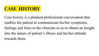
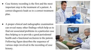
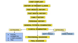
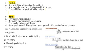
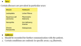
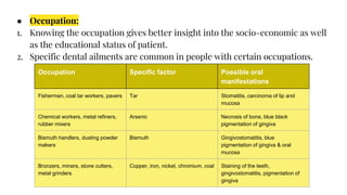
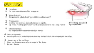
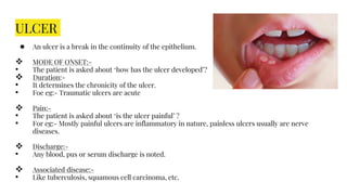
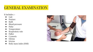
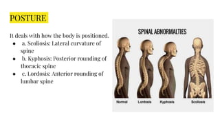
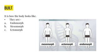
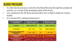
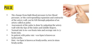
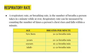
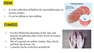
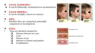
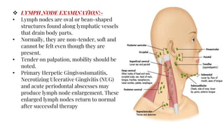
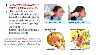
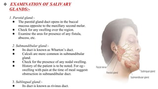
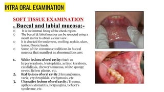
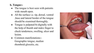
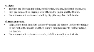
CASE HISTORY IN
PERIODONTICS
CASE HISTORY IN
PERIODONTICS
CASE HISTORY
Case history is a planned professional conversation that
enables the patient to communicate his/her symptoms,
feelings and fears to the clinician so as to obtain an insight
into the nature of patient’s illness and his/her attitude
towards them.
● Case history recording is the first and the most
important step in the treatment of a patient. A
correct diagnosis leads us to a correct treatment
plan.
● A proper clinical and radiographic examination
can reveal many other findings which help us to
find out associated problems in a particular case
thus helping us to provide a good periodontal
health and a functional occlusion to the patient.
Following figure describes the sequence of
various steps involved in the recording of case
history.
CHIEF COMPLAINT
HISTORY OF PRESENT ILLNESS
PAST MEDICAL HISTORY
PAST DENTAL HISTORY
FAMILY HISTORY
PERSONAL HISTORY
GENERAL PHYSICAL EXAMINATION
CLINICAL EXAMINATION INTRAORAL
EXTRAORAL
SOFT TISSUE HARD TISSUE
● FACIAL
SYMMETRY
● LYMPH NODES
● TMJ
PROVISIONAL DIAGNOSIS
FINAL DIAGNOSIS
DEMOGRAPHIC DETAILS
● OPD No:
1. For keeping record of patients (OPD-out patient department)
2. Billing purposes
3. Medicolegal aspect
● Date:
1. For tracking the duration of ailment.
2. For keeping a record and plan follow up visits.
● Name:
1. It is noted for addressing the patient.
2. It helps in better coordination and interaction.
3. To establish a rapport with the patient.
● Age:
1. For treatment planning.
2. Behavior management techniques.
3. To calculate dosage of drugs.
4. Some periodontal diseases are more prevalent in particular age groups.
E.g. ♦ Localized aggressive periodontitis
11-19 years.
♦ Generalised aggressive periodontitis
20-35 years.
♦ Chronic periodontitis
>35 years.
● Sex:
Certain diseases are prevalent in particular sexes
● Address:
1. Should be recorded for further communication with the patient.
2. Certain conditions are endemic in specific areas. e.g.,fluorosis.
MALES FEMALES
Leukoplakia Lichen Planus
Squamous cell
carcinoma
Pleomorphic
Adenoma
Hemophilia Iron deficiency
Anemia
Chronic
Periodontitis
Osteoporosis
● Occupation:
1. Knowing the occupation gives better insight into the socio-economic as well
as the educational status of patient.
2. Specific dental ailments are common in people with certain occupations.
Occupation
Specific factor
Possible oral manifestations
Occupation Specific factor Possible oral
manifestations
Fisherman, coal tar workers, pavers Tar Stomatitis, carcinoma of lip and
mucosa
Chemical workers, metal refiners,
rubber mixers
Arsenic Necrosis of bone, blue black
pigmentation of gingiva
Bismuth handlers, dusting powder
makers
Bismuth Gingivostomatitis, blue
pigmentation of gingiva & oral
mucosa
Bronzers, miners, stone cutters,
metal grinders
Copper, iron, nickel, chromium, coal Staining of the teeth,
gingivostomatitis, pigmentation of
gingiva
Chief Complaint:-
● The chief complaint is usually the reason for the
patient’s visit.
● It is stated in patient’s own words in chronological
order of their appearance and their severity.
● It aids in diagnosis and treatment. Therefore,
should be given utmost priority.
● The common chief complaints are:-
1. Pain
2. Swelling
3. Ulcer
4. Bleeding from gums
5. Dry mouth
6. Burning sensation
HISTORY OF PRESENT ILLNESS :-
● It is a chronological account of the chief complaint and associated symptoms from the
time of onset to the time the history is taken.
● The history commences from the beginning of the first symptom and extends to the
time of the examination.
● The questions can be asked in the manner:
– When did the problem start?
– What did you notice first?
– Did you have any problems or symptoms related to this?
– What makes the problem worse or better?
– Have any tests been performed before to diagnose this complaint?
– Have you consulted any other examiner for this problem?
– What have you done to treat this problem? Etc
● In general, the symptoms can be elaborated under:
– Mode of onset
– Cause of onset
– Duration
– Progress and referred
–Site
PAIN :
Pain is an unpleasant sensory and emotional experience associated with actual or potential tissue damage, or
described in terms of such damage
Factors considered in pain :-
 Site of pain:-
● The patient is asked about “where did the pain start”?
 Origin & mode of onset:-
● It determines the chronicity of the pain.
 Intensity of pain :-
• The pain can be mild, moderate or severe.
 Nature of the pain :-
• There are various type of pain. The most common are:-
1. Vague pain:- It is a mild continuous pain. Eg:- periodontal pain
2. Burning pain:- Pain occurring with the burning sensation. Eg:- Reflex oesophagitis
3. Throbbing pain:- Throbbing sensation is felt. Eg:- abscess
4. Stabbing pain:- Sudden, severe, sharp and short-lived pain. Eg:- Acute pulpal pain
5. Shooting pain:- Pain increases in severity in a short period. Eg:- Trigeminal neuralgia
 Progression of pain:-
• The patient should be asked ‘how it is progressing’?
• The pain may begin on a weak note and gradually reach a peak and then gradually declines.
 Duration of pain:-
• It is the period from the time of onset to the time of pain disappearance.
• It can be continuous or intermittent.
 Radiation of pain:-
• It is the extension of pain to another site.
• It can be referred, shifting or migration of pain
 Precipitating or aggravating factors:-
• Different factors may worsen the pain suggesting a specific diagnosis about the disease.
• For eg:- Trigeminal pain is increased while talking, brushing.
 Relieving factors:-
• Factors which reduce the severity or frequency of pain.
• For eg:- pain of chronic pulpitis gets relieved by cold application.
 Associated symptoms:-
• Pain can be associated with other factors like vomiting, sweating, flushing and increase in pulse rate
SWELLING
 Duration:-
● For how many days swelling is present.
 Mode of onset:-
● The patient is asked about ‘how did the swelling start’?
 Progression:-
● Swelling can increase gradually in size or rapidly.
● Eg:- bony swellings grows very slowly and remain static for a long period of time.
 Site of swelling:-
The original site where the swelling is started
 Other symptoms:-
• Include pain, fever, difficulty in swallowing, disfigurement, bleeding or pus discharge.
 Recurrence of the swelling:-
• Many swellings do recur after removal of the tissue.
• For eg:- Ranula
ULCER
● An ulcer is a break in the continuity of the epithelium.
 MODE OF ONSET:-
• The patient is asked about ‘how has the ulcer developed’?
 Duration:-
• It determines the chronicity of the ulcer.
• Foe eg:- Traumatic ulcers are acute
 Pain:-
• The patient is asked about ‘is the ulcer painful’ ?
• For eg:- Mostly painful ulcers are inflammatory in nature, painless ulcers usually are nerve
diseases.
 Discharge:-
• Any blood, pus or serum discharge is noted.
 Associated disease:-
• Like tuberculosis, squamous cell carcinoma, etc.
MEDICAL HISTORY
● All medical problems the patient has and is aware of should be recorded.A
variety of medical problems have varied interrelationship with oral ailments and
would require the modification of treatment.e.g.
♦ Diabetes and HIV infection increase the susceptibility to periodontal diseases.
♦ Leukaemia and mucocutaneous disorders can have periodontal manifestations.
♦ Drugs such as calcium channel blockers and phenytoin have periodontal side
effects.
♦ Use of medications such as anticoagulants or bisphosphonates warrant specific
precautions to avoid complications.
♦ Ultrasonic instrumentation should be avoided in patients with communicable
diseases such as tuberculosis to prevent the production of aerosols as they may
present risk to the dental team.
FAMILY HISTORY
• The patient should be asked about family history of periodontal problems.
• Certain diseases have genetic predilection,e.g:hemophilia, diabetes,
hypertension
• Prenatal, natal and postnatal history should be taken in case of pediatric
patients.
PAST DENTAL HISTORY
● It tells the dentist about the attitude of the patient towards dentistry.
● History of complications experienced by the patient.
● Eg:
1. Allergies to local anesthesia, syncope,
2. if the patient is a regular attende
3. in case the patient is undergoing periodontal treatment and
4. if the patient has undergone previous orthodontic treatment.
5. Recording the previous history of extraction must include the reasons for
extraction.
PERSONAL HABITS
ORAL HABITS
ORAL HYGIENE
HABITS
ADVERSE HABITS DIET HISTORY
• Thumb sucking
• Finger sucking
• Tongue
thrusting
• Lip biting
• Nail biting
•Cheek biting
• Mouth
breathing
•Bruxism
• Regularity of
brushing –
•Frequency and
method of brushing
• Type of brush and
how often it is
changed
• Use of other oral
hygiene aids.
• Smoking
• Alcohol
consumption
• Tobacco chewing
• Arecanut
chewing/Paan
chewing,
• vegetarian
• mixed
NOTE: CHECK FOR AMOUNT, FREQUENCY AND DURATION OF THE HABITS
GENERAL EXAMINATION
It includes :-
● Gait
● Posture
● Built
● Blood pressure
● Pulse
● Temperature
● Respiratory rate
● Pallor
● Cyanosis
● Edema
● Icterus
● Body mass index (BMI)
GAIT
It is the pattern of movement of the limbs during locomotion.
• Different types of gait are:-
a. Antalgic gait
b. Ataxic gait
c. Festinating gait
d. Four point gait
e. Hemiplegic gait
f. Spastic gait
POSTURE
It deals with how the body is positioned.
● a. Scoliosis: Lateral curvature of
spine
● b. Kyphosis: Posterior rounding of
thoracic spine
● c. Lordosis: Anterior rounding of
lumbar spine
BUILT
It is how the body looks like.
• They are:-
a. Endomorph
b. Mesomorph
c. Ectomorph
BLOOD PRESSURE
• It is the lateral pressure exerted by the blood flowing through the peripheral
arteries, as a result of the pumping action of the heart.
• Any variation in the BP from normal either low or high is indicator of poor
health.
• It is measured by sphygmomanometer.
PULSE
• The change from high blood pressure to low blood
pressure, or the corresponding expansion and contraction
of the artery wall, can be felt through palpation of an
artery called as pulse.
• Assessment of the pulse is done by palpating the artery
wall with the tips of the index and middle fingers.
• Normal rate is 60-100 beats/min and average rate is 72
beats/min.
• In patient with pulse rate >100 bpm is known as
tachycardia.
• Rate <60 bpm is known as bradycardia, seen in sinus
bradycardia.
RESPIRATORY RATE
● A respiratory rate, or breathing rate, is the number of breaths a person
takes in 1 minute while at rest. Respiratory rate can be measured by
counting the number of times a person’s chest rises and falls within a
minute.
AGE BREATHS PER MINUTE
New Born 30-50 breaths/min
3years 20-30 breaths/min
10years 16-22 breaths/min
Adult- 12-20 breaths/min
EDEMA
• It is the collection of fluid in the interstitial space or
serous cavities.
• It can be pitting or non-pitting
CYANOSIS
• It is the bluish discoloration of the skin and
mucous membranes due to low levels of oxygen
in the blood.
• Site of cyanosis are palate, tongue, lips, cheek,
nail bed, tip of nose, etc.
• Cyanosis can be central or peripheral.
BODY MASS INDEX(BMI)
• BMI is defined as the body mass divided by the square of the body height,
and is universally expressed in units of kg/(m)2.
CALCULATION
OF BMI
PALLOR
It is the paleness of the skin and mucous membrane caused as a result of diminished
blood supply.
• Causes of pallor:
● - Anemia
● - Shock
● - Exposure to cold
● - Heart disease
ICTERUS
• It is a yellowish pigmentation of skin, sclera
and other mucous membrane caused by
hyperbilirubinemia.
• It is seen in the condition of jaundice.
• Jaundice is often seen in liver diseases such
as hepatitis
CLUBBING
• It is bulbous enlargement of the soft parts
of terminal phalanges with both
transverse and longitudinal curving of
nails.
• Causes of clubbing are pulmonary
disease, lung abscess, etc.
.
BODY TEMPRATURE
Normal body temperature of a healthy individual is 37 degrees
Celsius
EXTRA ORAL EXAMINATION
 SKIN:-
● It is looked for-
- Appearance for any rashes, sores or itching
- Color- anemia patients have pale skin color, yellow tint is seen in jaundice
patients
- Pigmentation
- Edema
- temperature
 HEAD:-
• Any abnormality in size, shape and
symmetry of the head is to be examined.
• Different types of head are:-
● 1. Mesocephalic – average shape of head
● 2. Doliocephalic – long and narrow shaped
head
● 3. Brachycephalic – broad and short shaped
head
 FACIAL FORM:-
• It can be:-
1. Mesoprosopic:-average facial form
2. Euryprosopic:- broad and short
facial form
3. Leptoprosopic:- long and narrow
facial form
 FACIAL SYMMETRY:-
● It can be bilaterally symmetrical or asymmetrical.
 FACIAL PROFILE:-
• It can be straight, convex or concave.
 LIPS:-
• Whether they are competent, potenially
competent or incompetent.
 EYES:-
• These are should be checked for:
 Distance between two eyes
 Color
 Dryness of eye
 Indicator of anemia and jaundice
 Exophthalmos
STRAIGHT
PROFILE
CONVEX
PROFILE
CONCAVE
PROFILE
 LYMPH NODE EXAMINATION:-
• Lymph nodes are oval or bean-shaped
structures found along lymphatic vessels
that drain body parts.
• Normally, they are non-tender, soft and
cannot be felt even though they are
present.
• Tender on palpation, mobility should be
noted.
• Primary Herpetic Gingivostomatitis,
Necrotizing Ulcerative Gingivitis (NUG)
and acute periodontal abscesses may
produce lymph node enlargement. These
enlarged lymph nodes return to normal
after successful therapy
 If a node is palpable, record:-
• Site
• Size
• Texture – soft(infection), rubbery hard(Hodgkin’s lymphoma), stony
hard(secondary carcinoma)
• Tenderness on palpation
• Number of nodes- e.g. Multiple in case of glandular fever
• Whether fixed to surrounding tissues or not.
 Palpable node characteristics:
 Acute infection – large, soft, painful, mobile, discrete
 Chronic infection – large, firm, less tender, mobile
 Lymphoma – rubbery hard, matted, painless, multiple
 Metastatic cancer – stony hard, fixed to surrounding tissues, painless
 TEMPOROMANDIBULAR
JOINT EXAMINATION:-
● The importance is to
determine deviation of jaw
from the midline during the
opening and closing of jaws.
- Maximum mouth opening:
40-45mm
- Lateral mandibular range of
motion: 8-10mm
Muscles of mastication:- helps in the
determination of TMJ dysfunction and
in the discovery of other abnormalities.
EXTRA AURICULAR INTRA AURICULAR
 EXAMINATION OF SALIVARY
GLANDS:-
1. Parotid gland:-
● The parotid gland duct opens in the buccal
mucosa opposite to the maxillary second molar.
● Check for any swelling over the region.
● Examine the area for presence of any fistula,
abscess, etc.
2. Submandibular gland:-
● Its duct is known as Wharton’s duct.
● Calculi are more common in submandibular
gland.
● Check for the presence of any nodal swelling.
● History of the patient is to be noted. For eg:-
swelling with pain at the time of meal suggests
obstruction in submandibular duct.
3. Sublingual gland:-
● Its duct is known as rivinus duct.
INTRA ORAL EXAMINATION
SOFT TISSUE EXAMINATION
a. Buccal and labial mucosa:-
o It is the internal lining of the cheek region.
o The buccal & labial mucosa can be retracted using a
mouth mirror to obtain a clear view.
o It is checked for tenderness, swelling, nodule, ulcer,
lesion, fibrotic bands.
o Some of the common conditions in buccal
mucosa that manifest as abnormalities are:
1. White lesions of oral cavity: Such as
hyperkeratosis, leukoplakia, actinic keratosis,
candidiasis, chewer’s mucosa, white sponge
nevus, lichen planus, etc.
2. Red lesions of oral cavity: Hemangiomas,
varix, erythroplakia, ecchymosis, etc.
3. Ulcerative lesions of oral cavity: Trauma,
apthous stomatitis, herpangina, behcet’s
syndrome, etc.
b. Tongue:-
● The tongue is best seen with patients
wide mouth open.
● All the surface i.e. tip, dorsal, ventral
,base and lateral border of the tongue
should be examined thoroughly.
● Tongue is palpated bi-digitally with
the help of thumb and index finger to
check tenderness, swelling, ulcer and
lesion.
● Common manifestations:-
Geographic tongue, median
rhomboid glossitis, etc.
c. Lips:-
• The lips are checked for color, competency, texture, fissuring, shape, etc.
• Lips are palpated bi-digitally using the index finger and the thumb.
• Common manifestations are cleft lip, lip pits, angular cheilitis, etc.
d. Floor of mouth:-
• Palpation of floor of mouth is done by asking the patient to raise the tongue
to the roof of the mouth and then using a mouth mirror to further retract
the tongue.
• Common manifestations are ranula, sialolith, mandibular tori, etc.
e. Hard palate and soft palate:-
 It can be visualized when the patient’s head is tilted back as the patient lies in a
supine position with the mouth wide open.
 Common manifestations are cleft palate, smoker’s palate, etc.
● HARD TISSUE EXAMINATION
● Dental Examination:-
a. Teeth present:-
- Check for:- size, color, structural changes of teeth,
eruption status of teeth, retained deciduous teeth, any
trauma to tooth, etc.
b. Teeth missing:-
Reason for missing of teeth/ tooth.
● Correlation of the missing teeth as an oral
manifestation of systemic disease or genetic
abnormality.
● The causes for missing teeth may include supra
eruption, tilting, drifting or rotation, etc.
● History of removal. For eg:- in case of any sport
injury, accident, etc.
c. Carious teeth:-
Carious teeth are evaluated by visual inspection, using
explorer or probe, percussion, radiographs, etc.
d. Wasting Diseases of teeth:-
It is defined as the noncarious loss of tooth substance. It is the interaction of four processes:-
1. Attrition:- It is the physiologic wearing away of a tooth as a result of tooth to tooth contact, as
in mastication.
2. Abrasion:- Refers to the loss of tooth structure due to external agents which have an abrasive
effect on the teeth.
3. Erosion:- It is defined as an irreversible loss of dental hard tissue by a chemical process in the
absence of plaque. Erosive factors can be extrinsic or intrinsic. For e.g. Carbonated drinks,
alcoholic beverages, vomiting, gastro-esophageal reflux, etc.
4. Abfraction:- The pathological loss of enamel and dentin due to occlusal stresses. Common in
patients with poor tooth alignment.
ATTRITION EROSION
ABRASION ABFRACTION
f. Tooth Mobility:-
● Test for mobility of the tooth is carried out by
moving the tooth laterally in the socket or
preferably in the handles between two instruments.
● Types:-
1. Pathologic mobility:- It results from inflammatory
process, parafunctional habits, etc.
2. Adaptive mobility:- Occur due to anatomic factors
such as short roots or poor crown to root ratio.
● Causes of tooth mobility are trauma from
occlusion, periodontitis, endo-perio lesion, after
periodontal surgery
● Miller’s classification:-
1.Grade 0:- No detectable movement
2. Grade 1:- Mobility greater than normal
3.Grade 2:- Mobility upto 1mm in buccolingual
direction
4.Grade 3:- Mobility >1mm in buccolingual
direction in combination with ability to depress tooth
in vertical direction
g. Malocclusion:-
Class I:-The mesiobuccal cusp of the maxillary first permanent molar coincides
with the buccal groove of the mandibular permanent first molar.
Class II:- The distobuccal cusp of the upper first permanent molar coincides
with the buccal groove of the lower first permanent molar.
Class III:- The mesiobuccal cusp of maxillary first permanent molar coincides
with the interdental space between the mandibular first and second permanent
molar.
● Stains:-
It is of 2 types:-
1. Extrinsic stain
2. Intrinsic stain
• Extrinsic stain:- Discoloration located
on the outer surface of tooth structure
and caused by topical agent .for
e.g,.tobacco ,pan chewing,tea etc.
• Intrinsic stain:- Occurs following a
change in the structural composition
or thickness of dental Hard Tissues.
o Causes:-
• Amelogenesis imperfecta
• Dentinogenesis imperfecta
• Dental flourosis
Extrinsic stain
Intrinsic stain
● Trauma From Occlusion:
• Occlusal Traumatism: Functional loading of teeth, usually off-axis, that is of
sufficient magnitude to induce changes to the teeth (e.g., fractures, wear) or
supporting structures.
• Changes may be temporary or permanent.
• Fremitus test:
Fremitus: A palpable or visible movement of a tooth when subjected to occlusal
forces.
• It is the vibration of root surfaces as the patient “taps” his or her teeth together.
♦ Grading:
Class I: Mild vibrations detected.
Class II: Clearly palpable vibrations, no visible movement.
Class III: Clinically visible vibrations.
Pathological Migration:
● It refers to tooth displacement that results
when the balance among the factors that
maintain physiological tooth position is
disturbed by periodontal disease.
● It may accur in asoociation with gingival
inflammation and pocket formation as
disease progresses.
● It is cmmon in anterior teeth and is
accompanied by mobility and extrusion of
teeth.
● CAUSES:
• Unreplaced missing teeth
• Pressure from tongue
• Pressure form granulation tissue of
periodontal pocket
• Habits
PERIODONTAL EXAMINATION
 GINGIVA:
I. Color:- Color of healthy gingiva has been described as coral pink varied by complexion and
pigmentation.
II. Contour:- A healthy gingival contour follows the margin of underlying bone and has scalloped
contour with knife edge margin.
III. Consistency:- Firm and resilient
IV. Surface texture:- Normal gingiva shows an orange peel appearance which is termed as stippling.
Stippling is an adaptive specialization of an epithelium to withstand physical forces. It is lost in
edematous gingiva.
V. Size:- It correspond to sum total of cellular, intercellular and vascular components. In inflamed
gingiva, the size becomes enlarged.
VI. Position:- The gingiva is at the level of the attached periodontal tissue. It is determined by
probing. The normal position is 1mm above the CEJ.
 Pocket Depth:
● Pocket is a pathologically deepened gingival
sulcus.
● The depth of the pocket is of two types:
1. Biologic depth/histologic depth: it is the
distance between the gingival margin and
the base of the pocket (the coronal end of
the junctional epithelium). This can be
measured in carefully prepared and well
oriented histologic sections.
2. Clinical/Probing depth is the distance from
the gingival margin to the base of the
probable crevice/ pocket.
● When the sulcus depth is more than 3mm, it
is called a pocket. Probing depth is
measured in millimetres using graduated
probes.
Some of the periodontal probes routinely used are
♦ Williams probe: the markings are 1,2,3,5,7,8,9
and 10mm.
♦ UNC-15: The markings are from 1 to 15 each at
millimetre distance.
♦ Community periodontal index of treatment
needs (CPITN) probe:
a. CPITN- E (epidemiological): 0.5, 3.5, 5.5.
b. CPITN-C (clinical):0.5, 3.5, 5.5, 8.5, 11.5
♦ Nabers probe: It is curved and used in
examining pathological furcation involvement.
♦ Probing depth is recorded along six points
around each tooth: distofacial, facial,
mesiofacial, distolingual, lingual and
mesiolingual.
 Loss Of Attachment/ Clinical Attachment Level (CAL)
It is the distance from the cementoenamel junction (or a fixed point on the tooth) to
the base of the probeable crevice.
● It is the true clinical measure of amount of destruction. When recorded at
different times it helps in determining the progression of periodontitis.
● It helps in determining whether periodontitis is present at a particular site.
● It is measured in a vertical direction with reference to a fixed point on the tooth,
usually the CEJ.
● In health, the apical end of junctional epithelium is on the CEJ and hence, there
should be no CAL.
♦ In case of gingival recession:
Loss of attachment : Probing depth + recession.
♦ When the gingival margin is coronal to CEJ:
CAL: Probeable pocket depth-the distance between the gingival margin and the
cementoenamel junction.
♦ When the gingival margin is at the cementoenamel junction or slightly coronal:
CAL: Probing depth
 GINGIVAL RECESSION:
Gingival recession is the exposure of root surfaces due to apical migration of
the gingival tissue margins.
 FURCATION ASSESSMENT:-
● The point at which the root trunk on a multirooted tooth diverges to form more than
one root is called as Furcation.
● Furcation involvement:- The extension of inflammatory periodontal disease into the
interradicular area of multirooted teeth is known as furcation involvement.
● Naber’s probe are used for detection of furcation involvement.
1. Grade I:- Incipient stage
2. Grade II:- The furcation lesion involve one or more furcation of same tooth. Here,
the remains attached to the tooth. Involve horizontal component to the bone loss.
3. Grade 3:- The bone is no longer attached to the furcation of the root.
4. Grade 4:- Interdental bone is destroyed. Furcation opening is clinically visible.
PROVISIONAL DIAGNOSIS
● It is also called as tentative, clinical or working diagnosis
● It is formed after evaluating the case history and performing the physical
examination.
● All the records and clinical findings are clubbed together.
DIFFERENTIAL DIAGNOSIS
● It is the process of listing out two or diseases having similar signs and symptoms of
which only one could be attributed to the patient’s suffering.
● A final diagnosis is only possible after carrying out further investigations.
INVESTIGATION
 Chair side investigations:- Pulp vitality tests,
percussion tests, cytology, and aspiration
 Radiological investigations:-
- Intraoral radiographs:- IOPA, occlusal and bitewing
- Extraoral radiographs:- OPG view, PA view, Water’s
view, Submentovertex view, etc.
- Radiograph can be used to assess
♦ Degree of bone loss: Mild, moderate or severe
♦ Distribution of bone loss: Localized or
generalized.
♦ Pattern of bone loss : vertical, horizontal or both.
♦ Root morphology and pathology.
♦ Restorations: Overhanging margins, deficient
margins or recurrent caries.
FINAL DIAGNOSIS
● The final diagnosis can usually be reached following chronologic organization and
critical evaluation of the information obtained from patient history, physical
examination and the result of radiological and laboratory examination.
TREATMENT PLAN
● It includes following phase:-
1. Phase1:- Emergency phase
2. Phase 2:- Preventive phase
3. Phase 3:- Surgical phase
4. Phase 4:- Restorative phase
5. Phase 5:- Maintenance phase
PROGNOSIS
● The prognosis is the prediction of the probable course, duration and outcome of
disease based on general knowledge of the pathogenesis of disease and the presence
of risk factors for the disease.
● Types:
1. Excellent prognosis
2. Good prognosis
3. Fair prognosis
4. Poor prognosis
5. Questionable prognosis
6. Hopeless prognosis
THANK YOU!

CASE HISTORY (1802).pptx

  • 1.
    CASE HISTORY IN PERIODONTICS CASEHISTORY IN PERIODONTICS
  • 2.
    CASE HISTORY Case historyis a planned professional conversation that enables the patient to communicate his/her symptoms, feelings and fears to the clinician so as to obtain an insight into the nature of patient’s illness and his/her attitude towards them.
  • 3.
    ● Case historyrecording is the first and the most important step in the treatment of a patient. A correct diagnosis leads us to a correct treatment plan. ● A proper clinical and radiographic examination can reveal many other findings which help us to find out associated problems in a particular case thus helping us to provide a good periodontal health and a functional occlusion to the patient. Following figure describes the sequence of various steps involved in the recording of case history.
  • 4.
    CHIEF COMPLAINT HISTORY OFPRESENT ILLNESS PAST MEDICAL HISTORY PAST DENTAL HISTORY FAMILY HISTORY PERSONAL HISTORY GENERAL PHYSICAL EXAMINATION CLINICAL EXAMINATION INTRAORAL EXTRAORAL SOFT TISSUE HARD TISSUE ● FACIAL SYMMETRY ● LYMPH NODES ● TMJ PROVISIONAL DIAGNOSIS FINAL DIAGNOSIS
  • 5.
    DEMOGRAPHIC DETAILS ● OPDNo: 1. For keeping record of patients (OPD-out patient department) 2. Billing purposes 3. Medicolegal aspect ● Date: 1. For tracking the duration of ailment. 2. For keeping a record and plan follow up visits.
  • 6.
    ● Name: 1. Itis noted for addressing the patient. 2. It helps in better coordination and interaction. 3. To establish a rapport with the patient. ● Age: 1. For treatment planning. 2. Behavior management techniques. 3. To calculate dosage of drugs. 4. Some periodontal diseases are more prevalent in particular age groups. E.g. ♦ Localized aggressive periodontitis 11-19 years. ♦ Generalised aggressive periodontitis 20-35 years. ♦ Chronic periodontitis >35 years.
  • 7.
    ● Sex: Certain diseasesare prevalent in particular sexes ● Address: 1. Should be recorded for further communication with the patient. 2. Certain conditions are endemic in specific areas. e.g.,fluorosis. MALES FEMALES Leukoplakia Lichen Planus Squamous cell carcinoma Pleomorphic Adenoma Hemophilia Iron deficiency Anemia Chronic Periodontitis Osteoporosis
  • 8.
    ● Occupation: 1. Knowingthe occupation gives better insight into the socio-economic as well as the educational status of patient. 2. Specific dental ailments are common in people with certain occupations. Occupation Specific factor Possible oral manifestations Occupation Specific factor Possible oral manifestations Fisherman, coal tar workers, pavers Tar Stomatitis, carcinoma of lip and mucosa Chemical workers, metal refiners, rubber mixers Arsenic Necrosis of bone, blue black pigmentation of gingiva Bismuth handlers, dusting powder makers Bismuth Gingivostomatitis, blue pigmentation of gingiva & oral mucosa Bronzers, miners, stone cutters, metal grinders Copper, iron, nickel, chromium, coal Staining of the teeth, gingivostomatitis, pigmentation of gingiva
  • 9.
    Chief Complaint:- ● Thechief complaint is usually the reason for the patient’s visit. ● It is stated in patient’s own words in chronological order of their appearance and their severity. ● It aids in diagnosis and treatment. Therefore, should be given utmost priority. ● The common chief complaints are:- 1. Pain 2. Swelling 3. Ulcer 4. Bleeding from gums 5. Dry mouth 6. Burning sensation
  • 10.
    HISTORY OF PRESENTILLNESS :- ● It is a chronological account of the chief complaint and associated symptoms from the time of onset to the time the history is taken. ● The history commences from the beginning of the first symptom and extends to the time of the examination. ● The questions can be asked in the manner: – When did the problem start? – What did you notice first? – Did you have any problems or symptoms related to this? – What makes the problem worse or better?
  • 11.
    – Have anytests been performed before to diagnose this complaint? – Have you consulted any other examiner for this problem? – What have you done to treat this problem? Etc ● In general, the symptoms can be elaborated under: – Mode of onset – Cause of onset – Duration – Progress and referred –Site
  • 12.
    PAIN : Pain isan unpleasant sensory and emotional experience associated with actual or potential tissue damage, or described in terms of such damage Factors considered in pain :-  Site of pain:- ● The patient is asked about “where did the pain start”?  Origin & mode of onset:- ● It determines the chronicity of the pain.  Intensity of pain :- • The pain can be mild, moderate or severe.  Nature of the pain :- • There are various type of pain. The most common are:- 1. Vague pain:- It is a mild continuous pain. Eg:- periodontal pain 2. Burning pain:- Pain occurring with the burning sensation. Eg:- Reflex oesophagitis 3. Throbbing pain:- Throbbing sensation is felt. Eg:- abscess 4. Stabbing pain:- Sudden, severe, sharp and short-lived pain. Eg:- Acute pulpal pain 5. Shooting pain:- Pain increases in severity in a short period. Eg:- Trigeminal neuralgia
  • 13.
     Progression ofpain:- • The patient should be asked ‘how it is progressing’? • The pain may begin on a weak note and gradually reach a peak and then gradually declines.  Duration of pain:- • It is the period from the time of onset to the time of pain disappearance. • It can be continuous or intermittent.  Radiation of pain:- • It is the extension of pain to another site. • It can be referred, shifting or migration of pain  Precipitating or aggravating factors:- • Different factors may worsen the pain suggesting a specific diagnosis about the disease. • For eg:- Trigeminal pain is increased while talking, brushing.  Relieving factors:- • Factors which reduce the severity or frequency of pain. • For eg:- pain of chronic pulpitis gets relieved by cold application.  Associated symptoms:- • Pain can be associated with other factors like vomiting, sweating, flushing and increase in pulse rate
  • 14.
    SWELLING  Duration:- ● Forhow many days swelling is present.  Mode of onset:- ● The patient is asked about ‘how did the swelling start’?  Progression:- ● Swelling can increase gradually in size or rapidly. ● Eg:- bony swellings grows very slowly and remain static for a long period of time.  Site of swelling:- The original site where the swelling is started  Other symptoms:- • Include pain, fever, difficulty in swallowing, disfigurement, bleeding or pus discharge.  Recurrence of the swelling:- • Many swellings do recur after removal of the tissue. • For eg:- Ranula
  • 15.
    ULCER ● An ulceris a break in the continuity of the epithelium.  MODE OF ONSET:- • The patient is asked about ‘how has the ulcer developed’?  Duration:- • It determines the chronicity of the ulcer. • Foe eg:- Traumatic ulcers are acute  Pain:- • The patient is asked about ‘is the ulcer painful’ ? • For eg:- Mostly painful ulcers are inflammatory in nature, painless ulcers usually are nerve diseases.  Discharge:- • Any blood, pus or serum discharge is noted.  Associated disease:- • Like tuberculosis, squamous cell carcinoma, etc.
  • 16.
    MEDICAL HISTORY ● Allmedical problems the patient has and is aware of should be recorded.A variety of medical problems have varied interrelationship with oral ailments and would require the modification of treatment.e.g. ♦ Diabetes and HIV infection increase the susceptibility to periodontal diseases. ♦ Leukaemia and mucocutaneous disorders can have periodontal manifestations. ♦ Drugs such as calcium channel blockers and phenytoin have periodontal side effects. ♦ Use of medications such as anticoagulants or bisphosphonates warrant specific precautions to avoid complications. ♦ Ultrasonic instrumentation should be avoided in patients with communicable diseases such as tuberculosis to prevent the production of aerosols as they may present risk to the dental team.
  • 17.
    FAMILY HISTORY • Thepatient should be asked about family history of periodontal problems. • Certain diseases have genetic predilection,e.g:hemophilia, diabetes, hypertension • Prenatal, natal and postnatal history should be taken in case of pediatric patients.
  • 18.
    PAST DENTAL HISTORY ●It tells the dentist about the attitude of the patient towards dentistry. ● History of complications experienced by the patient. ● Eg: 1. Allergies to local anesthesia, syncope, 2. if the patient is a regular attende 3. in case the patient is undergoing periodontal treatment and 4. if the patient has undergone previous orthodontic treatment. 5. Recording the previous history of extraction must include the reasons for extraction.
  • 19.
    PERSONAL HABITS ORAL HABITS ORALHYGIENE HABITS ADVERSE HABITS DIET HISTORY • Thumb sucking • Finger sucking • Tongue thrusting • Lip biting • Nail biting •Cheek biting • Mouth breathing •Bruxism • Regularity of brushing – •Frequency and method of brushing • Type of brush and how often it is changed • Use of other oral hygiene aids. • Smoking • Alcohol consumption • Tobacco chewing • Arecanut chewing/Paan chewing, • vegetarian • mixed NOTE: CHECK FOR AMOUNT, FREQUENCY AND DURATION OF THE HABITS
  • 20.
    GENERAL EXAMINATION It includes:- ● Gait ● Posture ● Built ● Blood pressure ● Pulse ● Temperature ● Respiratory rate ● Pallor ● Cyanosis ● Edema ● Icterus ● Body mass index (BMI)
  • 21.
    GAIT It is thepattern of movement of the limbs during locomotion. • Different types of gait are:- a. Antalgic gait b. Ataxic gait c. Festinating gait d. Four point gait e. Hemiplegic gait f. Spastic gait
  • 22.
    POSTURE It deals withhow the body is positioned. ● a. Scoliosis: Lateral curvature of spine ● b. Kyphosis: Posterior rounding of thoracic spine ● c. Lordosis: Anterior rounding of lumbar spine
  • 23.
    BUILT It is howthe body looks like. • They are:- a. Endomorph b. Mesomorph c. Ectomorph
  • 24.
    BLOOD PRESSURE • Itis the lateral pressure exerted by the blood flowing through the peripheral arteries, as a result of the pumping action of the heart. • Any variation in the BP from normal either low or high is indicator of poor health. • It is measured by sphygmomanometer.
  • 25.
    PULSE • The changefrom high blood pressure to low blood pressure, or the corresponding expansion and contraction of the artery wall, can be felt through palpation of an artery called as pulse. • Assessment of the pulse is done by palpating the artery wall with the tips of the index and middle fingers. • Normal rate is 60-100 beats/min and average rate is 72 beats/min. • In patient with pulse rate >100 bpm is known as tachycardia. • Rate <60 bpm is known as bradycardia, seen in sinus bradycardia.
  • 26.
    RESPIRATORY RATE ● Arespiratory rate, or breathing rate, is the number of breaths a person takes in 1 minute while at rest. Respiratory rate can be measured by counting the number of times a person’s chest rises and falls within a minute. AGE BREATHS PER MINUTE New Born 30-50 breaths/min 3years 20-30 breaths/min 10years 16-22 breaths/min Adult- 12-20 breaths/min
  • 27.
    EDEMA • It isthe collection of fluid in the interstitial space or serous cavities. • It can be pitting or non-pitting CYANOSIS • It is the bluish discoloration of the skin and mucous membranes due to low levels of oxygen in the blood. • Site of cyanosis are palate, tongue, lips, cheek, nail bed, tip of nose, etc. • Cyanosis can be central or peripheral.
  • 28.
    BODY MASS INDEX(BMI) •BMI is defined as the body mass divided by the square of the body height, and is universally expressed in units of kg/(m)2. CALCULATION OF BMI
  • 29.
    PALLOR It is thepaleness of the skin and mucous membrane caused as a result of diminished blood supply. • Causes of pallor: ● - Anemia ● - Shock ● - Exposure to cold ● - Heart disease ICTERUS • It is a yellowish pigmentation of skin, sclera and other mucous membrane caused by hyperbilirubinemia. • It is seen in the condition of jaundice. • Jaundice is often seen in liver diseases such as hepatitis
  • 30.
    CLUBBING • It isbulbous enlargement of the soft parts of terminal phalanges with both transverse and longitudinal curving of nails. • Causes of clubbing are pulmonary disease, lung abscess, etc. . BODY TEMPRATURE Normal body temperature of a healthy individual is 37 degrees Celsius
  • 31.
    EXTRA ORAL EXAMINATION SKIN:- ● It is looked for- - Appearance for any rashes, sores or itching - Color- anemia patients have pale skin color, yellow tint is seen in jaundice patients - Pigmentation - Edema - temperature
  • 32.
     HEAD:- • Anyabnormality in size, shape and symmetry of the head is to be examined. • Different types of head are:- ● 1. Mesocephalic – average shape of head ● 2. Doliocephalic – long and narrow shaped head ● 3. Brachycephalic – broad and short shaped head  FACIAL FORM:- • It can be:- 1. Mesoprosopic:-average facial form 2. Euryprosopic:- broad and short facial form 3. Leptoprosopic:- long and narrow facial form
  • 33.
     FACIAL SYMMETRY:- ●It can be bilaterally symmetrical or asymmetrical.  FACIAL PROFILE:- • It can be straight, convex or concave.  LIPS:- • Whether they are competent, potenially competent or incompetent.  EYES:- • These are should be checked for:  Distance between two eyes  Color  Dryness of eye  Indicator of anemia and jaundice  Exophthalmos STRAIGHT PROFILE CONVEX PROFILE CONCAVE PROFILE
  • 34.
     LYMPH NODEEXAMINATION:- • Lymph nodes are oval or bean-shaped structures found along lymphatic vessels that drain body parts. • Normally, they are non-tender, soft and cannot be felt even though they are present. • Tender on palpation, mobility should be noted. • Primary Herpetic Gingivostomatitis, Necrotizing Ulcerative Gingivitis (NUG) and acute periodontal abscesses may produce lymph node enlargement. These enlarged lymph nodes return to normal after successful therapy
  • 35.
     If anode is palpable, record:- • Site • Size • Texture – soft(infection), rubbery hard(Hodgkin’s lymphoma), stony hard(secondary carcinoma) • Tenderness on palpation • Number of nodes- e.g. Multiple in case of glandular fever • Whether fixed to surrounding tissues or not.  Palpable node characteristics:  Acute infection – large, soft, painful, mobile, discrete  Chronic infection – large, firm, less tender, mobile  Lymphoma – rubbery hard, matted, painless, multiple  Metastatic cancer – stony hard, fixed to surrounding tissues, painless
  • 36.
     TEMPOROMANDIBULAR JOINT EXAMINATION:- ●The importance is to determine deviation of jaw from the midline during the opening and closing of jaws. - Maximum mouth opening: 40-45mm - Lateral mandibular range of motion: 8-10mm Muscles of mastication:- helps in the determination of TMJ dysfunction and in the discovery of other abnormalities. EXTRA AURICULAR INTRA AURICULAR
  • 37.
     EXAMINATION OFSALIVARY GLANDS:- 1. Parotid gland:- ● The parotid gland duct opens in the buccal mucosa opposite to the maxillary second molar. ● Check for any swelling over the region. ● Examine the area for presence of any fistula, abscess, etc. 2. Submandibular gland:- ● Its duct is known as Wharton’s duct. ● Calculi are more common in submandibular gland. ● Check for the presence of any nodal swelling. ● History of the patient is to be noted. For eg:- swelling with pain at the time of meal suggests obstruction in submandibular duct. 3. Sublingual gland:- ● Its duct is known as rivinus duct.
  • 38.
    INTRA ORAL EXAMINATION SOFTTISSUE EXAMINATION a. Buccal and labial mucosa:- o It is the internal lining of the cheek region. o The buccal & labial mucosa can be retracted using a mouth mirror to obtain a clear view. o It is checked for tenderness, swelling, nodule, ulcer, lesion, fibrotic bands. o Some of the common conditions in buccal mucosa that manifest as abnormalities are: 1. White lesions of oral cavity: Such as hyperkeratosis, leukoplakia, actinic keratosis, candidiasis, chewer’s mucosa, white sponge nevus, lichen planus, etc. 2. Red lesions of oral cavity: Hemangiomas, varix, erythroplakia, ecchymosis, etc. 3. Ulcerative lesions of oral cavity: Trauma, apthous stomatitis, herpangina, behcet’s syndrome, etc.
  • 39.
    b. Tongue:- ● Thetongue is best seen with patients wide mouth open. ● All the surface i.e. tip, dorsal, ventral ,base and lateral border of the tongue should be examined thoroughly. ● Tongue is palpated bi-digitally with the help of thumb and index finger to check tenderness, swelling, ulcer and lesion. ● Common manifestations:- Geographic tongue, median rhomboid glossitis, etc.
  • 40.
    c. Lips:- • Thelips are checked for color, competency, texture, fissuring, shape, etc. • Lips are palpated bi-digitally using the index finger and the thumb. • Common manifestations are cleft lip, lip pits, angular cheilitis, etc. d. Floor of mouth:- • Palpation of floor of mouth is done by asking the patient to raise the tongue to the roof of the mouth and then using a mouth mirror to further retract the tongue. • Common manifestations are ranula, sialolith, mandibular tori, etc.
  • 41.
    e. Hard palateand soft palate:-  It can be visualized when the patient’s head is tilted back as the patient lies in a supine position with the mouth wide open.  Common manifestations are cleft palate, smoker’s palate, etc.
  • 42.
    ● HARD TISSUEEXAMINATION ● Dental Examination:- a. Teeth present:- - Check for:- size, color, structural changes of teeth, eruption status of teeth, retained deciduous teeth, any trauma to tooth, etc. b. Teeth missing:- Reason for missing of teeth/ tooth. ● Correlation of the missing teeth as an oral manifestation of systemic disease or genetic abnormality. ● The causes for missing teeth may include supra eruption, tilting, drifting or rotation, etc. ● History of removal. For eg:- in case of any sport injury, accident, etc. c. Carious teeth:- Carious teeth are evaluated by visual inspection, using explorer or probe, percussion, radiographs, etc.
  • 43.
    d. Wasting Diseasesof teeth:- It is defined as the noncarious loss of tooth substance. It is the interaction of four processes:- 1. Attrition:- It is the physiologic wearing away of a tooth as a result of tooth to tooth contact, as in mastication. 2. Abrasion:- Refers to the loss of tooth structure due to external agents which have an abrasive effect on the teeth. 3. Erosion:- It is defined as an irreversible loss of dental hard tissue by a chemical process in the absence of plaque. Erosive factors can be extrinsic or intrinsic. For e.g. Carbonated drinks, alcoholic beverages, vomiting, gastro-esophageal reflux, etc. 4. Abfraction:- The pathological loss of enamel and dentin due to occlusal stresses. Common in patients with poor tooth alignment. ATTRITION EROSION ABRASION ABFRACTION
  • 44.
    f. Tooth Mobility:- ●Test for mobility of the tooth is carried out by moving the tooth laterally in the socket or preferably in the handles between two instruments. ● Types:- 1. Pathologic mobility:- It results from inflammatory process, parafunctional habits, etc. 2. Adaptive mobility:- Occur due to anatomic factors such as short roots or poor crown to root ratio. ● Causes of tooth mobility are trauma from occlusion, periodontitis, endo-perio lesion, after periodontal surgery ● Miller’s classification:- 1.Grade 0:- No detectable movement 2. Grade 1:- Mobility greater than normal 3.Grade 2:- Mobility upto 1mm in buccolingual direction 4.Grade 3:- Mobility >1mm in buccolingual direction in combination with ability to depress tooth in vertical direction
  • 45.
    g. Malocclusion:- Class I:-Themesiobuccal cusp of the maxillary first permanent molar coincides with the buccal groove of the mandibular permanent first molar. Class II:- The distobuccal cusp of the upper first permanent molar coincides with the buccal groove of the lower first permanent molar. Class III:- The mesiobuccal cusp of maxillary first permanent molar coincides with the interdental space between the mandibular first and second permanent molar.
  • 46.
    ● Stains:- It isof 2 types:- 1. Extrinsic stain 2. Intrinsic stain • Extrinsic stain:- Discoloration located on the outer surface of tooth structure and caused by topical agent .for e.g,.tobacco ,pan chewing,tea etc. • Intrinsic stain:- Occurs following a change in the structural composition or thickness of dental Hard Tissues. o Causes:- • Amelogenesis imperfecta • Dentinogenesis imperfecta • Dental flourosis Extrinsic stain Intrinsic stain
  • 47.
    ● Trauma FromOcclusion: • Occlusal Traumatism: Functional loading of teeth, usually off-axis, that is of sufficient magnitude to induce changes to the teeth (e.g., fractures, wear) or supporting structures. • Changes may be temporary or permanent. • Fremitus test: Fremitus: A palpable or visible movement of a tooth when subjected to occlusal forces. • It is the vibration of root surfaces as the patient “taps” his or her teeth together. ♦ Grading: Class I: Mild vibrations detected. Class II: Clearly palpable vibrations, no visible movement. Class III: Clinically visible vibrations.
  • 48.
    Pathological Migration: ● Itrefers to tooth displacement that results when the balance among the factors that maintain physiological tooth position is disturbed by periodontal disease. ● It may accur in asoociation with gingival inflammation and pocket formation as disease progresses. ● It is cmmon in anterior teeth and is accompanied by mobility and extrusion of teeth. ● CAUSES: • Unreplaced missing teeth • Pressure from tongue • Pressure form granulation tissue of periodontal pocket • Habits
  • 49.
    PERIODONTAL EXAMINATION  GINGIVA: I.Color:- Color of healthy gingiva has been described as coral pink varied by complexion and pigmentation. II. Contour:- A healthy gingival contour follows the margin of underlying bone and has scalloped contour with knife edge margin. III. Consistency:- Firm and resilient IV. Surface texture:- Normal gingiva shows an orange peel appearance which is termed as stippling. Stippling is an adaptive specialization of an epithelium to withstand physical forces. It is lost in edematous gingiva. V. Size:- It correspond to sum total of cellular, intercellular and vascular components. In inflamed gingiva, the size becomes enlarged. VI. Position:- The gingiva is at the level of the attached periodontal tissue. It is determined by probing. The normal position is 1mm above the CEJ.
  • 51.
     Pocket Depth: ●Pocket is a pathologically deepened gingival sulcus. ● The depth of the pocket is of two types: 1. Biologic depth/histologic depth: it is the distance between the gingival margin and the base of the pocket (the coronal end of the junctional epithelium). This can be measured in carefully prepared and well oriented histologic sections. 2. Clinical/Probing depth is the distance from the gingival margin to the base of the probable crevice/ pocket. ● When the sulcus depth is more than 3mm, it is called a pocket. Probing depth is measured in millimetres using graduated probes.
  • 52.
    Some of theperiodontal probes routinely used are ♦ Williams probe: the markings are 1,2,3,5,7,8,9 and 10mm. ♦ UNC-15: The markings are from 1 to 15 each at millimetre distance. ♦ Community periodontal index of treatment needs (CPITN) probe: a. CPITN- E (epidemiological): 0.5, 3.5, 5.5. b. CPITN-C (clinical):0.5, 3.5, 5.5, 8.5, 11.5 ♦ Nabers probe: It is curved and used in examining pathological furcation involvement. ♦ Probing depth is recorded along six points around each tooth: distofacial, facial, mesiofacial, distolingual, lingual and mesiolingual.
  • 53.
     Loss OfAttachment/ Clinical Attachment Level (CAL) It is the distance from the cementoenamel junction (or a fixed point on the tooth) to the base of the probeable crevice. ● It is the true clinical measure of amount of destruction. When recorded at different times it helps in determining the progression of periodontitis. ● It helps in determining whether periodontitis is present at a particular site. ● It is measured in a vertical direction with reference to a fixed point on the tooth, usually the CEJ. ● In health, the apical end of junctional epithelium is on the CEJ and hence, there should be no CAL. ♦ In case of gingival recession: Loss of attachment : Probing depth + recession. ♦ When the gingival margin is coronal to CEJ: CAL: Probeable pocket depth-the distance between the gingival margin and the cementoenamel junction. ♦ When the gingival margin is at the cementoenamel junction or slightly coronal: CAL: Probing depth
  • 54.
     GINGIVAL RECESSION: Gingivalrecession is the exposure of root surfaces due to apical migration of the gingival tissue margins.
  • 55.
     FURCATION ASSESSMENT:- ●The point at which the root trunk on a multirooted tooth diverges to form more than one root is called as Furcation. ● Furcation involvement:- The extension of inflammatory periodontal disease into the interradicular area of multirooted teeth is known as furcation involvement. ● Naber’s probe are used for detection of furcation involvement. 1. Grade I:- Incipient stage 2. Grade II:- The furcation lesion involve one or more furcation of same tooth. Here, the remains attached to the tooth. Involve horizontal component to the bone loss. 3. Grade 3:- The bone is no longer attached to the furcation of the root. 4. Grade 4:- Interdental bone is destroyed. Furcation opening is clinically visible.
  • 56.
    PROVISIONAL DIAGNOSIS ● Itis also called as tentative, clinical or working diagnosis ● It is formed after evaluating the case history and performing the physical examination. ● All the records and clinical findings are clubbed together.
  • 58.
    DIFFERENTIAL DIAGNOSIS ● Itis the process of listing out two or diseases having similar signs and symptoms of which only one could be attributed to the patient’s suffering. ● A final diagnosis is only possible after carrying out further investigations.
  • 59.
    INVESTIGATION  Chair sideinvestigations:- Pulp vitality tests, percussion tests, cytology, and aspiration  Radiological investigations:- - Intraoral radiographs:- IOPA, occlusal and bitewing - Extraoral radiographs:- OPG view, PA view, Water’s view, Submentovertex view, etc. - Radiograph can be used to assess ♦ Degree of bone loss: Mild, moderate or severe ♦ Distribution of bone loss: Localized or generalized. ♦ Pattern of bone loss : vertical, horizontal or both. ♦ Root morphology and pathology. ♦ Restorations: Overhanging margins, deficient margins or recurrent caries.
  • 61.
    FINAL DIAGNOSIS ● Thefinal diagnosis can usually be reached following chronologic organization and critical evaluation of the information obtained from patient history, physical examination and the result of radiological and laboratory examination.
  • 62.
    TREATMENT PLAN ● Itincludes following phase:- 1. Phase1:- Emergency phase 2. Phase 2:- Preventive phase 3. Phase 3:- Surgical phase 4. Phase 4:- Restorative phase 5. Phase 5:- Maintenance phase
  • 64.
    PROGNOSIS ● The prognosisis the prediction of the probable course, duration and outcome of disease based on general knowledge of the pathogenesis of disease and the presence of risk factors for the disease. ● Types: 1. Excellent prognosis 2. Good prognosis 3. Fair prognosis 4. Poor prognosis 5. Questionable prognosis 6. Hopeless prognosis
  • 65.