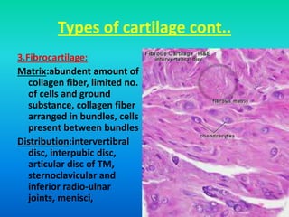
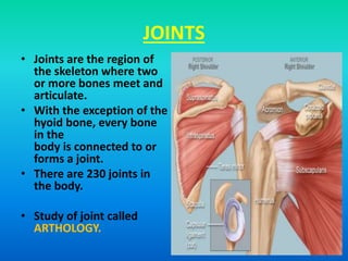
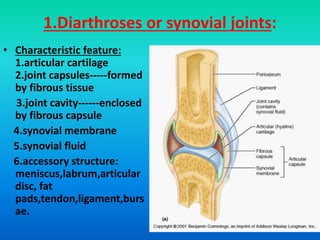
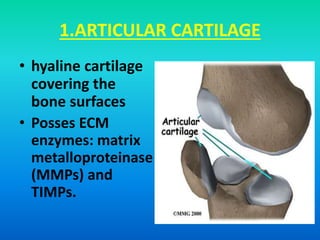
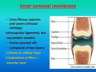
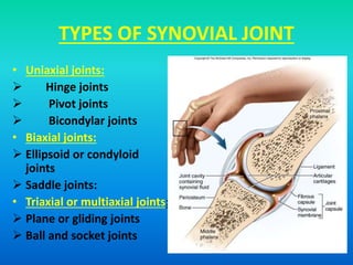
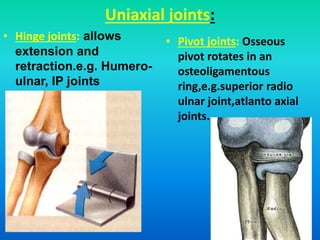
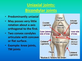
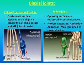
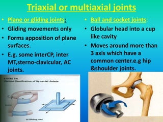
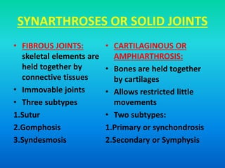
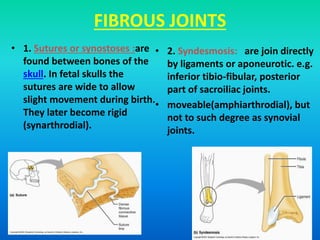
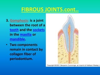
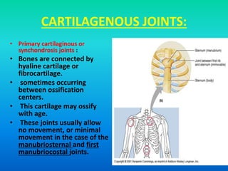
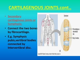
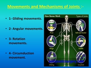
SEMINAR ON
CARTILAGE AND JOINTS
STRUCTURE AND FUNCTIONS
PRESENTER:DR. SIDDIQUE HUSSANUL RANNA
PGT,ORTHOPAEDICS
MODERATOR:DR.CHAO ROCHEK BURAGOHAIN
ASST.PROF,ORTHOPAEDICS
10TH JUNE 2015
INTRODUCTION
CARTILAGE
• Is an avascular, stiff, load bearing form of connective
tissue,
• consisting of extracellular fibers embedded in a
water filled ground substances.
Peculiarities of cartilage
•Avascular and non nervous
•Receives nutrition by diffusion
•Posses some cartilage canals
•Canals help osteogenic cells and vessels to grow in ossific
centers.
•Posses low metabolic rate than bone cells
•Most bones in intrauterine life are cartilage.
Peculiarities of cartilage cond..
•Chondrocytes are isolated in separate lacunae
•Has capacity of appositional and interstitial
growth
•Cartilaginous matrix has low antigenicity
•Homogenous transplantation is possible
•Repair of cartilage takes long time
Structure of cartilage
Consists of
 Cells:
* chondrocytes
*cells appear in the lacunae or in cell nest
 Matrix
*collagen fibers & elastin fibers
 Ground substance(gel like)
*water,
*proteoglycans&glycosamineglycans
Structure of ground substance
• Gel like substance rich in carbohydrate
• Consists mainly of water and dissolved salts
• meshwork of long proteoglycan molecules
• Proteoglycans consists of long polymers called
GAGS(glycosaminoglycans)
Proteoglycans and GAGS
•Proteoglycans consists of long polymers called
GAGS(glycosaminoglycans)
•GAGS are covalently bound with “core protein”
•Core protein may bear more than 100 GAGS
sideways.
•Several PG molecules bound along huge
Hyaluronate molecules to form aggregates.
Proteoglycans and GAGS,cont..
•Weak inter molecular forces hold aggregates in 3D
network with large water filled space between.
•GAGS posses acidic groups
•GAGS repel each other thus standing out stiffy
from core protein.
•PG aggregates appear to be compressed and posses
repulsion chains and subunits.
•PG may act as a minute spring, conferring elasticity.
Proteoglycans and GAGS,cont..
•Some GAGS found in PG aggregans are
condroitin-4 sulfate,condroitin-6 sulfate,dermatan
sulfate and keratan sulfate.
•Link protein and chondronectin,are important for
PG structure.
Proteoglycans and GAGS,cont..
Structure of collagen fiber
• Too small and thin
• Characteristic cross
banding and 3D
structure
• Tropocollagen posses
triple helices of
polypeptide
• Tensile strength
properties
Function of cartilage
• Supportive: soft tissue support
• Mechanical:response to
friction,compression,shear and tensile
loading
• Lubrication:lubricin plays major role in
biolubrication and protection of joints.
• Viscoelastic function: proteoglycans, GAGS
and elastic fiber
• Provides smooth gliding surface in joints
• Development and growth of bones
Types of cartilage
mainly three types
1.Hyaline cartilage: most of
the cartilage in body are
hyaline cartilage
Distribution: articular
cartilage,costal,tracheobr
onchial,nose,and
laryngeal
cartilage(except
epiglotis,cuniform,cornic
ulate,apex of arytenoids)
Matrix: hour glass like,
Chondroitin sulfate
predominance,
Few collagen fiber
Types of cartilage cont..
2.Elastic cartilage:
Matrix: contains collagen
fiber with large number
elastic fiber which
anastomose in all
direction except around
cartilage cells.
• Maintains shape of
cartilage.
Distribution:Pinna,eustachi
an,tubes,epiglottis,cornic
ulate,cuniform,apex of
arytenoids
Types of cartilage cont..
3.Fibrocartilage:
Matrix:abundent amount of
collagen fiber, limited no.
of cells and ground
substance, collagen fiber
arranged in bundles, cells
present between bundles
Distribution:intervertibral
disc, interpubic disc,
articular disc of TM,
sternoclavicular and
inferior radio-ulnar
joints, menisci,
JOINTS
• Joints are the region of
the skeleton where two
or more bones meet and
articulate.
• With the exception of the
hyoid bone, every bone
in the
body is connected to or
forms a joint.
• There are 230 joints in
the body.
• Study of joint called
ARTHOLOGY.
Classification of joints
mainly of two types
1.Diarthroses or synovial joints:
*skeletal elements are separated by a
cavity
*free movements are possible
2.Synarthroses or solid joints:
*skeletal elements are held together by
connective tissues or cartilage.
*allowed restricted little or no movements
1.Diarthroses or synovial joints:
• Characteristic feature:
1.articular cartilage
2.joint capsules-----formed
by fibrous tissue
3.joint cavity------enclosed
by fibrous capsule
4.synovial membrane
5.synovial fluid
6.accessory structure:
meniscus,labrum,articular
disc, fat
pads,tendon,ligament,burs
ae.
1.ARTICULAR CARTILAGE
• hyaline cartilage
covering the
bone surfaces
• Posses ECM
enzymes: matrix
metalloproteinase
(MMPs) and
TIMPs.
Functions of articular cartilages
• Extremely low
coefficient of friction
• Shock absorption
• Distribution of joint
loads
• Reduce contact stress
Injury and healing of articular
cartilage
• Limited healing potential due to avascularity.
• Three types injury of articular cartilage:
Type 1: microscopic damage to cells and ECM
Type 2: chondral fracture or fissuring
Type 3: deeper injury,penetration to subchondral
bone
Microscopic structure of articular
cartilage
• Four microscopic zones
1.Superficial or gliding
zone: 10—20%
2.Middle or transitional
zone : 40---60%
3.Deep or Radial zone:
30%
4. Calcified zone
MENISCUS
• Meniscus is an
incomplete rim of
white fibrous cartilage
between articular
cartilages.
• Distribution: knee
joints, articular disc of
sternoclavicular and
acromioclavicular
joints,triangularfibroca
rtilage, labra in
hip&shoulder.
Meniscus cont..
functions
• Tibiofemoral load transmission.
• Shock absorption
• Lubrication
• Prevent synovial impingement
• Distribute synovial fluid
• Contribute to joint stability, gliding motion.
Composition of meniscus
CELLS
• Primarily
fibrochondrocyte
• Responsible for
synthesis and
maintenance of ECM
ECM
• Water-70%
• Interlacing collagen
fibers.
• 95% type 1 collagen
• Smaller amount type
II,III,V,VI
• Elastin-0.06%
• Noncollagenous protein-
8-13%
Ultra structure of meniscus
• Three collagen fiber layer.
• Which convert compressive
loads to circumferential or
hoop stress.
• Superficial layer: gliding
movements
• Circumferential layer: resist
stress during weight
bearing
• Radial layer: between
above two layer serve as
“ties”.
Joint capsule
two layers:
• Outer fibrous membrane
• Complete enclose joints
• Interrupted by synovial
protrusion called bursae
• Bursae are routes for
spread of infection into
joints
• Form intrinsic ligaments.
Inner synovial membrane
• Lines fibrous capsules
and covers articular
cartilage,
Intracapsular ligaments, bur
-sae,tendon sheaths.
• Forms synovial villi
• Composed of two layers:
1.Intima or cellular layer
2.Subintimal or fibro –
-vascular layer
Synovial fluid
• Plasma transudate from synovial capillaries.
• Modified by secretory activities of type B
cells.
• Lack of fibrinogen
• Does not clot normally.
Laboratory Testing: Macroscopic
• Volume-0.2-4ml
• Color and Clarity-colorless
and clear or transparent.
• Inclusions-rice
bodies,ochronotic shards
• Viscosity-string test
• Clotting
• Mucin Clot
Laboratory Testing: Chemical
• Protein: 1-3 g/dl
• Glucose: no lower than
10mg/dl less than serum
glucose
• Uric Acid: 6 to 8 mg/dl
• Lactic Acid: less than 25
mg/dl
• Rheumatoid Factor:50% RA
+ve.
Laboratory Testing: Microscopic
• Cell Counts: Normal values:
RBCs = none
WBCs = 0 – 150/cumm.
• Differential: Normal values:
– Neutrophils 7%
– Lymphocytes 24%
– Monocyte (Histocytes) 48%
– Macrophages 10%
– Synovial lining cells 4%
• Crystals: uric acid
crystals,CPPD,cholester--
ol,steroid induced,
apatite crystals.
Laboratory Testing: Microbiology
• Infectious organisms
– Bacteria
– Fungi
– Mycobacteria
– Viruses
• Route of entry
– Bloodstream
– Penetrating wounds
– Osteomyelitis rupture
– Arthroscopy
– intra-articular steroid
injections
– prosthetic joint surgery
TYPES OF SYNOVIAL JOINT
• Uniaxial joints:
 Hinge joints
 Pivot joints
 Bicondylar joints
• Biaxial joints:
 Ellipsoid or condyloid
joints
 Saddle joints:
• Triaxial or multiaxial joints:
 Plane or gliding joints
 Ball and socket joints
Uniaxial joints:
• Hinge joints: allows
extension and
retraction.e.g. Humero-
ulnar, IP joints
• Pivot joints: Osseous
pivot rotates in an
osteoligamentous
ring,e.g.superior radio
ulnar joint,atlanto axial
joints.
Uniaxial joints:
Bicondylar joints
• Predominantly uniaxial
• May posses very little
rotation about a axis
orthogonal to the first.
• Two convex condyles
articulate with concave
or flat surface.
• Example: knee joints,
TM joints
Biaxial joints:
Ellipsoid or condyloid joints:
• Oval convex surface
apposed to an elliptical
concavity e.g. radio-carpal
and MP joints in wrist.
Saddle joints:
• Opposing surface are
reciprocally concavo-convex.
• Flexion- Extension, Abduction-
Adduction, May combined as
circumduction.
Triaxial or multiaxial joints
• Plane or gliding joints:
• Gliding movements only
• Forms apposition of plane
surfaces.
• E.g. some interCP, inter
MT,sterno-clavicular, AC
joints.
• Ball and socket joints:
• Globular head into a cup
like cavity
• Moves around more than
3 axis which have a
common center.e.g hip
&shoulder joints.
SYNARTHROSES OR SOLID JOINTS
• FIBROUS JOINTS:
skeletal elements are
held together by
connective tissues
• Immovable joints
• Three subtypes
1.Sutur
2.Gomphosis
3.Syndesmosis
• CARTILAGINOUS OR
AMPHIARTHROSIS:
• Bones are held together
by cartilages
• Allows restricted little
movements
• Two subtypes:
1.Primary or synchondrosis
2.Secondary or Symphysis
FIBROUS JOINTS
• 1. Sutures or synostoses :are
found between bones of the
skull. In fetal skulls the
sutures are wide to allow
slight movement during birth.
They later become rigid
(synarthrodial).
• 2. Syndesmosis: are join directly
by ligaments or aponeurotic. e.g.
inferior tibio-fibular, posterior
part of sacroiliac joints.
• moveable(amphiarthrodial), but
not to such degree as synovial
joints.
FIBROUS JOINTS.cont..
3. Gomphosis: is a joint
between the root of a
tooth and the sockets
in the maxilla or
mandible.
• Two components
remain in contact by
collagen fiber of
periodontium.
CARTILAGENOUS JOINTS:
• Primary cartilaginous or
synchondrosis joints :
• Bones are connected by
hyaline cartilage or
fibrocartilage.
• sometimes occurring
between ossification
centers.
• This cartilage may ossify
with age.
• These joints usually allow
no movement, or minimal
movement in the case of the
manubriosternal and first
manubriocostal joints.
CARTILAGENOUS JOINTS.cont..
• Secondary
cartilaginous joints or
Symphysis:
• Connect the two bones
by fibrocartilage.
• E.g. Symphysis
pubis,vertibral bodies
connected by
Intervertibral disc.
Movements and Mechanisms of Joints :-
• 1- Gliding movements.
• 2- Angular movements.
• 3- Rotation
movements.
• 4- Circumduction
movement.
1- Gliding movement
• It is simple movement
that occurs in plane
joint.
• In this movement one
articular surface will
glides over the other.
2- Angular movement :
increase or decrease of an angle between the articular
bones.
• A- Flexion & extension:
These movements occur around
transverse axis.
• B- Abduction & adduction:
Occur around antero-posterior
axis.
3- Rotation movements :
• It is the movement in
which the bone rotates
around .
1. Axis inside the bone itself
e.g. rotation of humerus.
2. Axis outside the bone e.g.
rotation of radius around
ulna in pronation &
supination
3. Axis in another bone e.g.
rotation of atlas around
axis e.g. rotation of head.
4- Circumduction
• It is a rotatory
movement of a limb
around a imaginary
cone.
• e.g. in circumduction of
upper limb the apex of
the imaginary cone lies
in axilla and hand
rotates around the
base of the cone
Factors Affecting Stability of joints
• 1- Shape & fitting of
surfaces.
• 2- Thickness &
elasticity of capsule.
• 3- Ligaments.
• 4- Surrounding
muscles.
Factors Affecting Rang of Movements
• 1- Tension of ligaments
and capsule.
• 2- Contraction of
antagonistic muscles.
• 3- Contact of soft tissue.
• 4- Shape of articulating
bones.
THANK YOU
ALL

Cartilage and joints

  • 1.
    SEMINAR ON CARTILAGE ANDJOINTS STRUCTURE AND FUNCTIONS PRESENTER:DR. SIDDIQUE HUSSANUL RANNA PGT,ORTHOPAEDICS MODERATOR:DR.CHAO ROCHEK BURAGOHAIN ASST.PROF,ORTHOPAEDICS 10TH JUNE 2015
  • 2.
    INTRODUCTION CARTILAGE • Is anavascular, stiff, load bearing form of connective tissue, • consisting of extracellular fibers embedded in a water filled ground substances.
  • 3.
    Peculiarities of cartilage •Avascularand non nervous •Receives nutrition by diffusion •Posses some cartilage canals •Canals help osteogenic cells and vessels to grow in ossific centers. •Posses low metabolic rate than bone cells •Most bones in intrauterine life are cartilage.
  • 4.
    Peculiarities of cartilagecond.. •Chondrocytes are isolated in separate lacunae •Has capacity of appositional and interstitial growth •Cartilaginous matrix has low antigenicity •Homogenous transplantation is possible •Repair of cartilage takes long time
  • 5.
    Structure of cartilage Consistsof  Cells: * chondrocytes *cells appear in the lacunae or in cell nest  Matrix *collagen fibers & elastin fibers  Ground substance(gel like) *water, *proteoglycans&glycosamineglycans
  • 6.
    Structure of groundsubstance • Gel like substance rich in carbohydrate • Consists mainly of water and dissolved salts • meshwork of long proteoglycan molecules • Proteoglycans consists of long polymers called GAGS(glycosaminoglycans)
  • 7.
    Proteoglycans and GAGS •Proteoglycansconsists of long polymers called GAGS(glycosaminoglycans) •GAGS are covalently bound with “core protein” •Core protein may bear more than 100 GAGS sideways. •Several PG molecules bound along huge Hyaluronate molecules to form aggregates.
  • 8.
    Proteoglycans and GAGS,cont.. •Weakinter molecular forces hold aggregates in 3D network with large water filled space between. •GAGS posses acidic groups •GAGS repel each other thus standing out stiffy from core protein. •PG aggregates appear to be compressed and posses repulsion chains and subunits. •PG may act as a minute spring, conferring elasticity.
  • 9.
    Proteoglycans and GAGS,cont.. •SomeGAGS found in PG aggregans are condroitin-4 sulfate,condroitin-6 sulfate,dermatan sulfate and keratan sulfate. •Link protein and chondronectin,are important for PG structure.
  • 10.
  • 11.
  • 12.
    Structure of collagenfiber • Too small and thin • Characteristic cross banding and 3D structure • Tropocollagen posses triple helices of polypeptide • Tensile strength properties
  • 13.
    Function of cartilage •Supportive: soft tissue support • Mechanical:response to friction,compression,shear and tensile loading • Lubrication:lubricin plays major role in biolubrication and protection of joints. • Viscoelastic function: proteoglycans, GAGS and elastic fiber • Provides smooth gliding surface in joints • Development and growth of bones
  • 14.
    Types of cartilage mainlythree types 1.Hyaline cartilage: most of the cartilage in body are hyaline cartilage Distribution: articular cartilage,costal,tracheobr onchial,nose,and laryngeal cartilage(except epiglotis,cuniform,cornic ulate,apex of arytenoids) Matrix: hour glass like, Chondroitin sulfate predominance, Few collagen fiber
  • 15.
    Types of cartilagecont.. 2.Elastic cartilage: Matrix: contains collagen fiber with large number elastic fiber which anastomose in all direction except around cartilage cells. • Maintains shape of cartilage. Distribution:Pinna,eustachi an,tubes,epiglottis,cornic ulate,cuniform,apex of arytenoids
  • 16.
    Types of cartilagecont.. 3.Fibrocartilage: Matrix:abundent amount of collagen fiber, limited no. of cells and ground substance, collagen fiber arranged in bundles, cells present between bundles Distribution:intervertibral disc, interpubic disc, articular disc of TM, sternoclavicular and inferior radio-ulnar joints, menisci,
  • 17.
    JOINTS • Joints arethe region of the skeleton where two or more bones meet and articulate. • With the exception of the hyoid bone, every bone in the body is connected to or forms a joint. • There are 230 joints in the body. • Study of joint called ARTHOLOGY.
  • 18.
    Classification of joints mainlyof two types 1.Diarthroses or synovial joints: *skeletal elements are separated by a cavity *free movements are possible 2.Synarthroses or solid joints: *skeletal elements are held together by connective tissues or cartilage. *allowed restricted little or no movements
  • 19.
    1.Diarthroses or synovialjoints: • Characteristic feature: 1.articular cartilage 2.joint capsules-----formed by fibrous tissue 3.joint cavity------enclosed by fibrous capsule 4.synovial membrane 5.synovial fluid 6.accessory structure: meniscus,labrum,articular disc, fat pads,tendon,ligament,burs ae.
  • 20.
    1.ARTICULAR CARTILAGE • hyalinecartilage covering the bone surfaces • Posses ECM enzymes: matrix metalloproteinase (MMPs) and TIMPs.
  • 21.
    Functions of articularcartilages • Extremely low coefficient of friction • Shock absorption • Distribution of joint loads • Reduce contact stress
  • 22.
    Injury and healingof articular cartilage • Limited healing potential due to avascularity. • Three types injury of articular cartilage: Type 1: microscopic damage to cells and ECM Type 2: chondral fracture or fissuring Type 3: deeper injury,penetration to subchondral bone
  • 23.
    Microscopic structure ofarticular cartilage • Four microscopic zones 1.Superficial or gliding zone: 10—20% 2.Middle or transitional zone : 40---60% 3.Deep or Radial zone: 30% 4. Calcified zone
  • 24.
    MENISCUS • Meniscus isan incomplete rim of white fibrous cartilage between articular cartilages. • Distribution: knee joints, articular disc of sternoclavicular and acromioclavicular joints,triangularfibroca rtilage, labra in hip&shoulder.
  • 25.
    Meniscus cont.. functions • Tibiofemoralload transmission. • Shock absorption • Lubrication • Prevent synovial impingement • Distribute synovial fluid • Contribute to joint stability, gliding motion.
  • 26.
    Composition of meniscus CELLS •Primarily fibrochondrocyte • Responsible for synthesis and maintenance of ECM ECM • Water-70% • Interlacing collagen fibers. • 95% type 1 collagen • Smaller amount type II,III,V,VI • Elastin-0.06% • Noncollagenous protein- 8-13%
  • 27.
    Ultra structure ofmeniscus • Three collagen fiber layer. • Which convert compressive loads to circumferential or hoop stress. • Superficial layer: gliding movements • Circumferential layer: resist stress during weight bearing • Radial layer: between above two layer serve as “ties”.
  • 28.
    Joint capsule two layers: •Outer fibrous membrane • Complete enclose joints • Interrupted by synovial protrusion called bursae • Bursae are routes for spread of infection into joints • Form intrinsic ligaments.
  • 29.
    Inner synovial membrane •Lines fibrous capsules and covers articular cartilage, Intracapsular ligaments, bur -sae,tendon sheaths. • Forms synovial villi • Composed of two layers: 1.Intima or cellular layer 2.Subintimal or fibro – -vascular layer
  • 30.
    Synovial fluid • Plasmatransudate from synovial capillaries. • Modified by secretory activities of type B cells. • Lack of fibrinogen • Does not clot normally.
  • 31.
    Laboratory Testing: Macroscopic •Volume-0.2-4ml • Color and Clarity-colorless and clear or transparent. • Inclusions-rice bodies,ochronotic shards • Viscosity-string test • Clotting • Mucin Clot
  • 32.
    Laboratory Testing: Chemical •Protein: 1-3 g/dl • Glucose: no lower than 10mg/dl less than serum glucose • Uric Acid: 6 to 8 mg/dl • Lactic Acid: less than 25 mg/dl • Rheumatoid Factor:50% RA +ve.
  • 33.
    Laboratory Testing: Microscopic •Cell Counts: Normal values: RBCs = none WBCs = 0 – 150/cumm. • Differential: Normal values: – Neutrophils 7% – Lymphocytes 24% – Monocyte (Histocytes) 48% – Macrophages 10% – Synovial lining cells 4% • Crystals: uric acid crystals,CPPD,cholester-- ol,steroid induced, apatite crystals.
  • 34.
    Laboratory Testing: Microbiology •Infectious organisms – Bacteria – Fungi – Mycobacteria – Viruses • Route of entry – Bloodstream – Penetrating wounds – Osteomyelitis rupture – Arthroscopy – intra-articular steroid injections – prosthetic joint surgery
  • 35.
    TYPES OF SYNOVIALJOINT • Uniaxial joints:  Hinge joints  Pivot joints  Bicondylar joints • Biaxial joints:  Ellipsoid or condyloid joints  Saddle joints: • Triaxial or multiaxial joints:  Plane or gliding joints  Ball and socket joints
  • 36.
    Uniaxial joints: • Hingejoints: allows extension and retraction.e.g. Humero- ulnar, IP joints • Pivot joints: Osseous pivot rotates in an osteoligamentous ring,e.g.superior radio ulnar joint,atlanto axial joints.
  • 37.
    Uniaxial joints: Bicondylar joints •Predominantly uniaxial • May posses very little rotation about a axis orthogonal to the first. • Two convex condyles articulate with concave or flat surface. • Example: knee joints, TM joints
  • 38.
    Biaxial joints: Ellipsoid orcondyloid joints: • Oval convex surface apposed to an elliptical concavity e.g. radio-carpal and MP joints in wrist. Saddle joints: • Opposing surface are reciprocally concavo-convex. • Flexion- Extension, Abduction- Adduction, May combined as circumduction.
  • 39.
    Triaxial or multiaxialjoints • Plane or gliding joints: • Gliding movements only • Forms apposition of plane surfaces. • E.g. some interCP, inter MT,sterno-clavicular, AC joints. • Ball and socket joints: • Globular head into a cup like cavity • Moves around more than 3 axis which have a common center.e.g hip &shoulder joints.
  • 40.
    SYNARTHROSES OR SOLIDJOINTS • FIBROUS JOINTS: skeletal elements are held together by connective tissues • Immovable joints • Three subtypes 1.Sutur 2.Gomphosis 3.Syndesmosis • CARTILAGINOUS OR AMPHIARTHROSIS: • Bones are held together by cartilages • Allows restricted little movements • Two subtypes: 1.Primary or synchondrosis 2.Secondary or Symphysis
  • 41.
    FIBROUS JOINTS • 1.Sutures or synostoses :are found between bones of the skull. In fetal skulls the sutures are wide to allow slight movement during birth. They later become rigid (synarthrodial). • 2. Syndesmosis: are join directly by ligaments or aponeurotic. e.g. inferior tibio-fibular, posterior part of sacroiliac joints. • moveable(amphiarthrodial), but not to such degree as synovial joints.
  • 42.
    FIBROUS JOINTS.cont.. 3. Gomphosis:is a joint between the root of a tooth and the sockets in the maxilla or mandible. • Two components remain in contact by collagen fiber of periodontium.
  • 43.
    CARTILAGENOUS JOINTS: • Primarycartilaginous or synchondrosis joints : • Bones are connected by hyaline cartilage or fibrocartilage. • sometimes occurring between ossification centers. • This cartilage may ossify with age. • These joints usually allow no movement, or minimal movement in the case of the manubriosternal and first manubriocostal joints.
  • 44.
    CARTILAGENOUS JOINTS.cont.. • Secondary cartilaginousjoints or Symphysis: • Connect the two bones by fibrocartilage. • E.g. Symphysis pubis,vertibral bodies connected by Intervertibral disc.
  • 45.
    Movements and Mechanismsof Joints :- • 1- Gliding movements. • 2- Angular movements. • 3- Rotation movements. • 4- Circumduction movement.
  • 46.
    1- Gliding movement •It is simple movement that occurs in plane joint. • In this movement one articular surface will glides over the other.
  • 47.
    2- Angular movement: increase or decrease of an angle between the articular bones. • A- Flexion & extension: These movements occur around transverse axis. • B- Abduction & adduction: Occur around antero-posterior axis.
  • 48.
    3- Rotation movements: • It is the movement in which the bone rotates around . 1. Axis inside the bone itself e.g. rotation of humerus. 2. Axis outside the bone e.g. rotation of radius around ulna in pronation & supination 3. Axis in another bone e.g. rotation of atlas around axis e.g. rotation of head.
  • 49.
    4- Circumduction • Itis a rotatory movement of a limb around a imaginary cone. • e.g. in circumduction of upper limb the apex of the imaginary cone lies in axilla and hand rotates around the base of the cone
  • 50.
    Factors Affecting Stabilityof joints • 1- Shape & fitting of surfaces. • 2- Thickness & elasticity of capsule. • 3- Ligaments. • 4- Surrounding muscles.
  • 51.
    Factors Affecting Rangof Movements • 1- Tension of ligaments and capsule. • 2- Contraction of antagonistic muscles. • 3- Contact of soft tissue. • 4- Shape of articulating bones.
  • 52.