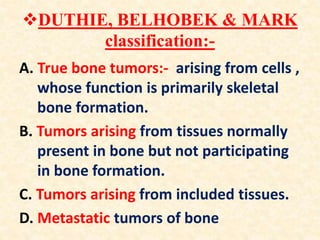
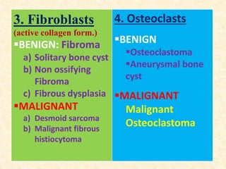
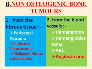
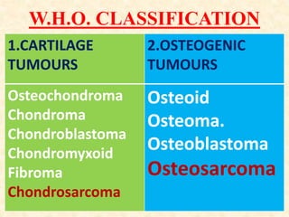
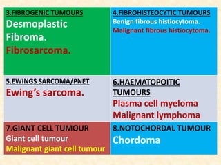
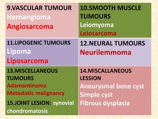
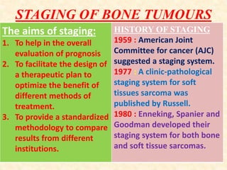
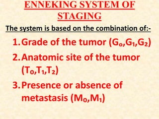
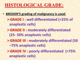
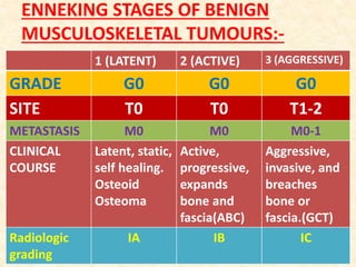
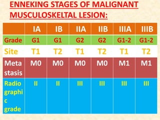
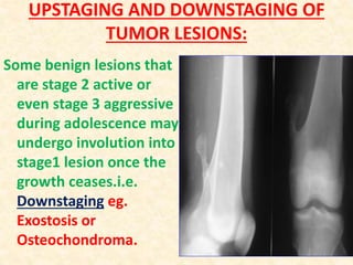
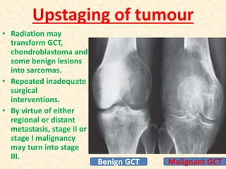
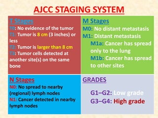
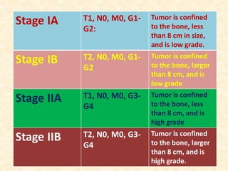
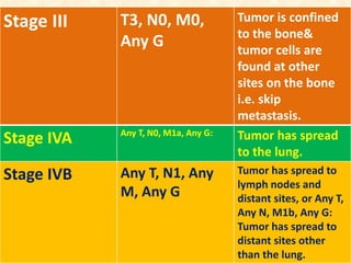
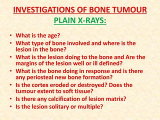
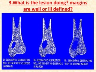
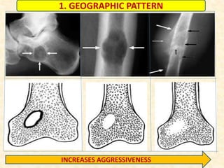
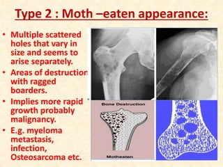
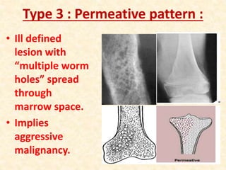
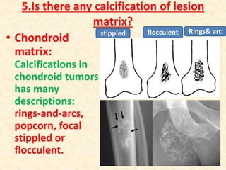
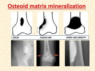
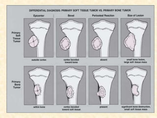
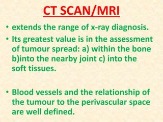
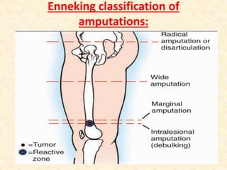
Russian Call Girl Brookfield - 7001305949 Escorts Service 50% Off with Cash O...
Â
Bone tumour
1. Bone Tumour
⢠Classification ,Investigations,
General Management .
Moderator : Dr. P. Tahbildar
Prof.&HOD, Orthopaedics
Presenter : Dr. S. H. Ranna
PGT, Orthopaedics
09/12/2015
2. INTRODUCTION
Defined by Willis, is "an
abnormal mass of tissue
the growth of which
exceeds and is
uncoordinated with that
of the normal tissues and
persists in the same
excessive manner after
the cessation of the
stimuli which evoked the
change.â
BURNET (1957) defined it
as âa primary change in
the character of cells,
which undergo a series of
mutations with an
increased growth
potential rendering them
insusceptible to normal
growth controlâ.
4. ďśIn bone along with
the true osteogenic
tissues, other
elements are present
like marrow, fat,
nerves etc.
5. Facts before classifying
a particular tumor:-
⢠To define the cell of origin.
⢠To identify the cytology of the
tumor cells and its stroma as
well as their degree of
differentiation.
⢠To observe the behavior and
growth of the tumors.
7. ďśDUTHIE, BELHOBEK & MARK
classification:-
A. True bone tumors:- arising from cells ,
whose function is primarily skeletal
bone formation.
B. Tumors arising from tissues normally
present in bone but not participating
in bone formation.
C. Tumors arising from included tissues.
D. Metastatic tumors of bone
9. 3. Fibroblasts
(active collagen form.)
ď§BENIGN: Fibroma
a) Solitary bone cyst
b) Non ossifying
Fibroma
c) Fibrous dysplasia
ď§MALIGNANT
a) Desmoid sarcoma
b) Malignant fibrous
histiocytoma
4. Osteoclasts
ď§BENIGN
ď§Osteoclastoma
ď§Aneurysmal bone
cyst
ď§MALIGNANT
Malignant
Osteoclastoma
10. B.NON OSTEOGENIC BONE
TUMOURS
1. from the
fibrous tissue :-
ďPeriosteal
Fibroma
ďPeriosteal
fibrosarcoma
ďMalignant fibrous
osteocytoma
2. from the blood
vessels :-
ďHemangioma
ďHemangioblas
toma
ďABC
ďAngiosarcoma
11. 3. from the
elements of bone
marrow :-
ďMyeloma
ďReticulum cell sarcoma
ďHodgkinâs disease of
bone
ďLymphosarcoma
ďEwingâs sarcoma
5. From nerves
Neurilemmoma
Neurofibroma
6. Tumors of
synovium
Synovial cell sarcoma
4. from adipose
tissues
ďLipoma
ďLiposarcoma
7. Striated muscle
Rhabdomyosarcoma
13. ďśTumour like lesions of bone
1. REACTIVE BONE
LESIONS
STIMULATING
TUMORS:
ď§Osteoid Osteoma
ď§Benign
osteoblastoma
ď§Non osteogenic
Fibroma
2. HAMARTOMAS
OF BONE
ď§Osteochondroma
ď§Enchondroma
ď§Fibrous dysplasia
3. CYSTIC LESIONS OF
BONE:
ď§Solitary bone cyst
ď§ABC
17. STAGING OF BONE TUMOURS
The aims of staging:
1. To help in the overall
evaluation of prognosis
2. To facilitate the design of
a therapeutic plan to
optimize the benefit of
different methods of
treatment.
3. To provide a standardized
methodology to compare
results from different
institutions.
HISTORY OF STAGING
1959 : American Joint
Committee for cancer (AJC)
suggested a staging system.
1977 : A clinic-pathological
staging system for soft
tissues sarcoma was
published by Russell.
1980 : Enneking, Spanier and
Goodman developed their
staging system for both bone
and soft tissue sarcomas.
18. ENNEKING SYSTEM OF
STAGING
The system is based on the combination of:-
1.Grade of the tumor (Gâ,Gâ,Gâ)
2.Anatomic site of the tumor
(Tâ,Tâ,Tâ)
3.Presence or absence of
metastasis (Mâ,Mâ)
19. HISTOLOGICAL GRADE:
⢠BRODERâS grading of malignancy is used:
ďGRADE I : well differentiated (<25% of
anaplastic cells)
ďGRADE II : moderately differentiated
(25- 50% anaplastic cells)
ďGRADE III : moderately differentiated (50
â75% anaplastic cells)
ďGRADE IV : poorly differentiated (>75%
anaplastic cells)
20. RADIOGRAPHIC ASSESSMENT
LODWICKâS IA well marginated without
deformation or expansion.
LODWICKâS IB well defined lesion, but
irregularly marginated .
Septated appearance.
Expansion, bulging or
deformation .
LODWICKâS IC ragged permeative interface
with adjacent bone
-Cortical
destruction,codmans triangle
etc
21. LODWICKâS II often have a generous
reactive rim of
cancellous bone
admixed with defects of
extra capsular &/or soft
tissue extension
LODWICKâs III poorly marginated
interface with a
diffuse permeative
border.
HIGH GRADE SARCOMA
23. ANATOMICAL SITE
The anatomic
location of a tumor
lesion directly
influences the
prognosis & also
the choice of
surgical
procedures.
NATURAL BARRIERS
TO TUMOR GROWTH
i. Cortical bone
ii. Growth plate
iii. Articular cartilage
iv. Fascia enclosing a
compartment
v. Joint capsule
vi. Tendon sheaths
vii. Nerve sheaths
viii.Ligaments
24. ENNEKING STAGES OF BENIGN
MUSCULOSKELETAL TUMOURS:-
1 (LATENT) 2 (ACTIVE) 3 (AGGRESSIVE)
GRADE G0 G0 G0
SITE T0 T0 T1-2
METASTASIS M0 M0 M0-1
CLINICAL
COURSE
Latent, static,
self healing.
Osteoid
Osteoma
Active,
progressive,
expands
bone and
fascia(ABC)
Aggressive,
invasive, and
breaches
bone or
fascia.(GCT)
Radiologic
grading
IA IB IC
25. ENNEKING STAGES OF MALIGNANT
MUSCULOSKELTAL LESION:
IA IB IIA IIB IIIA IIIB
Grade G1 G1 G2 G2 G1-2 G1-2
Site T1 T2 T1 T2 T1 T2
Meta
stasis
M0 M0 M0 M0 M1 M1
Radio
graphi
c
grade
II II III III III III
26. UPSTAGING AND DOWNSTAGING OF
TUMOR LESIONS:
Some benign lesions that
are stage 2 active or
even stage 3 aggressive
during adolescence may
undergo involution into
stage1 lesion once the
growth ceases.i.e.
Downstaging eg.
Exostosis or
Osteochondroma.
27. Upstaging of tumour
⢠Radiation may
transform GCT,
chondroblastoma and
some benign lesions
into sarcomas.
⢠Repeated inadequate
surgical
interventions.
⢠By virtue of either
regional or distant
metastasis, stage II or
stage I malignancy
may turn into stage
III.
Malignant GCTBenign GCT
28. AJCC STAGING SYSTEM
T Stages
T0: No evidence of the tumor
T1: Tumor is 8 cm (3 inches) or
less
T2: Tumor is larger than 8 cm
T3: Tumor cells detected at
another site(s) on the same
bone
M Stages
M0: No distant metastasis
M1: Distant metastasis
M1a: Cancer has spread
only to the lung
M1b: Cancer has spread
to other sites
N Stages
N0: No spread to nearby
(regional) lymph nodes
N1: Cancer detected in nearby
lymph nodes
GRADES
G1âG2: Low grade
G3âG4: High grade
29. Stage IA T1, N0, M0, G1-
G2:
Tumor is confined
to the bone, less
than 8 cm in size,
and is low grade.
Stage IB T2, N0, M0, G1-
G2
Tumor is confined
to the bone, larger
than 8 cm, and is
low grade
Stage IIA T1, N0, M0, G3-
G4
Tumor is confined
to the bone, less
than 8 cm, and is
high grade
Stage IIB T2, N0, M0, G3-
G4
Tumor is confined
to the bone, larger
than 8 cm, and is
high grade.
30. Stage III T3, N0, M0,
Any G
Tumor is confined
to the bone&
tumor cells are
found at other
sites on the bone
i.e. skip
metastasis.
Stage IVA Any T, N0, M1a, Any G: Tumor has spread
to the lung.
Stage IVB Any T, N1, Any
M, Any G
Tumor has spread to
lymph nodes and
distant sites, or Any T,
Any N, M1b, Any G:
Tumor has spread to
distant sites other
than the lung.
31. INVESTIGATIONS OF BONE TUMOUR
PLAIN X-RAYS:
⢠What is the age?
⢠What type of bone involved and where is the
lesion in the bone?
⢠What is the lesion doing to the bone and Are the
margins of the lesion well or ill defined?
⢠What is the bone doing in response and is there
any periosteal new bone formation?
⢠Is the cortex eroded or destroyed? Does the
tumour extent to soft tissue?
⢠Is there any calcification of lesion matrix?
⢠Is the lesion solitary or multiple?
33. 2.What type of bone involved and
where is the lesion?
In the transverse plane :
⢠Central: Enchondroma.
⢠Eccentric: GCT,
Osteosarcoma,
Chondromyxoid fibroid.
⢠Cortical : Osteoid
Osteoma, Non Ossifying
Fibroma
⢠Parosteal: Parosteal
Osteosarcoma,
Osteochondroma.
34. In the longitudinal plane
EPIPHYSEAL METAPHYSEAL DIAPHYSEAL
Chondroblastoma
Clear cell
Chondrosarcoma
GCT
ABC
Geode
(subchondral cyst)
Infection
Eosinophilic
granuloma
Non ossifying
Fibroma
-Chondromyxoid
Fibroma
-GCT, ABC, Solitary
bone cyst/Brodies
abscess
-Osteochondroma.
-osteogenic
sarcoma,
Chondrosarcoma.
Non ossifying
Fibroma
Ewing sarcoma
Osteoid Osteoma.
Osteoblastoma
Metastasis.
Reticulum cell
sarcoma.
Leukemia
Lymphoma.
Adamantinoma.
35. BONE TUMOURS&COMMONEST SITE
BONE TUMOURS COMMONEST SITE
ENCHONDROMA Metaphyses of small bone of hands &
feet
GCT,ABC,Osteosarcoma Lower end femur > upper end tibia
OSTEOCHONDROMA Lower end femur>prox. Tibia>prox.
humerus
CHONDROBLASTOMA,SBC Proximal humerus > prox. femur
EWINGS SARCOMA Femur > Fibula > tibia
OSTEOID OSTEOMA Femur > tibia
OSTEOBLASTOMA Posterior spine
Adamantinoma Mandible > tibia
Fibrous dysplasia Ribs > Upper Femur > Tibia > Lower
Femur.
Chordoma Sacrum > anterior vertebral body.
36. 3.What is the lesion doing? margins
are well or ill defined?
⢠Type 1: Geographic
pattern:
⢠1a. well defined lucency
with sclerotic margin.
⢠1b. well defined lytic
focus without sclerotic
rim, Enchondral
scalloping may seen.
⢠1c. lytic lesion with ill
defined margin.
38. Type 2 : Moth âeaten appearance:
⢠Multiple scattered
holes that vary in
size and seems to
arise separately.
⢠Areas of destruction
with ragged
boarders.
⢠Implies more rapid
growth probably
malignancy.
⢠E.g. myeloma
metastasis,
infection,
Osteosarcoma etc.
39. Type 3 : Permeative pattern :
⢠Ill defined
lesion with
âmultiple worm
holesâ spread
through
marrow space.
⢠Implies
aggressive
malignancy.
40. 4.What is the bone doing in
response?
ďś3 type of bone response:
⢠Lysis or lucency - bone destruction.
⢠Sclerosis -bone reaction.
⢠Periosteal reaction - remodeling of bone.
ďśRate of growth determines type of bone
response to the lesion.
⢠Slow progression sclerosis predominates.
⢠Rapid progression destruction
predominates.
41. PERIOSTEAL REACTION
⢠Two types of periosteal
reactions:
⢠Benign: solid type.
⢠Aggressive: Lamellated or
onion peel, Sunburst and
Codmanâs triangle types.
42. Benign: solid type.
⢠Slow-growing
tumors provoke
focal cortical
thickening
⢠A continuous
layer of new
bone that
attaches to
outer cortical
surface
OSTEOID OSTEOMA
45. Lamellated or onion peel
⢠lesion grows
unevenly in fits
and starts.
⢠Alternating layers
of opaque and
lucent densities
⢠Ewing's sarcoma,
infection.
46. Sunburst or hair on end
⢠lesion grows
rapidly but
steadily.
⢠Sharpey's
fibers become
stretched out
& ossify.
⢠Osteosarcoma
47. Codman's Triangle
⢠Too fast growth
for periosteum
to respond.
⢠only the edges
of raised
periosteum will
ossify forming a
small angle with
the surface of
bone.
48. Zone of Transition
⢠Most reliable
indicator for benign
versus malignant
osteolytic lesions.
⢠âNarrowâ, if it is so
well defined that it
can be drawn with a
fine-point pen.
⢠Wide : An aggressive
process should be
considered, Narrow zone Wide zone
49. two tumor like lesions which may mimic a
malignancy and have to be included in the
differential diagnosis.
50. 5.Is there any calcification of lesion
matrix?
⢠Chondroid
matrix:
Calcifications in
chondroid tumors
has many
descriptions:
rings-and-arcs,
popcorn, focal
stippled or
flocculent.
stippled flocculent Rings& arc
53. CT SCAN/MRI
⢠extends the range of x-ray diagnosis.
⢠Its greatest value is in the assessment
of tumour spread: a) within the bone
b)into the nearby joint c) into the
soft tissues.
⢠Blood vessels and the relationship of
the tumour to the perivascular space
are well defined.
54. PRINCIPLE OF BONE TUMOUR BIOPSY
1.After
evaluation
& imaging
complete
55. TYPES OF BIOPSY
⢠Fine needle aspiration biopsy (FNAB)
⢠Core needle biopsy (CNB)
⢠Surgical (open) biopsy
ďIncisional biopsy removes only part of
the suspicious area, enough to make a
diagnosis.
ďExcisional biopsy removes the entire
tumor or abnormal area,
56. GENERAL MANAGEMENT OF BONE
TUMOUR
ďMultidisciplinary team approach
ďBenign asymptomatic tumors
⢠If certain observe
⢠If in doubt biopsy
ďBenign symptomatic or enlarging tumors
⢠Biopsy
⢠Excision/ curettage
ďSuspected malignant tumors
⢠If primary admit for work-up
⢠Staging
⢠Choices; amputation, limb sparing surgery,
adjuvant therapy
57. TUMOUR EXCISION
Enneking classification of local procedures
⢠I-A - Wide excision
⢠I-B - Wide excision with
larger clearance
⢠II-A - Wide
excision/amputation
⢠II-B - Radical resection or
disarticulation
⢠III - Palliative treatment
⢠Low grade intra
compartmental lesions â
wide resection and
management of
metastases
58. LIMB SALVAGE
â˘A set of surgical procedures
designed to accomplish
removal of a malignant
tumor and reconstruction of
the limb with an acceptable
oncologic, functional, and
cosmetic result.
59. INDICATION
⢠Every patient with tumor of the extremity
should be considered for limb salvage if
the tumor can be removed with an
adequate margin and the resulting limb is
worth saving.
⢠certain that there are no skip lesions and
if a functional limb can be preserved.
⢠No justification for limiting the limb
salvage process based only on the
prognosis.
60. STEPS FOR LIMB SALVAGE
⢠wide excision of tumour with preservation of
neurovascular structures.
⢠The resulting gap is then dealt with several
ways:
ďShort diaphyseal segments - bone grafts.
ďLonger gaps may require custom made or
modular made implants.
ďOsteo-articular segments can be replaced by
large allograft, endoprostheses or allograft-
prosthetic composites.
ďIn growing children extendible implants used in
order to avoid repeated surgery.
61.
62.
63.
64.
65. BARRIERS TO LIMB SALVAGE:
⢠Poorly placed biopsy incisional
complications.
⢠Major Neurovascular involvement
which unable to repair.
⢠Grossly displaced pathologic fracture.
⢠Fungating and infected tumors.
⢠Recurrence of malignant tumors
⢠Inability to afford chemotherapy
67. GOALS OF TREATMENT WITH
CHEMOTHERAY:
â˘Cure
â˘Control â Stop growing &
spreading of the tumour.
â˘Palliation â In advance
stage, aim is to relieve
symptoms.
68. Before an operation (neo-adjuvant
therapy).
AIM DISADVANTAGE
ď§To shrink the tumour mass (
tumour necrosis)
ď§To make surgery less
destructive, more effective &
less post operative
metastasis.
ď§To kill micro metastatic.
ď§To prevent development of
resistant clones.
ď§Patients resistant to
chemotherapy get delayed
for surgery.
ď§Complication of chemo may
delay the surgery.
ď§Post op. Infection & wound
complication is common.
ď§Neo adjuvant chemo has to
be stopped 3-4 week prior to
surgery.
69. After an operation (adjuvant
therapy).
⢠To destroy the micro metastasis after total
resection of tumour mass ď recurrence
decreases.
⢠This reduces chances of resistance
developing.
⢠If total removal of tumour mass failed by
surgery, Started 3 wks after surgery so that
wound is healed.
70. Chemo irradiation : Palliative chemotherapy
:
ď§Chemotherapy
given same time
with radiotherapy.
ď§This to potentiate
radiation therapy.
ď§Given without
curative intent
relieves symptoms in
advanced cases.
ď§ Decrease tumour
load.
ď§ And increase life
expectancy.
71. Chemotherapy used in sarcoma
Sarcoma Drugs used Neoadjuvant/
adjuvant
palliative
Ewing's
sarcoma
Vincristine,
ifosfamide/
doxorubicin
Definite
survival
benefit
Useful with
salvage
Osteosarco
ma
cisplatin,doxor
ubicin,methot
rexate,ifosfam
ide,etoposide.
-do- Some benefit
with
salvage,rarely
curative
Leiomyosarc
oma/sunovi
al sarcoma
Doxorubicin,
ifosfamide,
No benefit on
survival,
improve local
control
25% modest
benefit
72. Criteria for describing response to
chemotherapy
Huvos et al Grading
Complete response:
Complete remission i.e.
disappearance of all detectable
malignant disease.
Partial response: Malignant
mass decreased by > 50%.
Stable disease: No change in
measurable tumour mass.
Progressive disease: Increase
by at least 25 % of the mass or
appearance of new lesions.
Gr - I- Necrosis in 0-50
% of field.
Gr -II- Necrosis in 50-
90 % of field.
Gr -III- Necrosis in
over 90 % of field.
Gr- IV- Necrosis in all
fields.
74. RADIATION THERAPY:
⢠Direct Damage ď produces
double-strand breaks in DNA to
kill a cell.
⢠Indirect Damage ď generates
intracellular free radicals ď
damages cell
membranes, proteins and
organelles.
75. Common Types of Ionizing
Radiation
⢠X-rays and Gamma rays are the most
commonly used forms of radiation for
cancer treatment.
⢠X-rays are generated by linear
accelerators.
⢠Gamma rays are generated from decay of
atomic nuclei in radioisotopes such as
Cobalt and Radium .
76. Radiosensitive bone tumours
⢠small blue cell tumour like multiple
myeloma, lymphoma and Ewingâs
sarcoma.
⢠Carcinomas metastatic to bone with the
exception of renal cell carcinoma are
radiosensitive.
⢠Radiation therapy as such has got no
role GCT, in fact it may lead to malignant
transformation.