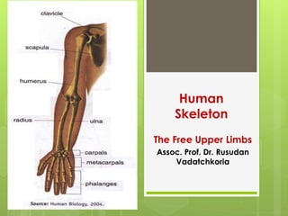
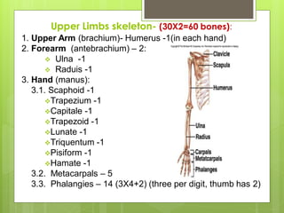
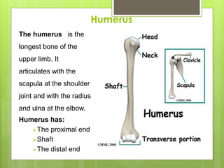
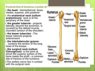
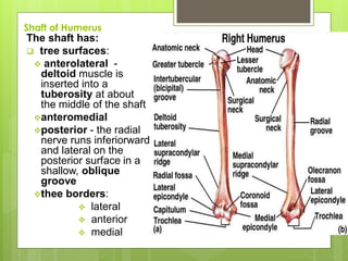
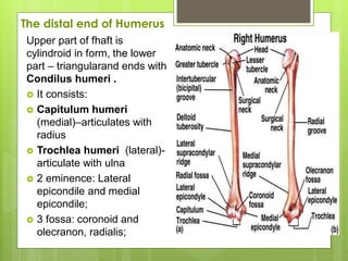
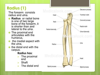
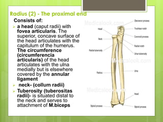
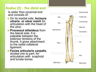
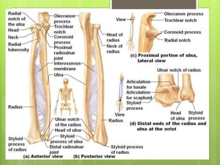
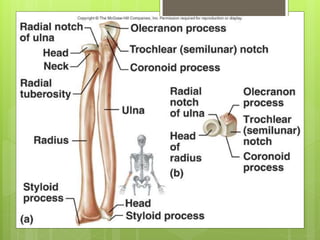
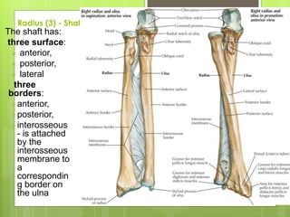
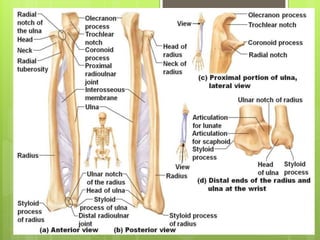
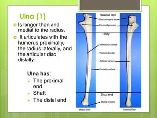
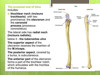
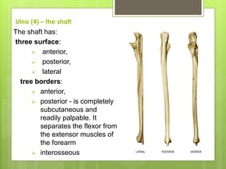
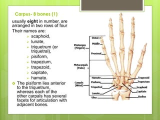
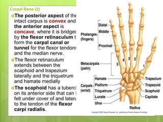
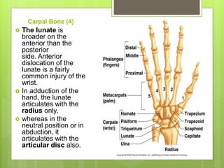
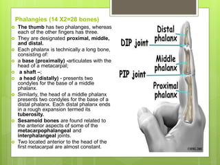
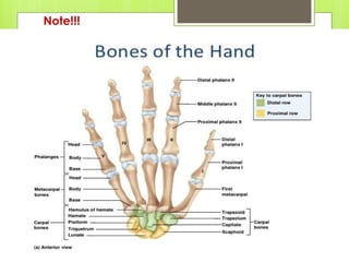
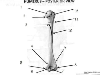
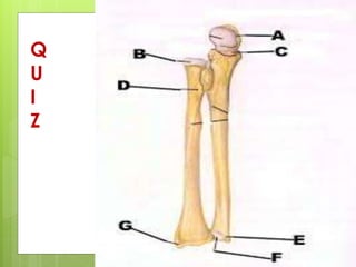
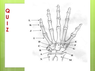
Human
Skeleton
The Free Upper Limbs
Assoc. Prof. Dr. Rusudan
Vadatchkoria
Upper Limbs skeleton- (30X2=60 bones):
1. Upper Arm (brachium)- Humerus -1(in each hand)
2. Forearm (antebrachium) – 2:
 Ulna -1
 Raduis -1
3. Hand (manus):
3.1. Scaphoid -1
Trapezium -1
Capitale -1
Trapezoid -1
Lunate -1
Triquentum -1
Pisiform -1
Hamate -1
3.2. Metacarpals – 5
3.3. Phalangies – 14 (3X4+2) (three per digit, thumb has 2)
Humerus
The humerus is the
longest bone of the
upper limb. It
articulates with the
scapula at the shoulder
joint and with the radius
and ulna at the elbow.
Humerus has:
The proximal end
Shaft
The distal end
Proximal End of Humerus consists of:
 the head - hemispherical, faces
medial, superior, and posterior
 the anatomical neck (collum
anatomicum)- neck is at the
periphery of the head
 the greater tubercle - projects
laterally, beyond the acromion, is
covered by the deltoid muscle
(rounded contour of the shoulder)
 the lesser tubercles - The
lesser tubercle projects
anteriorward
 the intertubercular groove-
contains the tendon of the long
head of the biceps.
 the surgical neck (collum
Chirurgicum)- is the point at
which the superior portion of the
bone meets the shaft - a common
site of fracture of the humerus.
The axillary nerve lies in contact
with the surgical neck
Shaft of Humerus
The shaft has:
 tree surfaces:
 anterolateral -
deltoid muscle is
inserted into a
tuberosity at about
the middle of the shaft
anteromedial
posterior - the radial
nerve runs inferiorward
and lateral on the
posterior surface in a
shallow, oblique
groove
thee borders:
 lateral
 anterior
 medial
The distal end of Humerus
Upper part of fhaft is
cylindroid in form, the lower
part – triangularand ends with
Condilus humeri .
 It consists:
 Capitulum humeri
(medial)–articulates with
radius
 Trochlea humeri (lateral)-
articulate with ulna
 2 eminence: Lateral
epicondile and medial
epicondile;
 3 fossa: coronoid and
olecranon, radialis;
Radius (1)
The forearm consists
radius and ulna.
 Radius or radial bone
is one of two large
bone of the forearm, it
is shorter than and
lateral to the ulna.
 The proximal end
articulates with the
humerus,
 the medial aspect with
the ulna,
 the distal end with the
carpus.
Radius has:
 The proximal
end
 Shaft
 The distal end
Radius (2) - The proximal end
Consists of:
 a head (caput radii) with
fovea articularis. The
superior, concave surface of
the head articulates with the
capitulum of the humerus.
The circumference
(circumferencia
articularis) of the head
articulates with the ulna
medially but is elsewhere
covered by the annular
ligament
 neck- (collum radii)
 Tuberosity (tuberositas
radii)- is situated distal to
the neck and serves to
attachment of M.biceps
Radius (2) - The distal end
Is wider than proximal end
and consists of:
 On its medial side, Incisura
ulnaris or ulnar notch for
articulation with the head of
the ulna;
 Processul stiloideus from
the lateral side; It is
palpable between the
extensor tendons of the
thumb. It gives attachment
to the radial collateral
ligament.
 Facies articularis carpalis,
divided into to part: for
articulation with scaphoid
and lunate bones
Radius (3) - Shaft
The shaft has:
three surface:
 anterior,
 posterior,
 lateral
three
borders:
anterior,
posterior,
interosseous
- is attached
by the
interosseous
membrane to
a
correspondin
g border on
the ulna
Ulna (1)
 Is longer than and
medial to the radius.
 It articulates with the
humerus proximally,
the radius laterally, and
the articular disc
distally.
Ulna has:
 The proximal
end
 Shaft
 The distal end
The proximal end of Ulna
includes:
 thochlear noch (incisura
trochlearis) with two
prominence: the olecranon and
the coronoid
process.(processus
coronoideus)
The lateral side has radial noch
(incisura radialis);
below it - the tuberositas ulna
The superior aspect of the
olecranon receives the insertion of
the M.triceps.
The posterior aspect, covered by
a bursa, is subcutaneous.
The antierior part of the olecranon
forms a part of the trochlear notch,
which articulates with the trochlea
of the humerus.
Ulna (4) – the shaft
The shaft has:
three surface:
 anterior,
 posterior,
 lateral
tree borders:
 anterior,
 posterior - is completely
subcutaneous and
readily palpable. It
separates the flexor from
the extensor muscles of
the forearm
 interosseous
Carpus- 8 bones (1)
usually eight in number, are
arranged in two rows of four
Their names are:
 scaphoid,
 lunate,
 triquetrum (or
triquetral),
 pisiform,
 trapezium,
 trapezoid,
 capitate,
 hamate.
 The pisiform lies anterior
to the triquetrum,
whereas each of the
other carpals has several
facets for articulation with
adjacent bones.
Carpal Bone (2)
The posterior aspect of the
intact carpus is convex and
the anterior aspect is
concave, where it is bridged
by the flexor retinaculum to
form the carpal canal or
tunnel for the flexor tendons
and the median nerve.
The flexor retinaculum
extends between the
scaphoid and trapezium
laterally and the triquetrum
and hamate medially
The scaphoid has a tubercle
on its anterior side that can be
felt under cover of and lateral
to the tendon of the flexor
carpi radialis.
Carpal Bone (4)
 The lunate is
broader on the
anterior than the
posterior
side. Anterior
dislocation of the
lunate is a fairly
common injury of the
wrist.
 In adduction of the
hand, the lunate
articulates with the
radius only,
 whereas in the
neutral position or in
abduction, it
articulates with the
articular disc also.
Carpal Bone (5)
 The pisiform, the
smallest of the
carpals and the
last to ossify, lies
anterior to the
triquetrum and
can be moved
passively from
side to side when
the flexor carpi
ulnaris is relaxed
Carpal Bone (6)
 The trapezium
supports the thumb
by means of a
saddle-shaped
facet for the first
metacarpal.
 Like the adjacent
scaphoid, it has an
anterior tubercle.
 The trapezoid is
associated with the
index finger.
Carpal Bone (7)
 The capitate - the
largest of the
carpals and the first
to ossify, is placed
centrally and is in
line with the third
metacarpal. It has a
prominent head on
its superior side.
 The hamate sends
a marked hook
anteriorly, which
gives attachment to
the flexor
retinaculum.
Metacarpus – 5 bones
 They are numbered from 1 to 5, from
the thumb to the little finger. The first
is the shortest and the second the
longest. They contribute to the palm,
and their posterior aspects can be felt
under cover of the extensor
tendons.
 Each metacarpal is technically a long
bone, consisting of:
 a base - (proximally)- articulates with
the carpus and, except for that of the
first, with the adjacent metacarpal(s)
also. The base of the first metacarpal
has a saddle-shaped facet for the
trapezium
 a shaft
 a head (distally) -of each metacarpal
articulates with a proximal phalanx
and forms a knuckle of the fist.
Phalangies (14 X2=28 bones)
 The thumb has two phalanges, whereas
each of the other fingers has three.
 They are designated proximal, middle,
and distal.
 Each phalanx is technically a long bone,
consisting of:
 a base (proximally) -articulates with the
head of a metacarpal;
 a shaft –;
 a head (distally) - presents two
condyles for the base of a middle
phalanx.
 Similarly, the head of a middle phalanx
presents two condyles for the base of a
distal phalanx. Each distal phalanx ends
in a rough expansion termed its
tuberosity.
 Sesamoid bones are found related to
the anterior aspects of some of the
metacarpophalangeal and
interphalangeal joints.
 Two located anterior to the head of the
first metacarpal are almost constant.
Note!!!
Q
U
I
Z
Q
U
I
Z
Reference
 Frazer's Anatomy of the Human Skeleton, 6th ed., rev.
by A. S. Breathnach, Churchill, London, 1965. A
detailed, regional synthesis of skeletal and muscular
anatomy.
 Pyle, S. I., Waterhouse, A. M., and Greulich, W. W.,
(eds.), A Radiographic Standard of Reference for the
Growing Hand and Wrist, Year Book Medical
Publishers, Chicago, 1971.
 Keith Moore Clinically. Oriented Anatomy. VII edition.
Wolter Kluwer, 2011, p. 672 -687

6.Upper limbs.pdf

  • 1.
    Human Skeleton The Free UpperLimbs Assoc. Prof. Dr. Rusudan Vadatchkoria
  • 2.
    Upper Limbs skeleton-(30X2=60 bones): 1. Upper Arm (brachium)- Humerus -1(in each hand) 2. Forearm (antebrachium) – 2:  Ulna -1  Raduis -1 3. Hand (manus): 3.1. Scaphoid -1 Trapezium -1 Capitale -1 Trapezoid -1 Lunate -1 Triquentum -1 Pisiform -1 Hamate -1 3.2. Metacarpals – 5 3.3. Phalangies – 14 (3X4+2) (three per digit, thumb has 2)
  • 3.
    Humerus The humerus isthe longest bone of the upper limb. It articulates with the scapula at the shoulder joint and with the radius and ulna at the elbow. Humerus has: The proximal end Shaft The distal end
  • 4.
    Proximal End ofHumerus consists of:  the head - hemispherical, faces medial, superior, and posterior  the anatomical neck (collum anatomicum)- neck is at the periphery of the head  the greater tubercle - projects laterally, beyond the acromion, is covered by the deltoid muscle (rounded contour of the shoulder)  the lesser tubercles - The lesser tubercle projects anteriorward  the intertubercular groove- contains the tendon of the long head of the biceps.  the surgical neck (collum Chirurgicum)- is the point at which the superior portion of the bone meets the shaft - a common site of fracture of the humerus. The axillary nerve lies in contact with the surgical neck
  • 5.
    Shaft of Humerus Theshaft has:  tree surfaces:  anterolateral - deltoid muscle is inserted into a tuberosity at about the middle of the shaft anteromedial posterior - the radial nerve runs inferiorward and lateral on the posterior surface in a shallow, oblique groove thee borders:  lateral  anterior  medial
  • 6.
    The distal endof Humerus Upper part of fhaft is cylindroid in form, the lower part – triangularand ends with Condilus humeri .  It consists:  Capitulum humeri (medial)–articulates with radius  Trochlea humeri (lateral)- articulate with ulna  2 eminence: Lateral epicondile and medial epicondile;  3 fossa: coronoid and olecranon, radialis;
  • 7.
    Radius (1) The forearmconsists radius and ulna.  Radius or radial bone is one of two large bone of the forearm, it is shorter than and lateral to the ulna.  The proximal end articulates with the humerus,  the medial aspect with the ulna,  the distal end with the carpus. Radius has:  The proximal end  Shaft  The distal end
  • 8.
    Radius (2) -The proximal end Consists of:  a head (caput radii) with fovea articularis. The superior, concave surface of the head articulates with the capitulum of the humerus. The circumference (circumferencia articularis) of the head articulates with the ulna medially but is elsewhere covered by the annular ligament  neck- (collum radii)  Tuberosity (tuberositas radii)- is situated distal to the neck and serves to attachment of M.biceps
  • 9.
    Radius (2) -The distal end Is wider than proximal end and consists of:  On its medial side, Incisura ulnaris or ulnar notch for articulation with the head of the ulna;  Processul stiloideus from the lateral side; It is palpable between the extensor tendons of the thumb. It gives attachment to the radial collateral ligament.  Facies articularis carpalis, divided into to part: for articulation with scaphoid and lunate bones
  • 12.
    Radius (3) -Shaft The shaft has: three surface:  anterior,  posterior,  lateral three borders: anterior, posterior, interosseous - is attached by the interosseous membrane to a correspondin g border on the ulna
  • 14.
    Ulna (1)  Islonger than and medial to the radius.  It articulates with the humerus proximally, the radius laterally, and the articular disc distally. Ulna has:  The proximal end  Shaft  The distal end
  • 15.
    The proximal endof Ulna includes:  thochlear noch (incisura trochlearis) with two prominence: the olecranon and the coronoid process.(processus coronoideus) The lateral side has radial noch (incisura radialis); below it - the tuberositas ulna The superior aspect of the olecranon receives the insertion of the M.triceps. The posterior aspect, covered by a bursa, is subcutaneous. The antierior part of the olecranon forms a part of the trochlear notch, which articulates with the trochlea of the humerus.
  • 16.
    Ulna (4) –the shaft The shaft has: three surface:  anterior,  posterior,  lateral tree borders:  anterior,  posterior - is completely subcutaneous and readily palpable. It separates the flexor from the extensor muscles of the forearm  interosseous
  • 17.
    Carpus- 8 bones(1) usually eight in number, are arranged in two rows of four Their names are:  scaphoid,  lunate,  triquetrum (or triquetral),  pisiform,  trapezium,  trapezoid,  capitate,  hamate.  The pisiform lies anterior to the triquetrum, whereas each of the other carpals has several facets for articulation with adjacent bones.
  • 18.
    Carpal Bone (2) Theposterior aspect of the intact carpus is convex and the anterior aspect is concave, where it is bridged by the flexor retinaculum to form the carpal canal or tunnel for the flexor tendons and the median nerve. The flexor retinaculum extends between the scaphoid and trapezium laterally and the triquetrum and hamate medially The scaphoid has a tubercle on its anterior side that can be felt under cover of and lateral to the tendon of the flexor carpi radialis.
  • 19.
    Carpal Bone (4) The lunate is broader on the anterior than the posterior side. Anterior dislocation of the lunate is a fairly common injury of the wrist.  In adduction of the hand, the lunate articulates with the radius only,  whereas in the neutral position or in abduction, it articulates with the articular disc also.
  • 20.
    Carpal Bone (5) The pisiform, the smallest of the carpals and the last to ossify, lies anterior to the triquetrum and can be moved passively from side to side when the flexor carpi ulnaris is relaxed
  • 21.
    Carpal Bone (6) The trapezium supports the thumb by means of a saddle-shaped facet for the first metacarpal.  Like the adjacent scaphoid, it has an anterior tubercle.  The trapezoid is associated with the index finger.
  • 22.
    Carpal Bone (7) The capitate - the largest of the carpals and the first to ossify, is placed centrally and is in line with the third metacarpal. It has a prominent head on its superior side.  The hamate sends a marked hook anteriorly, which gives attachment to the flexor retinaculum.
  • 23.
    Metacarpus – 5bones  They are numbered from 1 to 5, from the thumb to the little finger. The first is the shortest and the second the longest. They contribute to the palm, and their posterior aspects can be felt under cover of the extensor tendons.  Each metacarpal is technically a long bone, consisting of:  a base - (proximally)- articulates with the carpus and, except for that of the first, with the adjacent metacarpal(s) also. The base of the first metacarpal has a saddle-shaped facet for the trapezium  a shaft  a head (distally) -of each metacarpal articulates with a proximal phalanx and forms a knuckle of the fist.
  • 24.
    Phalangies (14 X2=28bones)  The thumb has two phalanges, whereas each of the other fingers has three.  They are designated proximal, middle, and distal.  Each phalanx is technically a long bone, consisting of:  a base (proximally) -articulates with the head of a metacarpal;  a shaft –;  a head (distally) - presents two condyles for the base of a middle phalanx.  Similarly, the head of a middle phalanx presents two condyles for the base of a distal phalanx. Each distal phalanx ends in a rough expansion termed its tuberosity.  Sesamoid bones are found related to the anterior aspects of some of the metacarpophalangeal and interphalangeal joints.  Two located anterior to the head of the first metacarpal are almost constant.
  • 25.
  • 27.
  • 28.
  • 29.
    Reference  Frazer's Anatomyof the Human Skeleton, 6th ed., rev. by A. S. Breathnach, Churchill, London, 1965. A detailed, regional synthesis of skeletal and muscular anatomy.  Pyle, S. I., Waterhouse, A. M., and Greulich, W. W., (eds.), A Radiographic Standard of Reference for the Growing Hand and Wrist, Year Book Medical Publishers, Chicago, 1971.  Keith Moore Clinically. Oriented Anatomy. VII edition. Wolter Kluwer, 2011, p. 672 -687