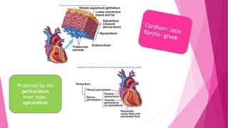
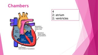
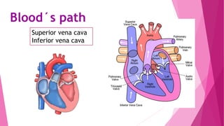
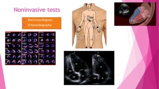
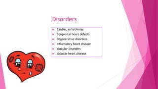
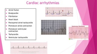
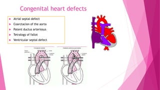
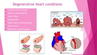
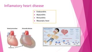
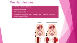
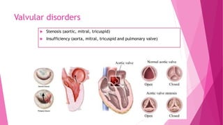
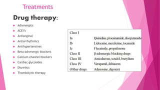
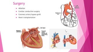
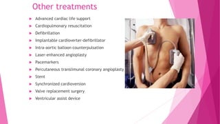
Cardiovascular system
Protected by the
pericardium
Inner layer:
epicardium
Chambers
4
2: atrium
2: ventricles
Blood´s path
Superior vena cava
Inferior vena cava
The heart’s rhythm
Contractions of the heart occur in a rhythm
Regulated by impulses that begin at the
sinoatrial node (SA) (pacemaker)
Atrioventricular node (AV)
Bundle of His
Trough the Purkinge fibers(ventricular
contaction)
Parasympathetic division
• Acts on the SA and AV
nodes
• Slows heart rate
• Reduces impulse
conduction
• Dilates coronary arteries
Sympathetic división
• Acts on the SA and AV nodes
• Increases heart rate and
impulse conduction
• Constricts and dilates the
coronary arteries
Autonomic nervous system
Cardiac cycle
 The period from the beginning of one
heart beat to the beginning of the
next
 Provide adequate blood flow to all
body parts
 Two phases:
Systole: period when the ventricles
contract
Diastole: when the heart relaxes
Vascular network…
Veins
• Carry blood toward
the heart
• Carry oxygen-
depleted blood
(*pulmonary vein)
Vital signs
The physiologic condition of a patient
Pulse and blood presure are directly related to the
cardiovascular system
Pulse: expansion and contraction of an
artery in a regular, rhythmic pattern
Blood pressure
♡ Influenced by the volumen of blood, the lumen
of the arteries and arterioles, and the force of
the cardiac contraction
♡ 120/80 mmHg
♡ Systolic blood pressure: 120
♡ Diastolic blood pressure: 80
Tools of the trade
Sphygmomanometer
Stethoscope
Physical examination
 Inspection: cyanosis, pallor, diaphoresis
 Palpation: edema
 Auscultation: murmur, blowing, fluttering and bruit
Diagnostic tests
 Blood tests: PTT (partial thromboplastin time), CK (creatine kinase),
cardiac troponin.
 Radiologic tests: cardiac catheterization, angiocardiography,
agiography, radionuclide scan, thellium stress test
 Electrophysiologic studies
 Pericardiocentesis
 Transesophageal echocardiography
Noninvasive tests
 Electrocardiogram
 Echocardiography
Disorders
 Cardiac arrhythmias
 Congenital heart defects
 Degenerative disorders
 Inflamatory heart disease
 Vascular disorders
 Valvular heart disease
Cardiac arrhythmias
 Atrial flutter
 Bradycardia
 Fibrillation
 Heart block
 Paroxysmal atrial tachycardia
 Premature atrial contraction
 Premature ventricular
contraction
 Tachycardia
 Ventricular tachycardia
Congenital heart defects
 Atrial septal defect
 Coarctacion of the aorta
 Patent ductus arteriosus
 Tetralogy of fallot
 Ventricular septal defect
Degenerative heart conditions
 Coronary artery disease
 Dilated cardiomyopathy
 Heart failure
 Hypertension
 Hypertrophic cardiomyopathy
 Heart attack
 Restrictive cardiomyopathy
Inflamatory heart disease
 Endocarditis
 Myocarditis
 Pericarditis
 Rheumatic fever
Vascular disorders
 Arterial occlusive disease
 Raynaud’s disease
 Thrombophlebitis
 Aneurysm (abdominal aorta, thoracic aorta, saccular, fusiform,
dissecting and false)
Valvular disorders
 Stenosis (aortic, mitral, tricuspid)
 Insufficiency (aorta, mitral, tricuspid and pulmonary valve)
Treatments
Drug therapy:
 Adrenergics
 ACEI’s
 Antianginal
 Antiarrhythmics
 Antihypertensives
 Beta-adrenergic blockers
 Calcium channel blockers
 Cardiac glycosides
 Diuretics
 Thombolytic therapy
Surgery
 Ablation
 Cardiac conduction surgery
 Coronary artery bypass graft
 Heart transplantation
Other treatments
 Advanced cardiac life support
 Cardiopulmonary resuscitation
 Defibrillation
 Implantable cardioverter-defibrillator
 Intra-aortic balloon counterpulsation
 Laser-enhanced angioplasty
 Pacemarkers
 Percutaneous translimunal coronary angioplasty
 Stent
 Synchronized cardioversion
 Valve replacement surgery
 Ventricular assist device
Cardiovascular System

Cardiovascular System

  • 1.
  • 2.
  • 3.
  • 4.
    Blood´s path Superior venacava Inferior vena cava
  • 5.
    The heart’s rhythm Contractionsof the heart occur in a rhythm Regulated by impulses that begin at the sinoatrial node (SA) (pacemaker) Atrioventricular node (AV) Bundle of His Trough the Purkinge fibers(ventricular contaction)
  • 6.
    Parasympathetic division • Actson the SA and AV nodes • Slows heart rate • Reduces impulse conduction • Dilates coronary arteries Sympathetic división • Acts on the SA and AV nodes • Increases heart rate and impulse conduction • Constricts and dilates the coronary arteries Autonomic nervous system
  • 7.
    Cardiac cycle  Theperiod from the beginning of one heart beat to the beginning of the next  Provide adequate blood flow to all body parts  Two phases: Systole: period when the ventricles contract Diastole: when the heart relaxes
  • 8.
    Vascular network… Veins • Carryblood toward the heart • Carry oxygen- depleted blood (*pulmonary vein)
  • 9.
    Vital signs The physiologiccondition of a patient Pulse and blood presure are directly related to the cardiovascular system Pulse: expansion and contraction of an artery in a regular, rhythmic pattern
  • 10.
    Blood pressure ♡ Influencedby the volumen of blood, the lumen of the arteries and arterioles, and the force of the cardiac contraction ♡ 120/80 mmHg ♡ Systolic blood pressure: 120 ♡ Diastolic blood pressure: 80
  • 11.
    Tools of thetrade Sphygmomanometer Stethoscope
  • 12.
    Physical examination  Inspection:cyanosis, pallor, diaphoresis  Palpation: edema  Auscultation: murmur, blowing, fluttering and bruit
  • 13.
    Diagnostic tests  Bloodtests: PTT (partial thromboplastin time), CK (creatine kinase), cardiac troponin.  Radiologic tests: cardiac catheterization, angiocardiography, agiography, radionuclide scan, thellium stress test  Electrophysiologic studies  Pericardiocentesis  Transesophageal echocardiography
  • 14.
  • 15.
    Disorders  Cardiac arrhythmias Congenital heart defects  Degenerative disorders  Inflamatory heart disease  Vascular disorders  Valvular heart disease
  • 16.
    Cardiac arrhythmias  Atrialflutter  Bradycardia  Fibrillation  Heart block  Paroxysmal atrial tachycardia  Premature atrial contraction  Premature ventricular contraction  Tachycardia  Ventricular tachycardia
  • 17.
    Congenital heart defects Atrial septal defect  Coarctacion of the aorta  Patent ductus arteriosus  Tetralogy of fallot  Ventricular septal defect
  • 18.
    Degenerative heart conditions Coronary artery disease  Dilated cardiomyopathy  Heart failure  Hypertension  Hypertrophic cardiomyopathy  Heart attack  Restrictive cardiomyopathy
  • 19.
    Inflamatory heart disease Endocarditis  Myocarditis  Pericarditis  Rheumatic fever
  • 20.
    Vascular disorders  Arterialocclusive disease  Raynaud’s disease  Thrombophlebitis  Aneurysm (abdominal aorta, thoracic aorta, saccular, fusiform, dissecting and false)
  • 21.
    Valvular disorders  Stenosis(aortic, mitral, tricuspid)  Insufficiency (aorta, mitral, tricuspid and pulmonary valve)
  • 22.
    Treatments Drug therapy:  Adrenergics ACEI’s  Antianginal  Antiarrhythmics  Antihypertensives  Beta-adrenergic blockers  Calcium channel blockers  Cardiac glycosides  Diuretics  Thombolytic therapy
  • 23.
    Surgery  Ablation  Cardiacconduction surgery  Coronary artery bypass graft  Heart transplantation
  • 24.
    Other treatments  Advancedcardiac life support  Cardiopulmonary resuscitation  Defibrillation  Implantable cardioverter-defibrillator  Intra-aortic balloon counterpulsation  Laser-enhanced angioplasty  Pacemarkers  Percutaneous translimunal coronary angioplasty  Stent  Synchronized cardioversion  Valve replacement surgery  Ventricular assist device