Downloaded 277 times
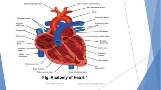


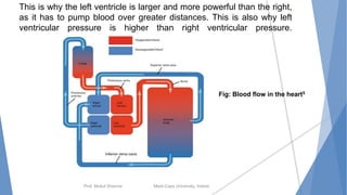


The document discusses the cardiovascular system and anatomy of the heart. It contains the following key points: 1. The cardiovascular system includes the heart, blood vessels, and blood. The heart acts as a pump to circulate blood throughout the body via blood vessels. 2. The heart has four chambers - two upper atria and two lower ventricles. It also contains valves that prevent backflow of blood. 3. Blood circulation occurs via two loops - pulmonary circulation oxygenates blood in the lungs, while systemic circulation delivers oxygenated blood to the entire body from the heart.


