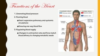
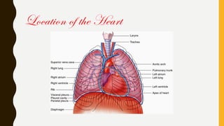
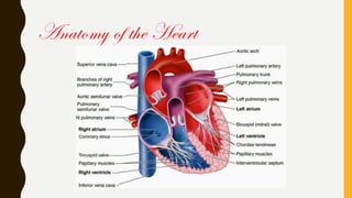
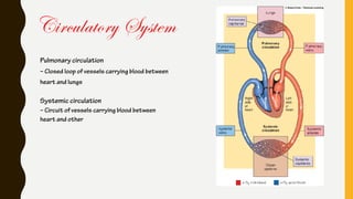
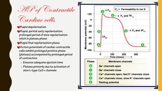
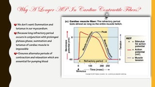
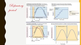
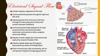
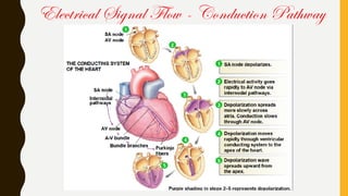
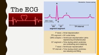
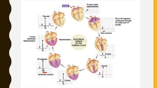
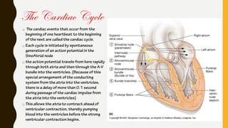
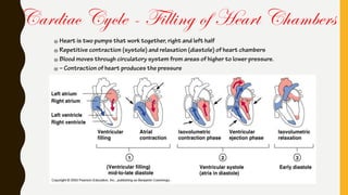
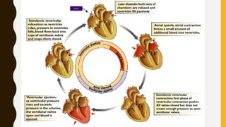
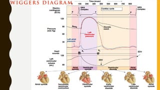
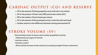
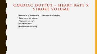
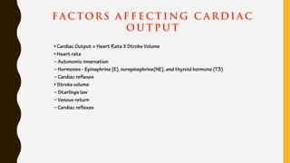
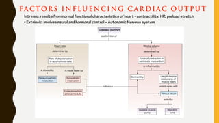
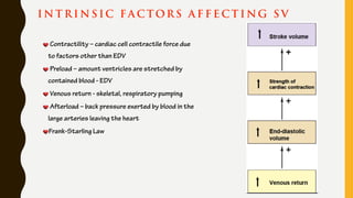
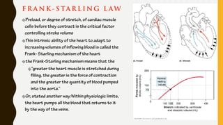
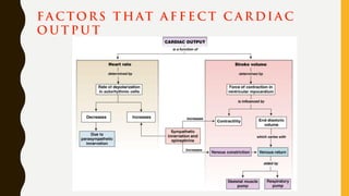
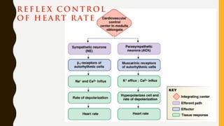
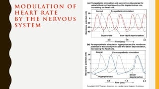
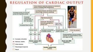
Joel G. Soria, MD, MBA-H
#puso
CARDIOPHYSIOLOGY
Functions of the Heart
1. Generating blood pressure
2. Routing blood
Heart separates pulmonary and systemic
circulations
Ensuring one-way blood flow
3. Regulating blood supply
Changes in contraction rate and force match
blood delivery to changing metabolic needs
Location of the Heart
Anatomy of the Heart
The heart is composed of three major types of cardiac muscle:
atrial muscle
ventricular muscle
specialized excitatory and conductive muscle fibers
The atrial and ventricular types of muscle contract in much the same way as skeletal
muscle, except that the duration of contraction is much longer.
Physiology of Cardiac Muscle
Circulatory System
Pulmonary circulation
– Closed loop of vessels carrying blood between
heart and lungs
Systemic circulation
– Circuit of vessels carrying blood between
heart and other
Cardiac Muscle as a Syncytium:
The dark areas crossing the cardiac muscle fibers are called intercalated discs;
Cardiac muscle fibers are made up of many individual cells connected in series and
in parallel with one another.
At each intercalated disc the cell membranes fuse with one another in such a way
that they form permeable “communicating” junctions (gap junctions) that allow
almost totally free diffusion of ions.
Cardiac muscle is a syncytium of many heart muscle cells in which the cardiac cells
are so interconnected that when one of these cells becomes excited, the action
potential spreads to all of them, spreading from cell to cell throughout the
latticework interconnections.
Atrial and Ventricular Syncytium
The atria are separated from the ventricles by fibrous tissue that surrounds the
atrioventricular (A-V) valvular openings between the atria and ventricles.
Normally, potentials are not conducted from the atrial syncytium into the
ventricular syncytium directly through this fibrous tissue.
Instead, they are conducted only by way of a specialized conductive system called
the A-V bundle
This division of the muscle of the heart into two
functional syncytiums allows the atria to contract a
short time ahead of ventricular contraction, which is
important for effectiveness of heart pumping.
Cardiac Muscle as a Syncytium:
The action potential recorded in a ventricular muscle
fiber about 105 millivolts,
the intracellular potential rises from a very negative value,
about - 85 millivolts, between beats to a slightly positive
value, about + 20 millivolts, during each beat. After the
initial spike
the membrane remains depolarized for about 0.2 second
exhibiting a plateau , followed at by abrupt repolarization.
The presence of this plateau in the action potential causes
ventricular contraction to last as much as 15 times as
long in cardiac muscle as in skeletal muscle.
Action Potentials in Cardiac Muscle
Heart beats rhythmically as result of action potentials it generates by itself
(autorhythmicity)
Two specialized types of cardiac muscle cells
– Contractile cells
• 99% of cardiac muscle cells
• Do mechanical work of pumping
• Normally do not initiate own action potentials
– Autorhythmic cells
• Do not contract
• Specialized for initiating and conducting action potentials responsible for
contraction of working cells
Electrical Activity of Heart
Intrinsic Conduction System
Autorhythmic cells:
Initiate action potentials
Have “drifting” resting potentials called
pacemaker potentials
Pacemaker potential - membrane slowly
depolarizes “drifts” to threshold, initiates
action potential, membrane repolarizes to -60
mV.
Use calcium influx (rather than sodium) for
rising phase of the action potential
Pacemaker Potential
Decreased efflux of K+, membrane permeability decreases between APs, they slowly close at negative potentials
Constant influx of Na+, no voltage-gated Na + channels
Gradual depolarization because K+ builds up and Na+ flows inward
As depolarization proceeds Ca++ channels (Ca2+ T) open influx of Ca++ further depolarizes to threshold (-40mV)
At threshold sharp depolarization due to activation of Ca2+ L channels allow large influx of Ca++
Falling phase at about +20 mV the Ca-L channels close, voltage-gated K channels open, repolarization due to normal K+
efflux
At -60mV K+ channels close
AP of Contractile
Cardiac cells
Rapid depolarization
Rapid, partial early repolarization,
prolonged period of slow repolarization
which is plateau phase
Rapid final repolarization phase
Action potentials of cardiac contractile
cells exhibit prolonged positive phase
(plateau) accompanied by prolonged period
of contraction
Ensures adequate ejection time
Plateau primarily due to activation of
slow L-type Ca2+ channels
Why A Longer AP In Cardiac Contractile Fibers?
We don’t want Summation and
tetanus in our myocardium.
Because long refractory period
occurs in conjunction with prolonged
plateau phase, summation and
tetanus of cardiac muscle is
impossible
• Ensures alternate periods of
contraction and relaxation which are
essential for pumping blood
Refractory
period
Cardiac impulse originates at SA node
Action potential spreads throughout right and
left atria
Impulse passes from atria into ventricles
through AV node (only point of electrical
contact between chambers)
Action potential briefly delayed at AV node
(ensures atrial contraction precedes
ventricular contraction to allow complete
ventricular filling)
Impulse travels rapidly down interventricular
septum by means of bundle of His
Impulse rapidly disperses throughout
myocardium by means of Purkinje fibers
Rest of ventricular cells activated by cell-to-
cell spread of impulse through gap junctions
Electrical Signal Flow
Electrical Signal Flow - Conduction Pathway
The ECG
E C G I N F OR M AT ION G A I N E D
(Non-invasive)
Heart Rate
Signal conduction
Heart tissue
Conditions
The cardiac events that occur from the
beginning of one heartbeat to the beginning
of the next are called the cardiac cycle.
Each cycle is initiated by spontaneous
generation of an action potential in the
SinoAtrial node
the action potential travels from here rapidly
through both atria and then through the A-V
bundle into the ventricles. (Because of this
special arrangement of the conducting
system from the atria into the ventricles,
there is a delay of more than 0.1 second
during passage of the cardiac impulse from
the atria into the ventricles)
This allows the atria to contract ahead of
ventricular contraction, thereby pumping
blood into the ventricles before the strong
ventricular contraction begins.
The Cardiac Cycle
Cardiac Cycle - Filling of Heart Chambers
Heart is two pumps that work together, right and left half
Repetitive contraction (systole) and relaxation (diastole) of heart chambers
Blood moves through circulatory system from areas of higher to lower pressure.
– Contraction of heart produces the pressure
START
Late diastole: both sets of
chambers are relaxed and
ventricles fill passively.
Atrial systole: atrial contraction
forces a small amount of
additional blood into ventricles.
Isovolumic ventricular
contraction: first phase of
ventricular contraction pushes
AV valves closed but does not
create enough pressure to open
semilunar valves.
Ventricular ejection:
as ventricular pressure
rises and exceeds
pressure in the arteries,
the semilunar valves
open and blood is
ejected.
Isovolumic ventricular
relaxation: as ventricles
relax, pressure in ventricles
falls, blood flows back into
cups of semilunar valves
and snaps them closed.
H E A RT S O U N D S
First heart sound or “lubb”
– AV valves close and surrounding fluid vibrations at systole
• Second heart sound or “dupp”
– Results from closure of aortic and pulmonary semilunar valves at diastole, lasts longer
W IG G E RS DI A G R A M
C A R DI A C O U T P U T ( C O ) A N D R E S E RV E
CO is the amount of blood pumped by each ventricle in one minute
CO is the product of heart rate (HR) and stroke volume (SV)
HR is the number of heart beats per minute
SV is the amount of blood pumped out by a ventricle with each beat
Cardiac reserve is the difference between resting and maximal CO
S T R OK E V OLUM E ( S V )
Determined by extent of venous return and by sympathetic activity
– Influenced by two types of controls
• Intrinsic control
• Extrinsic control
– Both controls increase stroke volume by increasing strength of heart contraction
C A R DI A C O U T P U T = H E A RT R AT E X
S T R OK E V OLUM E
• Around 5L : (70 beats/m ´ 70 ml/beat = 4900 ml)
• Rate: beats per minute
• Volume: ml per beat
– SV = EDV - ESV
– Residual (about 50%)
FA C TORS A F F E C T I NG C A R DI A C
O U T P U T
• Cardiac Output = Heart Rate X Stroke Volume
• Heart rate
– Autonomic innervation
– Hormones - Epinephrine (E), norepinephrine(NE), and thyroid hormone (T3)
– Cardiac reflexes
• Stroke volume
– Starlings law
– Venous return
– Cardiac reflexes
FA C TORS I N F LU E NC I NG C A R DI A C O U T P U T
Intrinsic: results from normal functional characteristics of heart - contractility, HR, preload stretch
• Extrinsic: involves neural and hormonal control – Autonomic Nervous system
I N T R I N S IC FA C TORS A F F E C T I NG S V
Contractility – cardiac cell contractile force due
to factors other than EDV
Preload – amount ventricles are stretched by
contained blood - EDV
Venous return - skeletal, respiratory pumping
Afterload – back pressure exerted by blood in the
large arteries leaving the heart
Frank-Starling Law
F R A N K - S TA R L I NG L AW
Preload, or degree of stretch, of cardiac muscle
cells before they contract is the critical factor
controlling stroke volume
This intrinsic ability of the heart to adapt to
increasing volumes of inflowing blood is called the
Frank- Starling mechanism of the heart
the Frank-Starling mechanism means that the
“greater the heart muscle is stretched during
filling, the greater is the force of contraction
and the greater the quantity of blood pumped
into the aorta.”
Or, stated another way:Within physiologic limits,
the heart pumps all the blood that returns to it
by the way of the veins.
E XT R I N S IC FA C TORS I N F LU E NC I NG S V
• Contractility is the increase in contractile strength, independent
of stretch
• Increase in contractility comes from
– Increased sympathetic stimuli
– Hormones - epinephrine and thyroxine
– Ca2+ and some drugs
– Intra- and extracellular ion concentrations must
be maintained for normal heart function
FA C TORS T H AT A F F E C T C A R DI A C
O U T P U T
R E F L E X C ON T R OL
OF H E A RT R AT E
M OD U L AT ION OF
H E A RT R AT E
BY T H E N E RV O U S
S Y S T E M
R E G U L AT ION OF C A R DI A C O U T P U T

Cardio physio

  • 1.
    Joel G. Soria,MD, MBA-H #puso CARDIOPHYSIOLOGY
  • 2.
    Functions of theHeart 1. Generating blood pressure 2. Routing blood Heart separates pulmonary and systemic circulations Ensuring one-way blood flow 3. Regulating blood supply Changes in contraction rate and force match blood delivery to changing metabolic needs
  • 3.
  • 4.
  • 5.
    The heart iscomposed of three major types of cardiac muscle: atrial muscle ventricular muscle specialized excitatory and conductive muscle fibers The atrial and ventricular types of muscle contract in much the same way as skeletal muscle, except that the duration of contraction is much longer. Physiology of Cardiac Muscle
  • 6.
    Circulatory System Pulmonary circulation –Closed loop of vessels carrying blood between heart and lungs Systemic circulation – Circuit of vessels carrying blood between heart and other
  • 7.
    Cardiac Muscle asa Syncytium: The dark areas crossing the cardiac muscle fibers are called intercalated discs; Cardiac muscle fibers are made up of many individual cells connected in series and in parallel with one another. At each intercalated disc the cell membranes fuse with one another in such a way that they form permeable “communicating” junctions (gap junctions) that allow almost totally free diffusion of ions. Cardiac muscle is a syncytium of many heart muscle cells in which the cardiac cells are so interconnected that when one of these cells becomes excited, the action potential spreads to all of them, spreading from cell to cell throughout the latticework interconnections. Atrial and Ventricular Syncytium
  • 8.
    The atria areseparated from the ventricles by fibrous tissue that surrounds the atrioventricular (A-V) valvular openings between the atria and ventricles. Normally, potentials are not conducted from the atrial syncytium into the ventricular syncytium directly through this fibrous tissue. Instead, they are conducted only by way of a specialized conductive system called the A-V bundle This division of the muscle of the heart into two functional syncytiums allows the atria to contract a short time ahead of ventricular contraction, which is important for effectiveness of heart pumping. Cardiac Muscle as a Syncytium:
  • 9.
    The action potentialrecorded in a ventricular muscle fiber about 105 millivolts, the intracellular potential rises from a very negative value, about - 85 millivolts, between beats to a slightly positive value, about + 20 millivolts, during each beat. After the initial spike the membrane remains depolarized for about 0.2 second exhibiting a plateau , followed at by abrupt repolarization. The presence of this plateau in the action potential causes ventricular contraction to last as much as 15 times as long in cardiac muscle as in skeletal muscle. Action Potentials in Cardiac Muscle
  • 10.
    Heart beats rhythmicallyas result of action potentials it generates by itself (autorhythmicity) Two specialized types of cardiac muscle cells – Contractile cells • 99% of cardiac muscle cells • Do mechanical work of pumping • Normally do not initiate own action potentials – Autorhythmic cells • Do not contract • Specialized for initiating and conducting action potentials responsible for contraction of working cells Electrical Activity of Heart
  • 11.
    Intrinsic Conduction System Autorhythmiccells: Initiate action potentials Have “drifting” resting potentials called pacemaker potentials Pacemaker potential - membrane slowly depolarizes “drifts” to threshold, initiates action potential, membrane repolarizes to -60 mV. Use calcium influx (rather than sodium) for rising phase of the action potential
  • 12.
    Pacemaker Potential Decreased effluxof K+, membrane permeability decreases between APs, they slowly close at negative potentials Constant influx of Na+, no voltage-gated Na + channels Gradual depolarization because K+ builds up and Na+ flows inward As depolarization proceeds Ca++ channels (Ca2+ T) open influx of Ca++ further depolarizes to threshold (-40mV) At threshold sharp depolarization due to activation of Ca2+ L channels allow large influx of Ca++ Falling phase at about +20 mV the Ca-L channels close, voltage-gated K channels open, repolarization due to normal K+ efflux At -60mV K+ channels close
  • 13.
    AP of Contractile Cardiaccells Rapid depolarization Rapid, partial early repolarization, prolonged period of slow repolarization which is plateau phase Rapid final repolarization phase Action potentials of cardiac contractile cells exhibit prolonged positive phase (plateau) accompanied by prolonged period of contraction Ensures adequate ejection time Plateau primarily due to activation of slow L-type Ca2+ channels
  • 14.
    Why A LongerAP In Cardiac Contractile Fibers? We don’t want Summation and tetanus in our myocardium. Because long refractory period occurs in conjunction with prolonged plateau phase, summation and tetanus of cardiac muscle is impossible • Ensures alternate periods of contraction and relaxation which are essential for pumping blood
  • 15.
  • 16.
    Cardiac impulse originatesat SA node Action potential spreads throughout right and left atria Impulse passes from atria into ventricles through AV node (only point of electrical contact between chambers) Action potential briefly delayed at AV node (ensures atrial contraction precedes ventricular contraction to allow complete ventricular filling) Impulse travels rapidly down interventricular septum by means of bundle of His Impulse rapidly disperses throughout myocardium by means of Purkinje fibers Rest of ventricular cells activated by cell-to- cell spread of impulse through gap junctions Electrical Signal Flow
  • 17.
    Electrical Signal Flow- Conduction Pathway
  • 18.
  • 20.
    E C GI N F OR M AT ION G A I N E D (Non-invasive) Heart Rate Signal conduction Heart tissue Conditions
  • 21.
    The cardiac eventsthat occur from the beginning of one heartbeat to the beginning of the next are called the cardiac cycle. Each cycle is initiated by spontaneous generation of an action potential in the SinoAtrial node the action potential travels from here rapidly through both atria and then through the A-V bundle into the ventricles. (Because of this special arrangement of the conducting system from the atria into the ventricles, there is a delay of more than 0.1 second during passage of the cardiac impulse from the atria into the ventricles) This allows the atria to contract ahead of ventricular contraction, thereby pumping blood into the ventricles before the strong ventricular contraction begins. The Cardiac Cycle
  • 22.
    Cardiac Cycle -Filling of Heart Chambers Heart is two pumps that work together, right and left half Repetitive contraction (systole) and relaxation (diastole) of heart chambers Blood moves through circulatory system from areas of higher to lower pressure. – Contraction of heart produces the pressure
  • 23.
    START Late diastole: bothsets of chambers are relaxed and ventricles fill passively. Atrial systole: atrial contraction forces a small amount of additional blood into ventricles. Isovolumic ventricular contraction: first phase of ventricular contraction pushes AV valves closed but does not create enough pressure to open semilunar valves. Ventricular ejection: as ventricular pressure rises and exceeds pressure in the arteries, the semilunar valves open and blood is ejected. Isovolumic ventricular relaxation: as ventricles relax, pressure in ventricles falls, blood flows back into cups of semilunar valves and snaps them closed.
  • 24.
    H E ART S O U N D S First heart sound or “lubb” – AV valves close and surrounding fluid vibrations at systole • Second heart sound or “dupp” – Results from closure of aortic and pulmonary semilunar valves at diastole, lasts longer
  • 25.
    W IG GE RS DI A G R A M
  • 26.
    C A RDI A C O U T P U T ( C O ) A N D R E S E RV E CO is the amount of blood pumped by each ventricle in one minute CO is the product of heart rate (HR) and stroke volume (SV) HR is the number of heart beats per minute SV is the amount of blood pumped out by a ventricle with each beat Cardiac reserve is the difference between resting and maximal CO S T R OK E V OLUM E ( S V ) Determined by extent of venous return and by sympathetic activity – Influenced by two types of controls • Intrinsic control • Extrinsic control – Both controls increase stroke volume by increasing strength of heart contraction
  • 27.
    C A RDI A C O U T P U T = H E A RT R AT E X S T R OK E V OLUM E • Around 5L : (70 beats/m ´ 70 ml/beat = 4900 ml) • Rate: beats per minute • Volume: ml per beat – SV = EDV - ESV – Residual (about 50%)
  • 28.
    FA C TORSA F F E C T I NG C A R DI A C O U T P U T • Cardiac Output = Heart Rate X Stroke Volume • Heart rate – Autonomic innervation – Hormones - Epinephrine (E), norepinephrine(NE), and thyroid hormone (T3) – Cardiac reflexes • Stroke volume – Starlings law – Venous return – Cardiac reflexes
  • 29.
    FA C TORSI N F LU E NC I NG C A R DI A C O U T P U T Intrinsic: results from normal functional characteristics of heart - contractility, HR, preload stretch • Extrinsic: involves neural and hormonal control – Autonomic Nervous system
  • 30.
    I N TR I N S IC FA C TORS A F F E C T I NG S V Contractility – cardiac cell contractile force due to factors other than EDV Preload – amount ventricles are stretched by contained blood - EDV Venous return - skeletal, respiratory pumping Afterload – back pressure exerted by blood in the large arteries leaving the heart Frank-Starling Law
  • 31.
    F R AN K - S TA R L I NG L AW Preload, or degree of stretch, of cardiac muscle cells before they contract is the critical factor controlling stroke volume This intrinsic ability of the heart to adapt to increasing volumes of inflowing blood is called the Frank- Starling mechanism of the heart the Frank-Starling mechanism means that the “greater the heart muscle is stretched during filling, the greater is the force of contraction and the greater the quantity of blood pumped into the aorta.” Or, stated another way:Within physiologic limits, the heart pumps all the blood that returns to it by the way of the veins.
  • 32.
    E XT RI N S IC FA C TORS I N F LU E NC I NG S V • Contractility is the increase in contractile strength, independent of stretch • Increase in contractility comes from – Increased sympathetic stimuli – Hormones - epinephrine and thyroxine – Ca2+ and some drugs – Intra- and extracellular ion concentrations must be maintained for normal heart function
  • 33.
    FA C TORST H AT A F F E C T C A R DI A C O U T P U T
  • 34.
    R E FL E X C ON T R OL OF H E A RT R AT E
  • 35.
    M OD UL AT ION OF H E A RT R AT E BY T H E N E RV O U S S Y S T E M
  • 36.
    R E GU L AT ION OF C A R DI A C O U T P U T