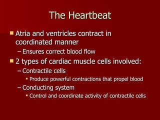
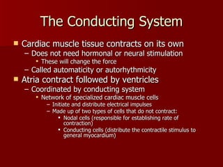
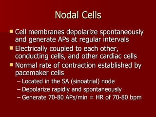
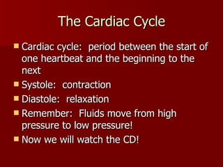
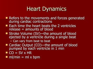
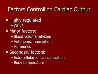
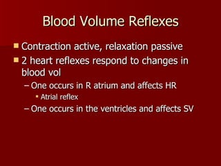
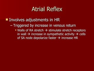
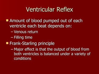
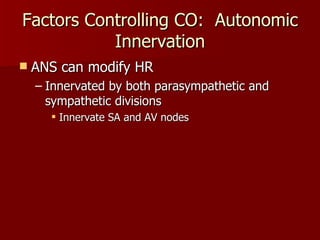
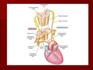
The Heart
Overview Introduction The Heart and The Circulation System The Heartbeat Contractile cells The conducting system The cardiac cycle Heart Dynamics Factors controlling cardiac output
 
 
The Heartbeat Atria and ventricles contract in coordinated manner Ensures correct blood flow 2 types of cardiac muscle cells involved: Contractile cells Produce powerful contractions that propel blood Conducting system Control and coordinate activity of contractile cells
The Heartbeat:  Contractile Cells Differs from skeletal muscle: Action potential Source of calcium ions Duration of resulting contraction
Cardiac vs. Skeletal Muscle Contraction Tetanus occurs because short refractory period Long refractory period which continues while relaxing = no tetanus! Type of contraction Initiate contraction Delay repolarization & initiate contraction Extracellular Calcium Ions Duration:  10 msec Duration:  250-300 msec Action Potential Skeletal Muscle Cardiac Muscle
 
The Conducting System Cardiac muscle tissue contracts on its own Does not need hormonal or neural stimulation These will change the force Called automaticity or autorhythmicity Atria contract followed by ventricles Coordinated by conducting system Network of specialized cardiac muscle cells  Initiate and distribute electrical impulses Made up of two types of cells that do not contract: Nodal cells (responsible for establishing rate of contraction) Conducting cells (distribute the contractile stimulus to general myocardium)
Nodal Cells Cell membranes depolarize spontaneously and generate APs at regular intervals Electrically coupled to each other, conducting cells, and other cardiac cells Normal rate of contraction established by pacemaker cells Located in the SA (sinoatrial) node Depolarize rapidly and spontaneously Generate 70-80 APs/min = HR of 70-80 bpm
Conducting Cells Stimulus for contraction generated by SA node, but must be distributed so that: The atria contract together before the ventricles The ventricles contract together  Wave must begin at apex and spread toward base This pushes blood toward base into the aorta and pulmonary trunk Now we will watch the CD!
The Cardiac Cycle Cardiac cycle:  period between the start of one heartbeat and the beginning to the next Systole:  contraction Diastole:  relaxation Remember:  Fluids move from high pressure to low pressure! Now we will watch the CD!
 
Heart Dynamics Refers to the movements and forces generated during cardiac contractions Each time the heart beats the 2 ventricles release = amounts of blood Stroke Volume (SV)—the amount of blood ejected by a ventricle during a single beat Can vary from beat to beat Cardiac Output (CO)—the amount of blood pumped by each ventricle in 1 min CO = SV x HR ml/min = ml x bpm
Factors Controlling Cardiac Output Highly regulated Why? Major factors: Blood volume reflexes Autonomic innervation Hormones  Secondary factors: Extracellular ion concentration Body temperature
Blood Volume Reflexes Contraction active, relaxation passive 2 heart reflexes respond to changes in blood vol One occurs in R atrium and affects HR Atrial reflex One occurs in the ventricles and affects SV
Atrial Reflex Involves adjustments in HR Triggered by increase in venous return Walls of RA stretch    stimulate stretch receptors in wall    increase in sympathetic activity    cells of SA node depolarize faster    increase HR
Ventricular Reflex Amount of blood pumped out of each ventricle each beat depends on: Venous return Filling time Frank-Starling principle Major effect is that the output of blood from both ventricles is balanced under a variety of conditions
Factors Controlling CO:  Autonomic Innervation ANS can modify HR Innervated by both parasympathetic and sympathetic divisions Innervate SA and AV nodes
 
Factors Affecting CO:  Hormones Adrenal medulla NE and E Result? Thyroid hormones and glucagon Secreted by pancreas Produce similar effects to NE and E
Coordination of Autonomic Activity Cardiac centers in medulla Cardioacceleratory center Which neurons are activated? Cardioinhibitory center Which neurons are activated? Gets from heart to medulla through vagus nerve Both respond to changes in bp and arterial concentrations of oxygen and carbon dioxide Monitored by baroreceptors

The Heart

  • 1.
  • 2.
    Overview Introduction TheHeart and The Circulation System The Heartbeat Contractile cells The conducting system The cardiac cycle Heart Dynamics Factors controlling cardiac output
  • 3.
  • 4.
  • 5.
    The Heartbeat Atriaand ventricles contract in coordinated manner Ensures correct blood flow 2 types of cardiac muscle cells involved: Contractile cells Produce powerful contractions that propel blood Conducting system Control and coordinate activity of contractile cells
  • 6.
    The Heartbeat: Contractile Cells Differs from skeletal muscle: Action potential Source of calcium ions Duration of resulting contraction
  • 7.
    Cardiac vs. SkeletalMuscle Contraction Tetanus occurs because short refractory period Long refractory period which continues while relaxing = no tetanus! Type of contraction Initiate contraction Delay repolarization & initiate contraction Extracellular Calcium Ions Duration: 10 msec Duration: 250-300 msec Action Potential Skeletal Muscle Cardiac Muscle
  • 8.
  • 9.
    The Conducting SystemCardiac muscle tissue contracts on its own Does not need hormonal or neural stimulation These will change the force Called automaticity or autorhythmicity Atria contract followed by ventricles Coordinated by conducting system Network of specialized cardiac muscle cells Initiate and distribute electrical impulses Made up of two types of cells that do not contract: Nodal cells (responsible for establishing rate of contraction) Conducting cells (distribute the contractile stimulus to general myocardium)
  • 10.
    Nodal Cells Cellmembranes depolarize spontaneously and generate APs at regular intervals Electrically coupled to each other, conducting cells, and other cardiac cells Normal rate of contraction established by pacemaker cells Located in the SA (sinoatrial) node Depolarize rapidly and spontaneously Generate 70-80 APs/min = HR of 70-80 bpm
  • 11.
    Conducting Cells Stimulusfor contraction generated by SA node, but must be distributed so that: The atria contract together before the ventricles The ventricles contract together Wave must begin at apex and spread toward base This pushes blood toward base into the aorta and pulmonary trunk Now we will watch the CD!
  • 12.
    The Cardiac CycleCardiac cycle: period between the start of one heartbeat and the beginning to the next Systole: contraction Diastole: relaxation Remember: Fluids move from high pressure to low pressure! Now we will watch the CD!
  • 13.
  • 14.
    Heart Dynamics Refersto the movements and forces generated during cardiac contractions Each time the heart beats the 2 ventricles release = amounts of blood Stroke Volume (SV)—the amount of blood ejected by a ventricle during a single beat Can vary from beat to beat Cardiac Output (CO)—the amount of blood pumped by each ventricle in 1 min CO = SV x HR ml/min = ml x bpm
  • 15.
    Factors Controlling CardiacOutput Highly regulated Why? Major factors: Blood volume reflexes Autonomic innervation Hormones Secondary factors: Extracellular ion concentration Body temperature
  • 16.
    Blood Volume ReflexesContraction active, relaxation passive 2 heart reflexes respond to changes in blood vol One occurs in R atrium and affects HR Atrial reflex One occurs in the ventricles and affects SV
  • 17.
    Atrial Reflex Involvesadjustments in HR Triggered by increase in venous return Walls of RA stretch  stimulate stretch receptors in wall  increase in sympathetic activity  cells of SA node depolarize faster  increase HR
  • 18.
    Ventricular Reflex Amountof blood pumped out of each ventricle each beat depends on: Venous return Filling time Frank-Starling principle Major effect is that the output of blood from both ventricles is balanced under a variety of conditions
  • 19.
    Factors Controlling CO: Autonomic Innervation ANS can modify HR Innervated by both parasympathetic and sympathetic divisions Innervate SA and AV nodes
  • 20.
  • 21.
    Factors Affecting CO: Hormones Adrenal medulla NE and E Result? Thyroid hormones and glucagon Secreted by pancreas Produce similar effects to NE and E
  • 22.
    Coordination of AutonomicActivity Cardiac centers in medulla Cardioacceleratory center Which neurons are activated? Cardioinhibitory center Which neurons are activated? Gets from heart to medulla through vagus nerve Both respond to changes in bp and arterial concentrations of oxygen and carbon dioxide Monitored by baroreceptors