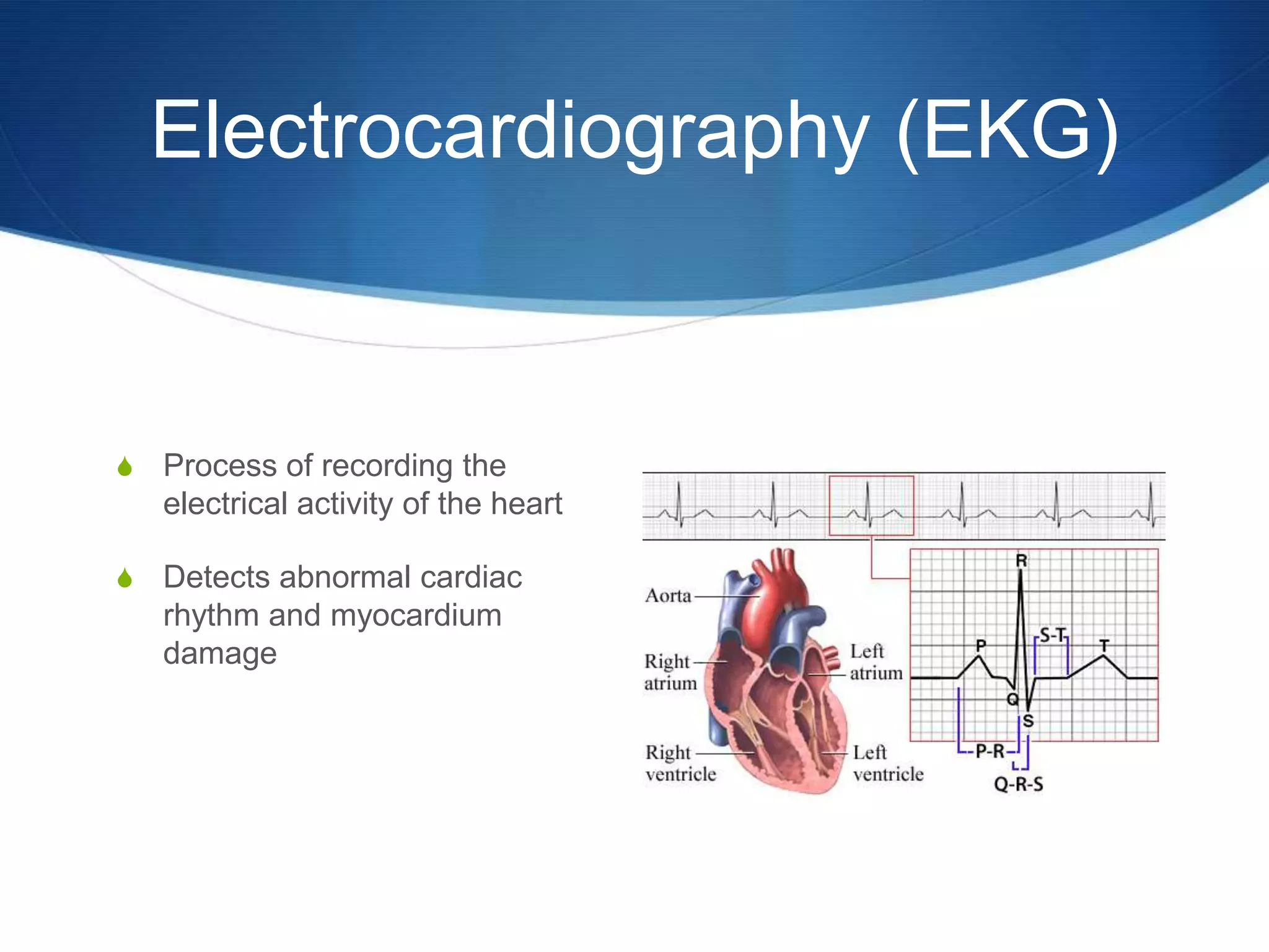
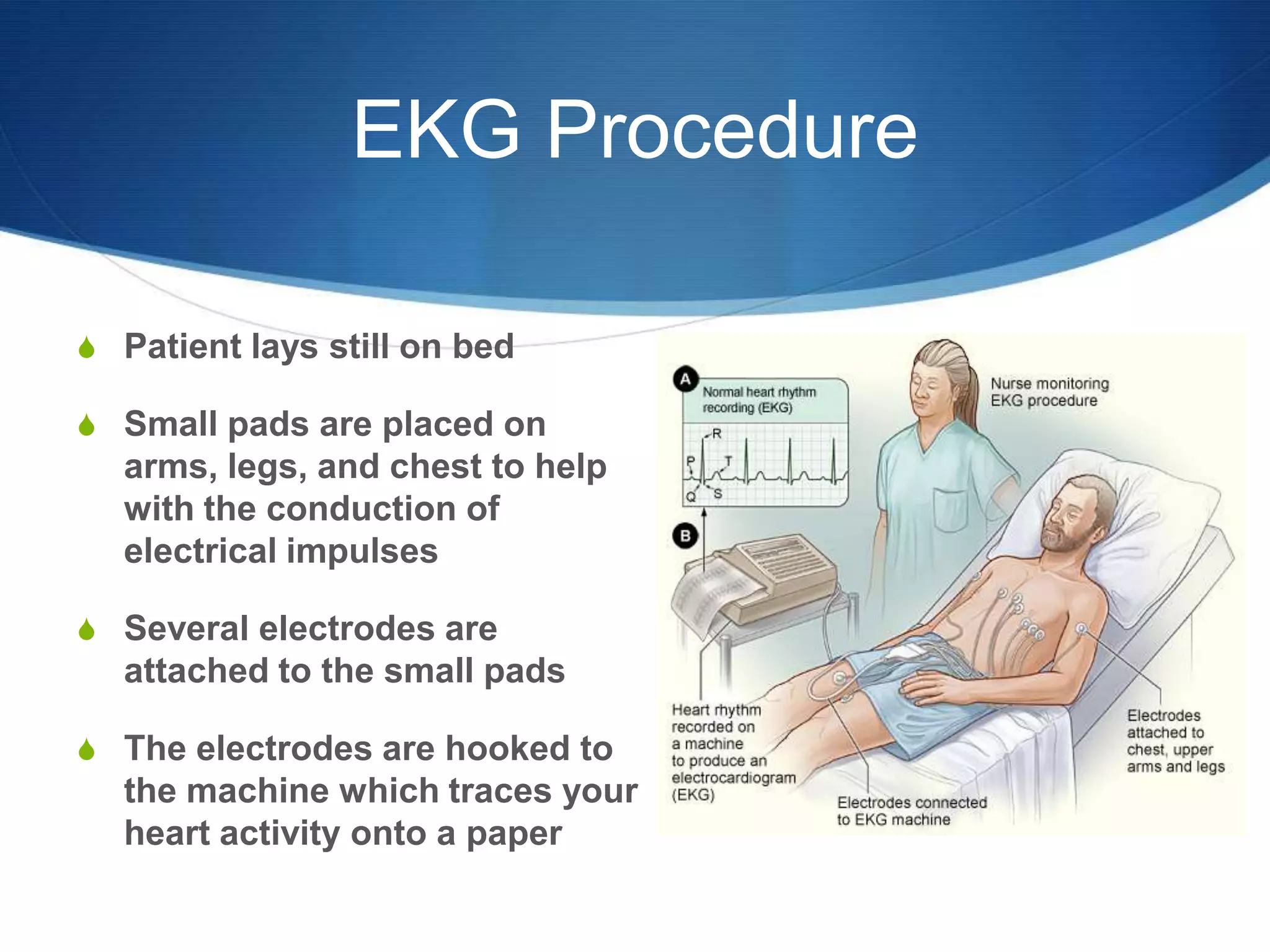
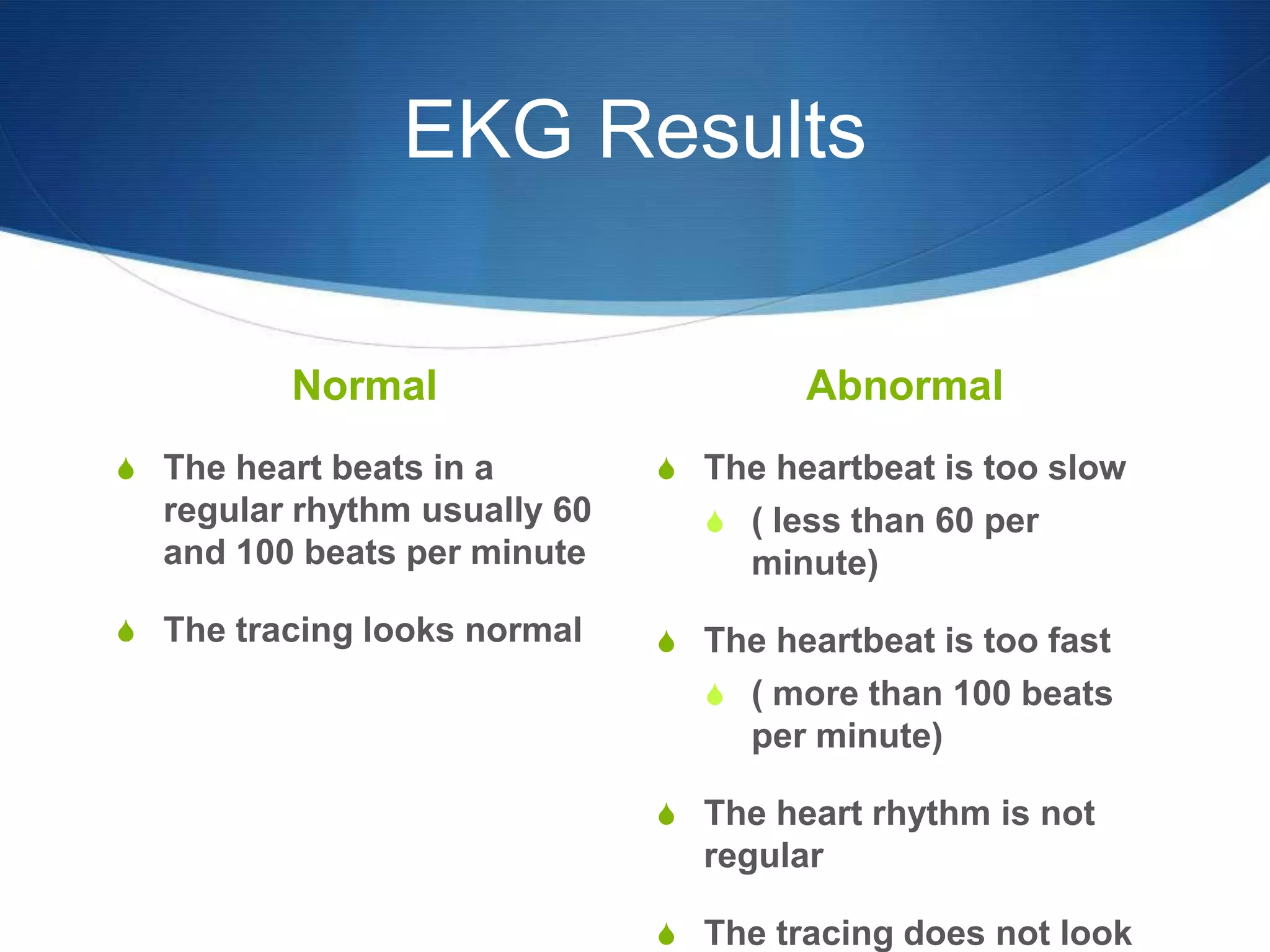
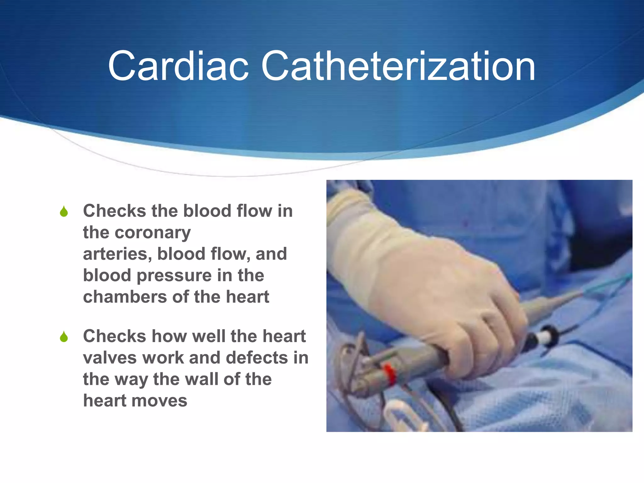
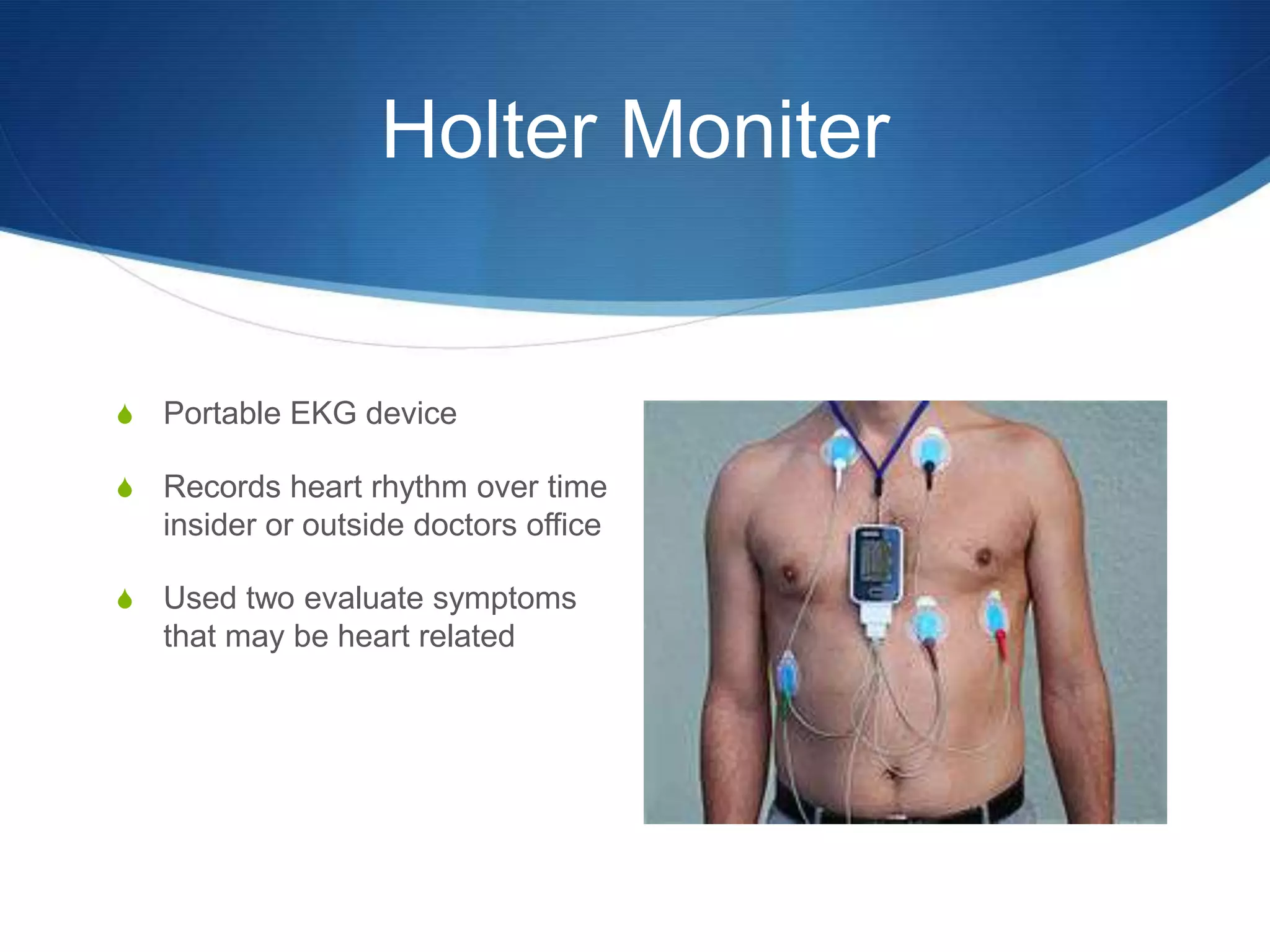
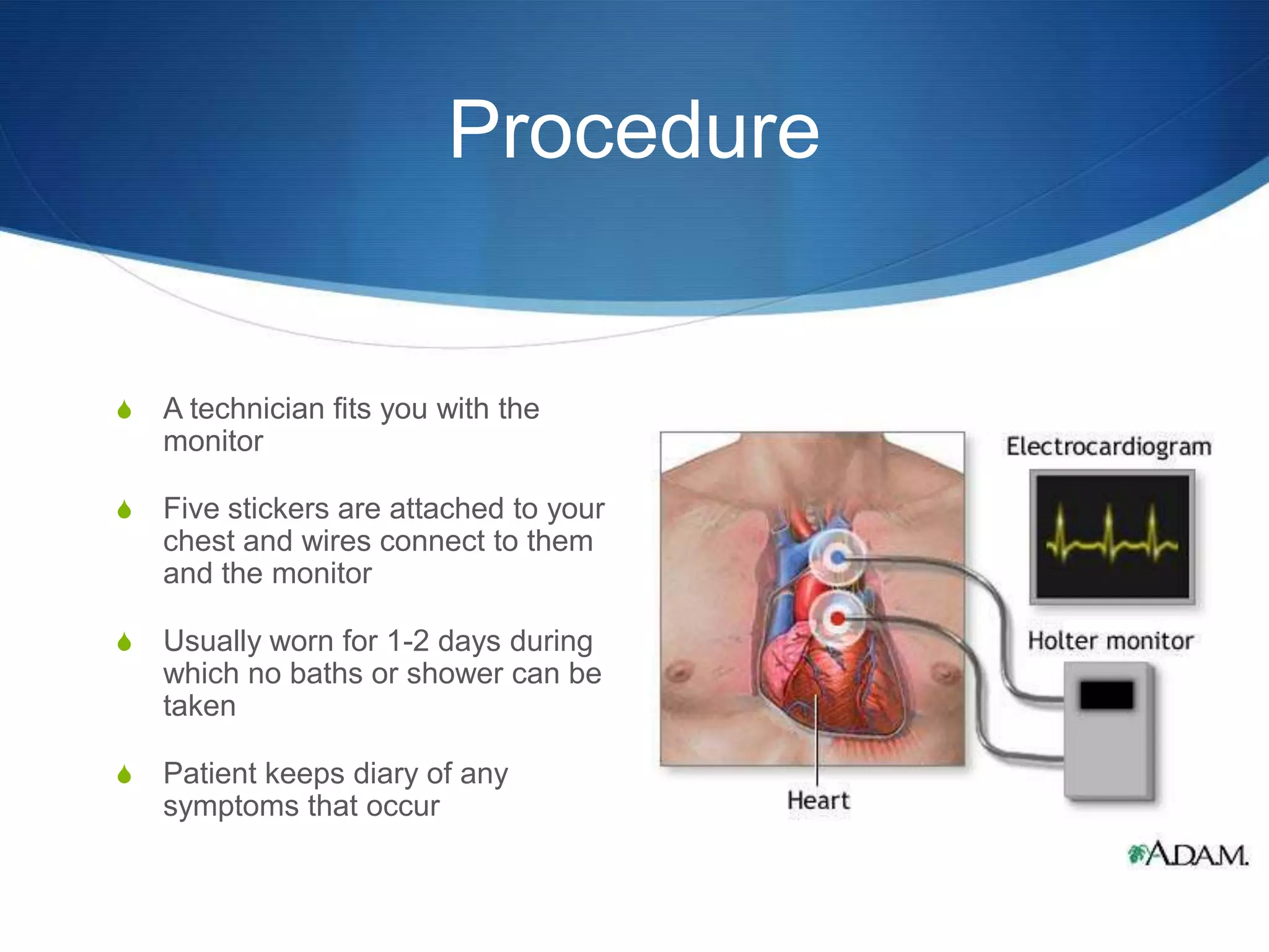
Cardiac Function Tests


            By: Krystal Roberson
                                    S
             By: Krystal Roberson
Electrocardiography (EKG)


S   Process of recording the
    electrical activity of the heart

S   Detects abnormal cardiac
    rhythm and myocardium
    damage
EKG Procedure

S Patient lays still on bed

S Small pads are placed on
  arms, legs, and chest to help
  with the conduction of
  electrical impulses

S Several electrodes are
  attached to the small pads

S The electrodes are hooked to
  the machine which traces your
  heart activity onto a paper
EKG Results

          Normal                      Abnormal
S The heart beats in a        S The heartbeat is too slow
  regular rhythm usually 60     S ( less than 60 per
  and 100 beats per minute         minute)
S The tracing looks normal    S The heartbeat is too fast
                                S ( more than 100 beats
                                   per minute)

                              S The heart rhythm is not
                                regular

                              S The tracing does not look
Cardiac Catheterization


S Checks the blood flow in
  the coronary
  arteries, blood flow, and
  blood pressure in the
  chambers of the heart

S Checks how well the heart
  valves work and defects in
  the way the wall of the
  heart moves
Procedure


S Patient lays still on table and are connected to an EKG
  machine so that heart activity is recorded during the test

S Sedative is given via IV line for relaxation

S Cardiac catheter is inserted in groin and advances to
  different parts of the hearts vessels and chambers

S Dye is injected into the heart and pictures of the arteries
  are taken as the dye moves through them
Results


S A cardiologist reviews the results

S The results will tell whether:
  S The coronary arteries are normal or have narrowing or
    blockage
  S The hearts pumping action and pressures are normal or
    abnormal
  S If the heart valves are working properly or not
Holter Moniter


S   Portable EKG device

S   Records heart rhythm over time
    insider or outside doctors office

S   Used two evaluate symptoms
    that may be heart related
Procedure


S   A technician fits you with the
    monitor

S   Five stickers are attached to your
    chest and wires connect to them
    and the monitor

S   Usually worn for 1-2 days during
    which no baths or shower can be
    taken

S   Patient keeps diary of any
    symptoms that occur
Results


S Doctor looks over the monitor recorder and what you’ve
  written in your activity diary

S The monitor may reveal a heart condition or your doctor
  may need to do more tests to find out what are causing
  the symptoms

Cardiac Function Test

  • 1.
    Cardiac Function Tests By: Krystal Roberson S By: Krystal Roberson
  • 2.
    Electrocardiography (EKG) S Process of recording the electrical activity of the heart S Detects abnormal cardiac rhythm and myocardium damage
  • 3.
    EKG Procedure S Patientlays still on bed S Small pads are placed on arms, legs, and chest to help with the conduction of electrical impulses S Several electrodes are attached to the small pads S The electrodes are hooked to the machine which traces your heart activity onto a paper
  • 4.
    EKG Results Normal Abnormal S The heart beats in a S The heartbeat is too slow regular rhythm usually 60 S ( less than 60 per and 100 beats per minute minute) S The tracing looks normal S The heartbeat is too fast S ( more than 100 beats per minute) S The heart rhythm is not regular S The tracing does not look
  • 5.
    Cardiac Catheterization S Checksthe blood flow in the coronary arteries, blood flow, and blood pressure in the chambers of the heart S Checks how well the heart valves work and defects in the way the wall of the heart moves
  • 6.
    Procedure S Patient laysstill on table and are connected to an EKG machine so that heart activity is recorded during the test S Sedative is given via IV line for relaxation S Cardiac catheter is inserted in groin and advances to different parts of the hearts vessels and chambers S Dye is injected into the heart and pictures of the arteries are taken as the dye moves through them
  • 7.
    Results S A cardiologistreviews the results S The results will tell whether: S The coronary arteries are normal or have narrowing or blockage S The hearts pumping action and pressures are normal or abnormal S If the heart valves are working properly or not
  • 8.
    Holter Moniter S Portable EKG device S Records heart rhythm over time insider or outside doctors office S Used two evaluate symptoms that may be heart related
  • 9.
    Procedure S A technician fits you with the monitor S Five stickers are attached to your chest and wires connect to them and the monitor S Usually worn for 1-2 days during which no baths or shower can be taken S Patient keeps diary of any symptoms that occur
  • 10.
    Results S Doctor looksover the monitor recorder and what you’ve written in your activity diary S The monitor may reveal a heart condition or your doctor may need to do more tests to find out what are causing the symptoms