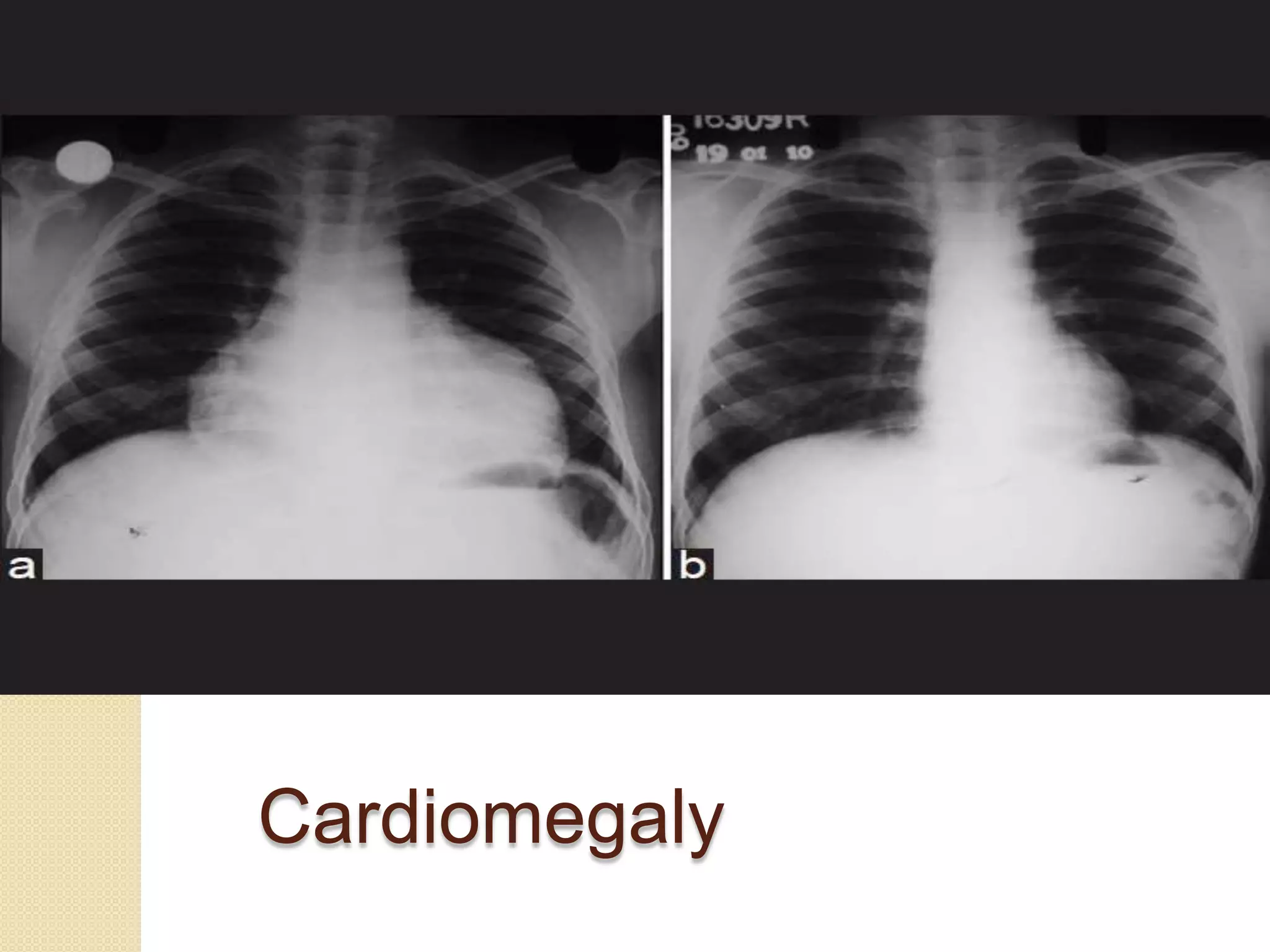
The document summarizes various cardiac function tests including electrocardiography (ECG), chest X-ray, cardiac enzyme tests, and coronary angiography. An ECG is a quick and easy way to assess heart function by studying electrical activities. Chest X-rays can detect abnormalities like heart enlargement. Cardiac enzyme tests like CK-MB and cardiac troponin detect heart muscle damage by measuring levels in blood. Coronary angiography uses dye and x-rays to examine blood flow through heart arteries.
![Certain factors or conditions may interfere
with or affect the results of the ECG.
Obesity
Pregnancy
Ascites (fluid buildup in the abdomen
[belly])
Anatomical considerations
Movement during the procedure
Exercise or smoking prior to the procedure
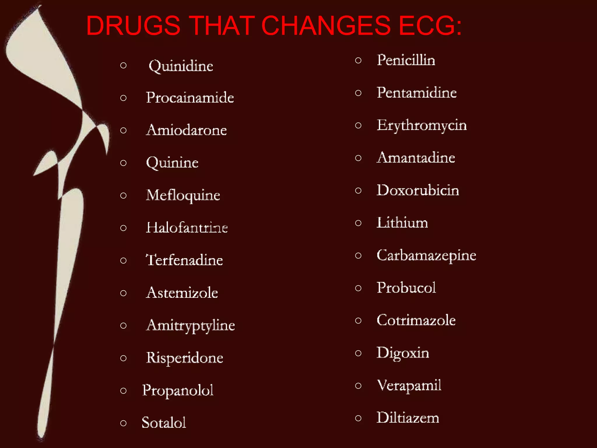
Certain medications
Electrolyte imbalances, such as too much
or too little potassium, magnesium, and/or
calcium in the blood.](https://image.slidesharecdn.com/cardiac-function-test-200207152925/75/Cardiac-function-test-8-2048.jpg)