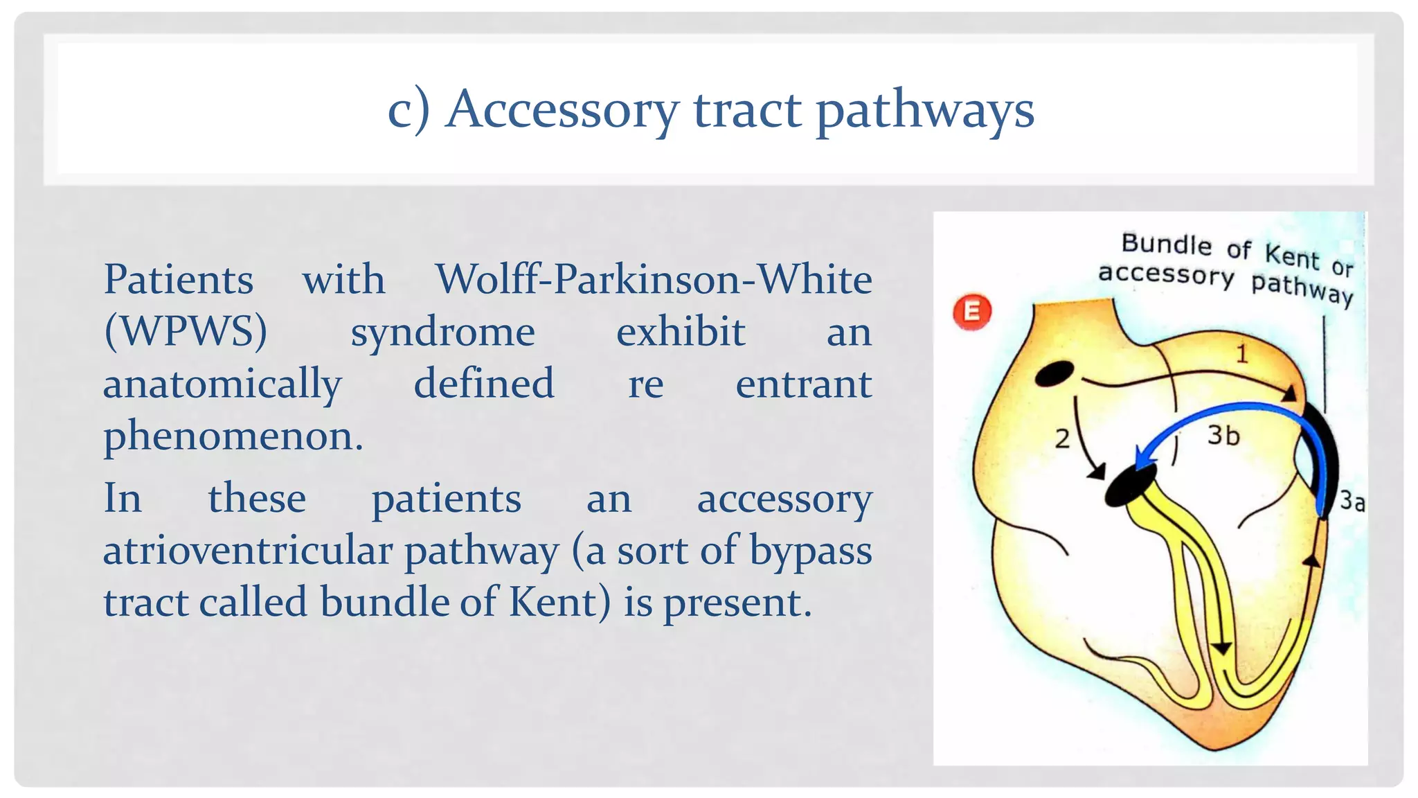
PH A R M ACO T H E R A PE U T I C S
CARDIAC ARRHYTHMIA-II
INTRODUCTION
• Cardiac arrhythmia refers to a group of conditions that
cause the heart to beat irregular, too slowly, or too quickly.
• There are several categories of arrhythmia, including:
bradycardia, or a slow heartbeat. tachycardia, or a fast
heartbeat. irregular heartbeat, also known as a flutter or
fibrillation.
GENESIS OF CARDIAC ARRHYTHMIAS
• The abnormalities in cardiac rhythm, and in the
conduction of cardiac impulse, may result in either
bradyarrhythmias or tachyarrhythmia.
• Bradyarrhythmias result from abnormality of SA node or
AV node. So can only be treated by cardiac pacemaker.
• Tachyarrhythmia can be satisfactorily managed by drugs.
THREE MECHANISMS
Cardiac arrhythmias, due to any reason, lead to
• Abnormal impulse generation,
• Triggered activity and/or
• Abnormal impulse conduction.
1) Abnormal Impulse Generation
• Abnormal impulse generation may stem from depressed
automaticity of SA node which leads to escape beats and
bradycardia.
• Conversely, there may be enhanced automaticity of SA
node leading to sinus tachycardia.
• AV node, Purkinje fibres, atrial and ventricular cells also
show spontaneous diastolic depolarisation and repetitive
firing, if their membrane potential comes to about -60 mV.
1) Abnormal Impulse Generation
• This leads to ectopic pacemakers in conducting system
causing AV nodal rhythm, idioventricular rhythm or
ectopic beats. (Ectopic foci)
• Such ectopic pacemaker activity is encouraged by:
(a) faster phase 4 depolarisation due to ischemic, digitalis,
Catecholamines, acidosis, hypokalaemia or stretching of
cardiac cells,
(b) less negative resting membrane potential or
(c) more negative threshold potential due to ischemic.
2) Triggered Activity (After-depolarization)
• Triggered activity (or after-depolarization) occur when a
normal AP triggers extra-abnormal depolarisations.
• These result from abnormal Ca+2 influxes into cardiac
myocyte during or immediately after phase 3 of the
ventricular AP and manifest as ventricular tachycardia.
• Two major forms of triggered rhythms are recognised:
"delayed after-depolarization (DAD)"
"early after-depolarisation (EAD)"
Delayed after Depolarization (DAD)
• Under the conditions of increased intracellular Ca+ ions,
as seen in digitalis intoxication, a normal AP may be
followed by a "delayed after depolarisation“.
• If this DAD reaches the threshold potential (TP) another
triggered beat (or beats) may occur.
Early after Depolarization (EAD)
• In this type of triggered activity, phase 3 (repolarisation) of
normal cardiac potential is interrupted (or plateau may be
prolonged).
• EADs markedly prolong cardiac repolarization phase and
therefore are associated with the development of
polymorphic ventricular tachycardia with a long QT
interval, known as torsade de pointes syndrome (French;
meaning 'twisting of the points‘).
3. Abnormal Impulse Conduction
• The third type of electrical disturbance of the heart
involves defects in impulse conduction which can result
from either one or combination of any of the three
mechanisms:
(a) conduction block,
(b) re-entry phenomenon and
(c) accessory tract path ways
a) Conduction Block
• Incomplete or complete block of conduction can lead to
partial heart block (dropped beats) or complete heart
block (idioventricular rhythm).
• Severely depressed conduction may result in AV nodal
block or His bundle branch block.
b) Re entry Phenomenon
(Circus movement of Rhythm)
It has been estimated
that 80 to 90% of
Clinical arrhythmias
occur due to re entry
phenomenon.
c) Accessory tract pathways
Patients with Wolff-Parkinson-White
(WPWS) syndrome exhibit an
anatomically defined re entrant
phenomenon.
In these patients an accessory
atrioventricular pathway (a sort of bypass
tract called bundle of Kent) is present.
Possible Mechanisms Of
Antiarrhythmic Drugs
A. Suppressing the enhanced automaticity of pacemaker or non-pacemaker
tissues by:
i) decreasing the rate of phase 0 depolarisation
ii) decreasing the slope of phase 0 depolarisation
iii) increasing the duration of Effective Refractory Period
iv) making resting membrane potential more negative
v) making threshold potential less negative.
The drugs which terminate cardiac arrhythmias may act by:
B. Abolishing re entry by further slowing the impulse conduction and
thus converting the unidirectional block to bidirectional block.
As implied, the anti - arrhythmic drugs can also prevent re entry by increasing
the ERP of cardiac fibres surrounding the region of re-entrant circuit.
Alternatively, the shorter the ERP in the depressed region, the less likely it is
that unidirectional block will occur.
Theoretically, accelerating the conduction, to make bidirectional conduction in
place of unidirectional block, would also be effective in combating arrhythmias
(see above). But no such drugs are available as yet.
Cardiac arrhythmias pathophysiology

Cardiac arrhythmias pathophysiology

  • 1.
    PH A RM ACO T H E R A PE U T I C S CARDIAC ARRHYTHMIA-II
  • 2.
    INTRODUCTION • Cardiac arrhythmiarefers to a group of conditions that cause the heart to beat irregular, too slowly, or too quickly. • There are several categories of arrhythmia, including: bradycardia, or a slow heartbeat. tachycardia, or a fast heartbeat. irregular heartbeat, also known as a flutter or fibrillation.
  • 3.
    GENESIS OF CARDIACARRHYTHMIAS • The abnormalities in cardiac rhythm, and in the conduction of cardiac impulse, may result in either bradyarrhythmias or tachyarrhythmia. • Bradyarrhythmias result from abnormality of SA node or AV node. So can only be treated by cardiac pacemaker. • Tachyarrhythmia can be satisfactorily managed by drugs.
  • 4.
    THREE MECHANISMS Cardiac arrhythmias,due to any reason, lead to • Abnormal impulse generation, • Triggered activity and/or • Abnormal impulse conduction.
  • 5.
    1) Abnormal ImpulseGeneration • Abnormal impulse generation may stem from depressed automaticity of SA node which leads to escape beats and bradycardia. • Conversely, there may be enhanced automaticity of SA node leading to sinus tachycardia. • AV node, Purkinje fibres, atrial and ventricular cells also show spontaneous diastolic depolarisation and repetitive firing, if their membrane potential comes to about -60 mV.
  • 6.
    1) Abnormal ImpulseGeneration • This leads to ectopic pacemakers in conducting system causing AV nodal rhythm, idioventricular rhythm or ectopic beats. (Ectopic foci) • Such ectopic pacemaker activity is encouraged by: (a) faster phase 4 depolarisation due to ischemic, digitalis, Catecholamines, acidosis, hypokalaemia or stretching of cardiac cells, (b) less negative resting membrane potential or (c) more negative threshold potential due to ischemic.
  • 7.
    2) Triggered Activity(After-depolarization) • Triggered activity (or after-depolarization) occur when a normal AP triggers extra-abnormal depolarisations. • These result from abnormal Ca+2 influxes into cardiac myocyte during or immediately after phase 3 of the ventricular AP and manifest as ventricular tachycardia. • Two major forms of triggered rhythms are recognised: "delayed after-depolarization (DAD)" "early after-depolarisation (EAD)"
  • 8.
    Delayed after Depolarization(DAD) • Under the conditions of increased intracellular Ca+ ions, as seen in digitalis intoxication, a normal AP may be followed by a "delayed after depolarisation“. • If this DAD reaches the threshold potential (TP) another triggered beat (or beats) may occur.
  • 9.
    Early after Depolarization(EAD) • In this type of triggered activity, phase 3 (repolarisation) of normal cardiac potential is interrupted (or plateau may be prolonged). • EADs markedly prolong cardiac repolarization phase and therefore are associated with the development of polymorphic ventricular tachycardia with a long QT interval, known as torsade de pointes syndrome (French; meaning 'twisting of the points‘).
  • 11.
    3. Abnormal ImpulseConduction • The third type of electrical disturbance of the heart involves defects in impulse conduction which can result from either one or combination of any of the three mechanisms: (a) conduction block, (b) re-entry phenomenon and (c) accessory tract path ways
  • 12.
    a) Conduction Block •Incomplete or complete block of conduction can lead to partial heart block (dropped beats) or complete heart block (idioventricular rhythm). • Severely depressed conduction may result in AV nodal block or His bundle branch block.
  • 13.
    b) Re entryPhenomenon (Circus movement of Rhythm) It has been estimated that 80 to 90% of Clinical arrhythmias occur due to re entry phenomenon.
  • 14.
    c) Accessory tractpathways Patients with Wolff-Parkinson-White (WPWS) syndrome exhibit an anatomically defined re entrant phenomenon. In these patients an accessory atrioventricular pathway (a sort of bypass tract called bundle of Kent) is present.
  • 15.
  • 16.
    A. Suppressing theenhanced automaticity of pacemaker or non-pacemaker tissues by: i) decreasing the rate of phase 0 depolarisation ii) decreasing the slope of phase 0 depolarisation iii) increasing the duration of Effective Refractory Period iv) making resting membrane potential more negative v) making threshold potential less negative. The drugs which terminate cardiac arrhythmias may act by:
  • 17.
    B. Abolishing reentry by further slowing the impulse conduction and thus converting the unidirectional block to bidirectional block. As implied, the anti - arrhythmic drugs can also prevent re entry by increasing the ERP of cardiac fibres surrounding the region of re-entrant circuit. Alternatively, the shorter the ERP in the depressed region, the less likely it is that unidirectional block will occur. Theoretically, accelerating the conduction, to make bidirectional conduction in place of unidirectional block, would also be effective in combating arrhythmias (see above). But no such drugs are available as yet.