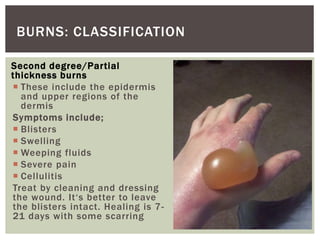
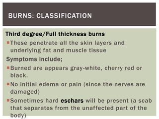
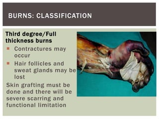
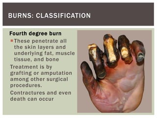
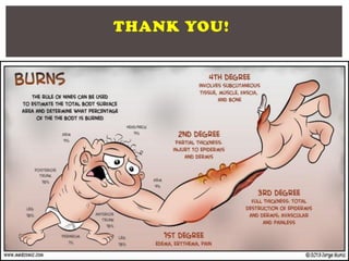
BURNSEVELYN CHARLOTTE NAKACHWA; BSN, MSN
BURNS: SKIN
Sensation
Thermoregulation
Protection
Excretion
Secretion
Vitamin D
production
Wound repair
Physical appearance
Absorption
Prevents
dehydration while
allowing a certain
level of water
evaporation to occur
Storage of fat
Pigmentation
BURNS: SKIN FUNCTIONS
 Scald burn: Most
frequent in homes from
hot liquids and foods. If
above 65 degrees, can
lead to cell death.
 Flame burn: Due to
gasoline, kerosene, and
other flammable gases.
 Chemical burn:
Common in industries
and labs and be due to
acids or alkalis
BURNS: KINDS
Electrical burn: Worse
that other kinds. It
has both entrance and
exit wounds. May stop
the heart and depress
the respiratory center.
May cause thrombosis
and cataracts.
Radiation burn: From
X-rays, radioactive
radiation and nuclear
bombs
First degree/Superficial
burns
 These are superficial burns
involving the epidermis e.g
sun burns
Symptoms include;
 Redness
 Mild tenderness
 Swelling
 Pain
Treat by cooling under tap
water and some analgesics.
Healing 3-6 days, no scarring
For sun burns, there’s risk of
skin cancer later in life
BURNS: CLASSIFICATION
Second degree/Partial
thickness burns
 These include the epidermis
and upper regions of the
dermis
Symptoms include;
 Blisters
 Swelling
 Weeping fluids
 Severe pain
 Cellulitis
Treat by cleaning and dressing
the wound. It‘s better to leave
the blisters intact. Healing is 7-
21 days with some scarring
BURNS: CLASSIFICATION
Third degree/Full thickness burns
These penetrate all the skin layers and
underlying fat and muscle tissue
Symptoms include;
Burned are appears gray-white, cherry red or
black.
No initial edema or pain (since the nerves are
damaged)
Sometimes hard eschars will be present (a scab
that separates from the unaffected part of the
body)
BURNS: CLASSIFICATION
Third degree/Full
thickness burns
 Contractures may
occur
 Hair follicles and
sweat glands may be
lost
Skin grafting must be
done and there will be
severe scarring and
functional limitation
BURNS: CLASSIFICATION
Fourth degree burn
These penetrate all
the skin layers and
underlying fat, muscle
tissue, and bone
Treatment is by
grafting or amputation
among other surgical
procedures.
Contractures and even
death can occur
BURNS: CLASSIFICATION
 When burned, proteins lose their shape they break
down leading to cell and tissue damage. This leads
to skin dysfunction like the ability to prevent water
loss through evaporation, temperature control and
sensation.
 Distraction of cell membranes cause loss of K+ to the
spaces outside the cells and uptake of Na+ and H2O
thus edema which is worsened by the inflammatory
response where there is increased fluid leakage from
the capillaries.
 This causes overall blood volume loss. The remaining
blood suffers significant plasma loss making blood
more concentrated leading to poor perfusion of
organs like kidneys (failure) and GIT (ulceration)
BURNS: PHYSIOLOGY
Rule of nines helps in showing
the percentage of the body
that is burned which is
essential in medical
management of burns
BURNS: RULE OF NINES
BURNS: ADMISSION IN ICU
 Total 2o and 3o burns >10% TBSA in patients <10
or >50yrs old
 Total 2o and 3o burns >20% TBSA in patients any
age.
 3o burns >5% TBSA in patients any age
 2o or 3o burns with a threat of serious functional or
cosmetic impairment (i.e face, hands, feet,
genitalia, perineum, major joints)
 Inhalation injury may lead to respiratory distress
 Electrical burns since internal injury may ne
underestimated by TBSA
 Burns associated with major trauma
BURNS: MANAGEMENT OF BURNS
Burn care must be planned according to burn
depth and local response, the extent of the
injury, and the presence of systemic response.
Burn care then proceeds through three
phases;
a) Emergent/Resuscitative phase
b) Acute/intermediate phase
c) Rehabilitation phase
Although priorities exist for each phase, these
phases can overlap.
BURNS: MANAGEMENT OF BURNS
EMERGENT/RESUSCITATIVE PHASE:
Starts from onset of injury to completion of
fluid resuscitation
Do the ABCs
Assess for inhalation injury
Initial wound care
 Management of fluid loss and shock
Signs and symptoms
Hoarseness
Lacrimation/Conjun
ctivitis
Brassy cough
Carbonaceous
mucus
Facial burns
Singed nasal hairs
Wheezing
Stridor
Bronchorrhea
(secretion of mucus
by the bronchi)
Dyspnea
Anxiety
Disorientation
Obtundation/Coma
BURNS: INHALATION INJURY
BURNS: FLUID RESUSCITATION
In first 24hrs, use Isotonic Crystalloids like
Hartmann's fluid (compound sodium lactate ~
CSL), Normal saline and Ringer’s lactate.
These pass through capillary walls easily
In the next 30-48hrs, use colloids plasma,
Dextrans, Haemaccel to compensate plasma
loss
After 48hrs, blood transfusion can be done.
BURNS: PARKALAND’S FORMULAR FOR
FLUID RESUSCITATION
For the first 24hrs:
4mls X degree of burns X Kgs = Volume
(mls)
 For the first 8hrs, half of the volume has
to be given
For the next 16hrs, give the rest of the fluid
volume
For the next 24hrs, only half of the first day
fluids volume is given.
BURNS: PARKALAND’S FORMULAR FOR
FLUID RESUSCITATION
 But children require additional
maintenance fluid that included
glucose.
 Time is determined from when the
burn occurred and not from when the
fluid resuscitation started.
 Remember that over fluid
resuscitation is equally detrimental
BURNS: EMERGENCY ESCHAROTOMY
Full thickness burns result in formation of eschar
(tough, inelastic mass of burnt tissue)
Due to the inelasticity, this results in burn-
induced compartment syndrome/torniquet effect.
There is accumulation of extracellullar and
extravascular fluids within confined anatomical
spaces.
This causes an increase in intracompartmental
pressure resulting in to collapse of contained
vascular and lymphatic structures and hence
loss of tissue viability/tissue death.
BURNS: EMERGENCY ESCHAROTOMY
Indications of emergency echarotomy
Presence of circumferential eschar with one of
the following;
o Impending or established vascular compromise
to the extremities
o Impending or established respiratory
compromise due to circumferential torso burns
 The incision must avoid all major nerves,
vessels, and all tendons and should extend
through the eschar down to the subcutaneous
fat.
 Escharotomy is rarely required within the first
6hrs post burn.
BURNS: EMERGENCY ESCHAROTOMY
Monitoring from compartment syndrome
Neurovascular integrity should be monitored
frequently
Capillary refill time, Doppler signals, Pulse
oximetry, Sensation distal to the burned area
should be checked every hour
Limb deep compartment pressures should be
checked initially to establish baseline.
BURNS: MANAGEMENT OF BURNS
ACUTE/INTERMEDIATE PHASE:
This marks the beginning of diuresis to near
completion of wound closure.
There’s maintenance of respiratory and
circulatory status, fluid and electrolyte
balance, GI function
Infection prevention, wound care, pain
management, and nutritional support
BURNS: ACUTE/INTERMEDIATE PHASE
Re-evaluate
 Give tetanus prophylaxis to a patient who
has not received one in the last 5 years
 Remove all clothing and rings
 Monitor fluid output and note urine color
 Labs: Hematocrit, ABG, Electrolytes, ECG,
Urea/Creatinine, Urine microscopy
 Chemical burns require extensive irrigation
BURNS: APPLICATION OF TOPICAL
AGENTS
There are two methods of management of burn
wounds with topical agents like Silver
Sulfadiazine/Mebo;
Exposure therapy:
 No dressing are applied over the wound after
application of a topical agent to the wound
twice or thrice daily. This approach is used
on face and head
 But there is increase in pain and heat loss
and risk of cross infection.
BURNS: APPLICATION OF TOPICAL
AGENTS
Closed management
 An occlusive dressing is applied over the agent
and is usually changed twice daily e.g Vaseline
gauze
 There is less pain, less heat loss and less cross
contamination
 But there is a potential for increased bacterial
growth if dressing is not changed twice daily
especially when there is thick eschar.
This is the preferred method in application of
topical agents
BURNS: ACUTE/INTERMEDIATE PHASE
Pain management
 First assess how much analgesia the patient
has receive before admission and confirm
the pain scale and do history check whether
patient uses illicit drugs or alcohol
 No intramuscular, subcutaneous injections
 Small burns can use paracetamol, NSAIDS
 Large burns can use I.V opiates like
Morphine + Paracetamol
BURNS: ACUTE/INTERMEDIATE PHASE
Pain management
 Use sedatives like Midazolam, Ketamine
 Administer antihistamines
 Anti-emetics are necessary when narcotics
are given
 Benzodiazepines help with anxiety
BURNS: ACUTE/INTERMEDIATE PHASE
Infection prevention
 Burns patients are immunocompromised
 Sterile precautions must be rigorous
 All patients who have facial burns with not dressing,
exudating wounds, inhalation injury or burns >20%
TBSA, should be nursed in isolation rooms to reduce
the risk of infection from cross contamination.
 Use protective gear for all producers
 Keep door closed at all times
 All procedures should be sterile
 Swabs should be taken regularly
 Assess blisters for potential infection
 Sometimes blisters are used as biological dressing if
there is acceptable capillary refill time in the wound
bed
BURNS: ACUTE/INTERMEDIATE PHASE
Prevent Hypothermia
 Skin loss and burn exposure impairs the body’s
ability to regulate body temperature
 Patients with major burns readily lose body heat
 Increase room temperature
 No burns patient should have a warming
blanket/Bair hugger directly over the burns or
dressings. Use a space blanket
 Use warm IV fluids
 Cover patients head if no facial burns
 Keep patient warm during all procedures
BURNS: ACUTE/INTERMEDIATE PHASE
Debriding burn wounds
 Debridement reduces bacterial colonization and
ensures a moist environment for cell growth
 Use sterile equipment
 Leave small blisters intact
 Aspirate or debride large blisters
 Debride all blisters if burn is from a chemical
 Debride blisters if over a joint
 Debride all loose skin if blisters burst
 Cease debriding is bleeding occurs
BURNS: ACUTE/INTERMEDIATE PHASE
Positioning
 Use splints to reduce contractures
 No pillow under head to reduce risk of neck
and ear contractures
 Elevate edematous limbs
 Position foot at 900 to prevent Achilles's
tendon shortening
BURNS: ACUTE/INTERMEDIATE PHASE
Nutrition
 Burn patients have hypermetabolism, increased
oxygen consumption, negative nitrogen balance,
high caloric requirements and weight loss
further associated with delayed wound healing,
cellular dysfunction and decreased immune
competence. Therefore, burns patients need
extra feeding
 RBS monitoring
 Nutritional requirements are still debatable but
a diet rich in proteins is very essential.
 READ: HARRIS-BENEDICT’S EQUATION FOR
BASAL ENERGY ESTIMATES
BURNS: ACUTE/INTERMEDIATE PHASE
Bed bath
 Ensure patient has analgesia before
procedure
 Use warm clean water
 Use Chlorhexidine 0.05%
 Avoid washing too hard to prevent pain,
bleeding or injuring healing skin
 Wash firmly enough to remove any debris.
Loose skin, exudate, and applied creams.
BURNS: ACUTE/INTERMEDIATE PHASE
Areas requiring specific management
 Facial burns: Maintain airway, eye care, dressing,
elevation, plastic surgeon’s review
 Eye care: Apply TEO or cover eyes, Clean with N/S
regularly, Trim eyelashes if edematous to prevent
corneal abrasions
 Ear care: Place sterile gauze between ear and head to
prevent webbing, position head up to minimize swelling
 Hand and finger care: Elevation, splint and optimize
dressing
 Perineal care: Remove all hairs, dressing must be
changed promptly of contaminated by urine of feces
 Circumferential care: Apply loose bandage to ensure
there is no constriction of the limb, Elevation
 Splinting burns
BURNS: MANAGEMENT OF BURNS
REHABILITATION PHASE:
Begins from wound closure to optimal
functional level
Wound healing
Restoring maximal functional activity
Alteration in self-image and lifestyle
BURNS: MANAGEMENT OF BURNS
BURNS: COMPLICATIONS
 Burn shock due to coagulation necrosis of tissue initiating
multiple organ response. There’s increased capillary
permeability, evaporative water loss and thus hypovolemic
shock
 Pulmonary distress due to inhalation injury
 Acute renal failure
 Infections and sepsis
 Keloids
 Clot formation
 PTSD
 Social isolation
 Curling’s ulcer in large urns over 30% usually after the 9th day
 Extensive and disabling scarring
 Psychological trauma
 Squamous cell carcinoma called Marjolin’s ulcer, may take up to
21yrs to develop.
MARJOLIN’S
ULCER; a
SQUAMOUS
CELL
CARCINOMA
BURNS:
COMPLICATIONS
THANK YOU!

BURNS

  • 1.
  • 2.
  • 3.
    Sensation Thermoregulation Protection Excretion Secretion Vitamin D production Wound repair Physicalappearance Absorption Prevents dehydration while allowing a certain level of water evaporation to occur Storage of fat Pigmentation BURNS: SKIN FUNCTIONS
  • 4.
     Scald burn:Most frequent in homes from hot liquids and foods. If above 65 degrees, can lead to cell death.  Flame burn: Due to gasoline, kerosene, and other flammable gases.  Chemical burn: Common in industries and labs and be due to acids or alkalis BURNS: KINDS Electrical burn: Worse that other kinds. It has both entrance and exit wounds. May stop the heart and depress the respiratory center. May cause thrombosis and cataracts. Radiation burn: From X-rays, radioactive radiation and nuclear bombs
  • 5.
    First degree/Superficial burns  Theseare superficial burns involving the epidermis e.g sun burns Symptoms include;  Redness  Mild tenderness  Swelling  Pain Treat by cooling under tap water and some analgesics. Healing 3-6 days, no scarring For sun burns, there’s risk of skin cancer later in life BURNS: CLASSIFICATION
  • 6.
    Second degree/Partial thickness burns These include the epidermis and upper regions of the dermis Symptoms include;  Blisters  Swelling  Weeping fluids  Severe pain  Cellulitis Treat by cleaning and dressing the wound. It‘s better to leave the blisters intact. Healing is 7- 21 days with some scarring BURNS: CLASSIFICATION
  • 7.
    Third degree/Full thicknessburns These penetrate all the skin layers and underlying fat and muscle tissue Symptoms include; Burned are appears gray-white, cherry red or black. No initial edema or pain (since the nerves are damaged) Sometimes hard eschars will be present (a scab that separates from the unaffected part of the body) BURNS: CLASSIFICATION
  • 8.
    Third degree/Full thickness burns Contractures may occur  Hair follicles and sweat glands may be lost Skin grafting must be done and there will be severe scarring and functional limitation BURNS: CLASSIFICATION
  • 9.
    Fourth degree burn Thesepenetrate all the skin layers and underlying fat, muscle tissue, and bone Treatment is by grafting or amputation among other surgical procedures. Contractures and even death can occur BURNS: CLASSIFICATION
  • 10.
     When burned,proteins lose their shape they break down leading to cell and tissue damage. This leads to skin dysfunction like the ability to prevent water loss through evaporation, temperature control and sensation.  Distraction of cell membranes cause loss of K+ to the spaces outside the cells and uptake of Na+ and H2O thus edema which is worsened by the inflammatory response where there is increased fluid leakage from the capillaries.  This causes overall blood volume loss. The remaining blood suffers significant plasma loss making blood more concentrated leading to poor perfusion of organs like kidneys (failure) and GIT (ulceration) BURNS: PHYSIOLOGY
  • 11.
    Rule of nineshelps in showing the percentage of the body that is burned which is essential in medical management of burns BURNS: RULE OF NINES
  • 12.
  • 13.
  • 14.
    BURNS: ADMISSION INICU  Total 2o and 3o burns >10% TBSA in patients <10 or >50yrs old  Total 2o and 3o burns >20% TBSA in patients any age.  3o burns >5% TBSA in patients any age  2o or 3o burns with a threat of serious functional or cosmetic impairment (i.e face, hands, feet, genitalia, perineum, major joints)  Inhalation injury may lead to respiratory distress  Electrical burns since internal injury may ne underestimated by TBSA  Burns associated with major trauma
  • 15.
    BURNS: MANAGEMENT OFBURNS Burn care must be planned according to burn depth and local response, the extent of the injury, and the presence of systemic response. Burn care then proceeds through three phases; a) Emergent/Resuscitative phase b) Acute/intermediate phase c) Rehabilitation phase Although priorities exist for each phase, these phases can overlap.
  • 16.
    BURNS: MANAGEMENT OFBURNS EMERGENT/RESUSCITATIVE PHASE: Starts from onset of injury to completion of fluid resuscitation Do the ABCs Assess for inhalation injury Initial wound care  Management of fluid loss and shock
  • 17.
    Signs and symptoms Hoarseness Lacrimation/Conjun ctivitis Brassycough Carbonaceous mucus Facial burns Singed nasal hairs Wheezing Stridor Bronchorrhea (secretion of mucus by the bronchi) Dyspnea Anxiety Disorientation Obtundation/Coma BURNS: INHALATION INJURY
  • 19.
    BURNS: FLUID RESUSCITATION Infirst 24hrs, use Isotonic Crystalloids like Hartmann's fluid (compound sodium lactate ~ CSL), Normal saline and Ringer’s lactate. These pass through capillary walls easily In the next 30-48hrs, use colloids plasma, Dextrans, Haemaccel to compensate plasma loss After 48hrs, blood transfusion can be done.
  • 20.
    BURNS: PARKALAND’S FORMULARFOR FLUID RESUSCITATION For the first 24hrs: 4mls X degree of burns X Kgs = Volume (mls)  For the first 8hrs, half of the volume has to be given For the next 16hrs, give the rest of the fluid volume For the next 24hrs, only half of the first day fluids volume is given.
  • 21.
    BURNS: PARKALAND’S FORMULARFOR FLUID RESUSCITATION  But children require additional maintenance fluid that included glucose.  Time is determined from when the burn occurred and not from when the fluid resuscitation started.  Remember that over fluid resuscitation is equally detrimental
  • 22.
    BURNS: EMERGENCY ESCHAROTOMY Fullthickness burns result in formation of eschar (tough, inelastic mass of burnt tissue) Due to the inelasticity, this results in burn- induced compartment syndrome/torniquet effect. There is accumulation of extracellullar and extravascular fluids within confined anatomical spaces. This causes an increase in intracompartmental pressure resulting in to collapse of contained vascular and lymphatic structures and hence loss of tissue viability/tissue death.
  • 23.
    BURNS: EMERGENCY ESCHAROTOMY Indicationsof emergency echarotomy Presence of circumferential eschar with one of the following; o Impending or established vascular compromise to the extremities o Impending or established respiratory compromise due to circumferential torso burns  The incision must avoid all major nerves, vessels, and all tendons and should extend through the eschar down to the subcutaneous fat.  Escharotomy is rarely required within the first 6hrs post burn.
  • 27.
    BURNS: EMERGENCY ESCHAROTOMY Monitoringfrom compartment syndrome Neurovascular integrity should be monitored frequently Capillary refill time, Doppler signals, Pulse oximetry, Sensation distal to the burned area should be checked every hour Limb deep compartment pressures should be checked initially to establish baseline.
  • 28.
    BURNS: MANAGEMENT OFBURNS ACUTE/INTERMEDIATE PHASE: This marks the beginning of diuresis to near completion of wound closure. There’s maintenance of respiratory and circulatory status, fluid and electrolyte balance, GI function Infection prevention, wound care, pain management, and nutritional support
  • 29.
    BURNS: ACUTE/INTERMEDIATE PHASE Re-evaluate Give tetanus prophylaxis to a patient who has not received one in the last 5 years  Remove all clothing and rings  Monitor fluid output and note urine color  Labs: Hematocrit, ABG, Electrolytes, ECG, Urea/Creatinine, Urine microscopy  Chemical burns require extensive irrigation
  • 30.
    BURNS: APPLICATION OFTOPICAL AGENTS There are two methods of management of burn wounds with topical agents like Silver Sulfadiazine/Mebo; Exposure therapy:  No dressing are applied over the wound after application of a topical agent to the wound twice or thrice daily. This approach is used on face and head  But there is increase in pain and heat loss and risk of cross infection.
  • 32.
    BURNS: APPLICATION OFTOPICAL AGENTS Closed management  An occlusive dressing is applied over the agent and is usually changed twice daily e.g Vaseline gauze  There is less pain, less heat loss and less cross contamination  But there is a potential for increased bacterial growth if dressing is not changed twice daily especially when there is thick eschar. This is the preferred method in application of topical agents
  • 33.
    BURNS: ACUTE/INTERMEDIATE PHASE Painmanagement  First assess how much analgesia the patient has receive before admission and confirm the pain scale and do history check whether patient uses illicit drugs or alcohol  No intramuscular, subcutaneous injections  Small burns can use paracetamol, NSAIDS  Large burns can use I.V opiates like Morphine + Paracetamol
  • 34.
    BURNS: ACUTE/INTERMEDIATE PHASE Painmanagement  Use sedatives like Midazolam, Ketamine  Administer antihistamines  Anti-emetics are necessary when narcotics are given  Benzodiazepines help with anxiety
  • 35.
    BURNS: ACUTE/INTERMEDIATE PHASE Infectionprevention  Burns patients are immunocompromised  Sterile precautions must be rigorous  All patients who have facial burns with not dressing, exudating wounds, inhalation injury or burns >20% TBSA, should be nursed in isolation rooms to reduce the risk of infection from cross contamination.  Use protective gear for all producers  Keep door closed at all times  All procedures should be sterile  Swabs should be taken regularly  Assess blisters for potential infection  Sometimes blisters are used as biological dressing if there is acceptable capillary refill time in the wound bed
  • 36.
    BURNS: ACUTE/INTERMEDIATE PHASE PreventHypothermia  Skin loss and burn exposure impairs the body’s ability to regulate body temperature  Patients with major burns readily lose body heat  Increase room temperature  No burns patient should have a warming blanket/Bair hugger directly over the burns or dressings. Use a space blanket  Use warm IV fluids  Cover patients head if no facial burns  Keep patient warm during all procedures
  • 37.
    BURNS: ACUTE/INTERMEDIATE PHASE Debridingburn wounds  Debridement reduces bacterial colonization and ensures a moist environment for cell growth  Use sterile equipment  Leave small blisters intact  Aspirate or debride large blisters  Debride all blisters if burn is from a chemical  Debride blisters if over a joint  Debride all loose skin if blisters burst  Cease debriding is bleeding occurs
  • 38.
    BURNS: ACUTE/INTERMEDIATE PHASE Positioning Use splints to reduce contractures  No pillow under head to reduce risk of neck and ear contractures  Elevate edematous limbs  Position foot at 900 to prevent Achilles's tendon shortening
  • 39.
    BURNS: ACUTE/INTERMEDIATE PHASE Nutrition Burn patients have hypermetabolism, increased oxygen consumption, negative nitrogen balance, high caloric requirements and weight loss further associated with delayed wound healing, cellular dysfunction and decreased immune competence. Therefore, burns patients need extra feeding  RBS monitoring  Nutritional requirements are still debatable but a diet rich in proteins is very essential.  READ: HARRIS-BENEDICT’S EQUATION FOR BASAL ENERGY ESTIMATES
  • 40.
    BURNS: ACUTE/INTERMEDIATE PHASE Bedbath  Ensure patient has analgesia before procedure  Use warm clean water  Use Chlorhexidine 0.05%  Avoid washing too hard to prevent pain, bleeding or injuring healing skin  Wash firmly enough to remove any debris. Loose skin, exudate, and applied creams.
  • 41.
    BURNS: ACUTE/INTERMEDIATE PHASE Areasrequiring specific management  Facial burns: Maintain airway, eye care, dressing, elevation, plastic surgeon’s review  Eye care: Apply TEO or cover eyes, Clean with N/S regularly, Trim eyelashes if edematous to prevent corneal abrasions  Ear care: Place sterile gauze between ear and head to prevent webbing, position head up to minimize swelling  Hand and finger care: Elevation, splint and optimize dressing  Perineal care: Remove all hairs, dressing must be changed promptly of contaminated by urine of feces  Circumferential care: Apply loose bandage to ensure there is no constriction of the limb, Elevation  Splinting burns
  • 42.
    BURNS: MANAGEMENT OFBURNS REHABILITATION PHASE: Begins from wound closure to optimal functional level Wound healing Restoring maximal functional activity Alteration in self-image and lifestyle
  • 43.
  • 44.
    BURNS: COMPLICATIONS  Burnshock due to coagulation necrosis of tissue initiating multiple organ response. There’s increased capillary permeability, evaporative water loss and thus hypovolemic shock  Pulmonary distress due to inhalation injury  Acute renal failure  Infections and sepsis  Keloids  Clot formation  PTSD  Social isolation  Curling’s ulcer in large urns over 30% usually after the 9th day  Extensive and disabling scarring  Psychological trauma  Squamous cell carcinoma called Marjolin’s ulcer, may take up to 21yrs to develop.
  • 45.
  • 46.