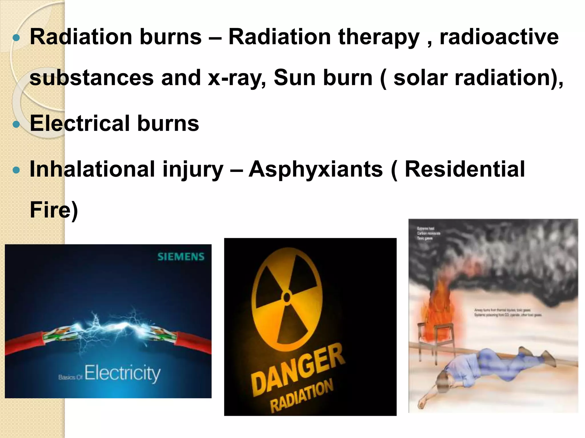
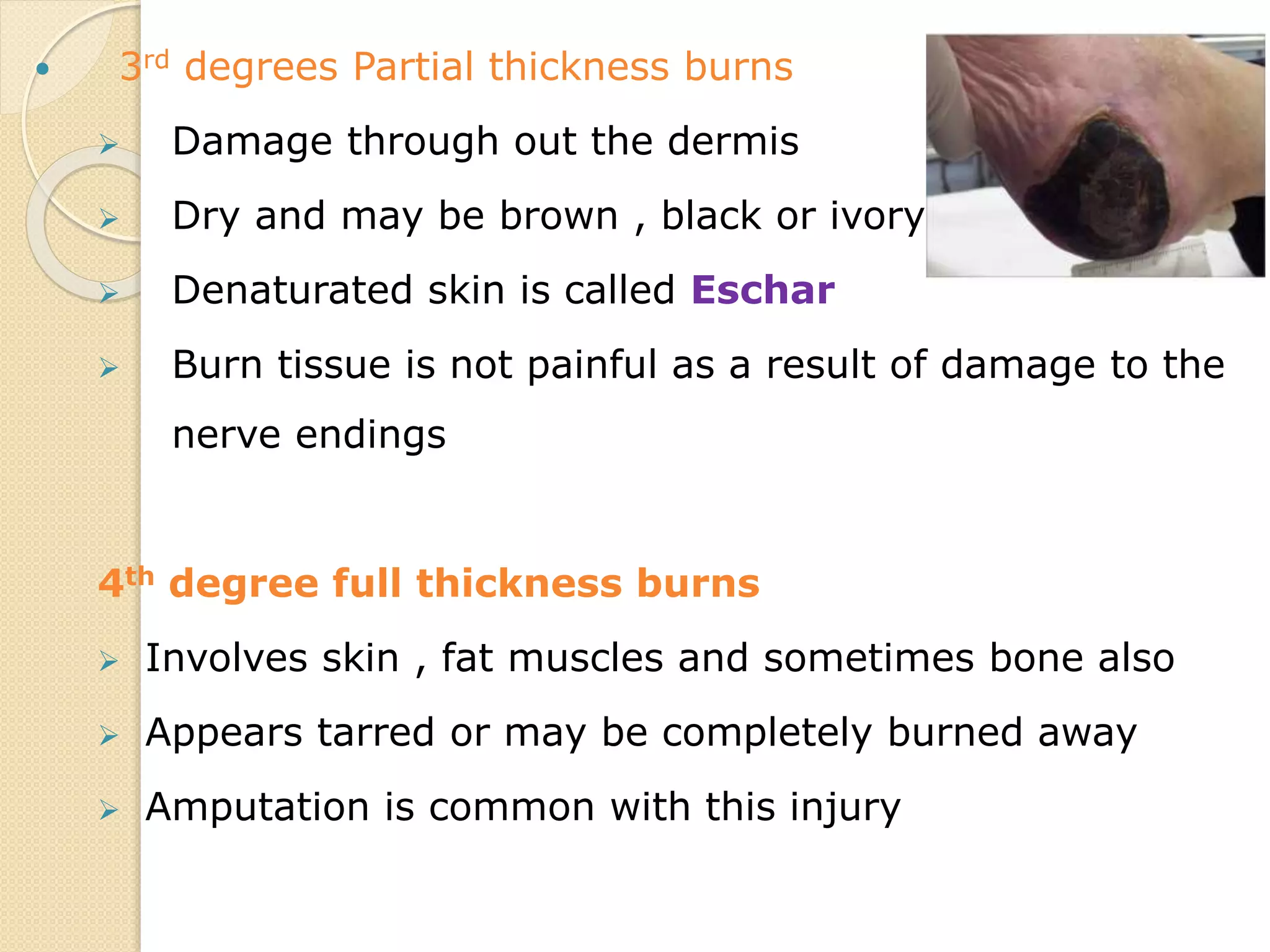
BURNS AND ITS MANAGEMENT
PROF. Dr.PUVANESWARI. K
NHCON
BANAGALORE
Anatomy of skin
Functions of Skin
 Skin is the largest organ of the body
 Essential for:
- Thermoregulation
- Prevention of fluid loss by evaporation
- Barrier against infection
- Protection against environment
provided by sensory information
Burn and Scalds
 Burn
A burn is a type of injury
results from direct contact or
exposure to thermal , electrical,
chemical or radiation source are
termed Burns.
 Scalds
Injuries results from moist heat
are termed as scalds
ETIOLOGY
 Thermal burns
 Chemical burns
 Radiation burns
 Electrical burns
 Inhalational injury
 Thermal burns – Flame , hot liquid, semi
liquids , residential fires , explosion
 Chemical burns- Acid, alkali or organic
compounds
 Radiation burns – Radiation therapy , radioactive
substances and x-ray, Sun burn ( solar radiation),
 Electrical burns
 Inhalational injury – Asphyxiants ( Residential
Fire)
PATHOPHYSIOLOGY- Skin
Direct injury to skin devitalises the cells
( 40 O-44OC)
Cellular system Infarction
Sodium Potassium pump fails
Cellular edema
3 Zones of tissue injury
 Zone of Coagulation – Directly damaged
skin is coagulated and fully destroyed (
Inner)
 Zone of stasis- Surrounding tissue exposed
to heat is edematous and has impaired
blood flow ( Middle zone)
 Zone of Hyperemia- It consists of the tissue
that is inflammed and vasodilated ( Outer)
Pathophysiology - Fluid shifts
Following burn injury
Release of vasoactive substances ( histamine, kinins
catacholamines ,serotonin , leukotrins, prostaglandins)
Alters cell permeability( Na enters the cell and K exits the
cell)
Increases intercellualr and interstitial fluid further deplets
intra vascular fluid volume
Hypovolemia
Hypovolemia
Vital organs gets lack of blood supply
Decreased Blood supply to mesentric bed →
Intestitial ileus → Curling’s ulcer
Decreased renal blood → Oliguria
flow (Renal failure)
Toxins released from the wound along with sepsis
causes acute tubular necrosis.
Myoglobin released from muscles (in case of electric
injury or often from Eschar) is most injurious to
kidneys.
Pulmonary system
Inhalational injury by exposure to asphyxiants
Oxygen molecule are displaced and combined
of Hb to form carboxy haemoglobin ( CO
have 200 times more affinity towards Hb than
O2)
Injury to URT Leads to Erythema, ulceration ,
edema etc
 Altered pulmonary resistance causing
pulmonary edema
Myo cardial depression
Liberation of Myocardial depressant factor
Decreased cardiac → Decreased
myocardial
output function
Impaired skin integrity
 Disruption of skin nerve endings , sweat glands
and hair follicles
 Barrier function of skin is last
Immuno supression
 Decreased Lymphocyte activity , Decrease in
immunoglobulin production ,suppression of
complement activity and an alteration of neutrophil
and macrophages function
 Increase risk of infection
Metabolic
 Hyper metabolic rate (BMR).
 Negative nitrogen balance.
 Electrolyte imbalance.
 Deficiencies of vitamins and essential
elements.
 Metabolic acidosis due to hypoxia and
lactic
Psychological response
 Can vary from fear to psychosis
 In addition separation from family
during admission in hospital
Infections
 Streptococci (Beta haemolytic—most
common)
 Pseudomonas
 Staphylococci
 Other gram-negative organisms
 Candida albicans
Classification of burn injury
 Partial thickness burns
Ist degree
IInd degree
IIIrd degree
 Full thickness burns
 IVth degree
Classification According to Depth
 First-degree partial thickness Burns (mild): ( Superficial )
epidermis is involved . Eg . Sun burn
 Pain, erythema & slight swelling, no blisters
 Tissue damage usually minimal, no scarring
 Pain resolves in 48-72 hours
 Second degree partial thickness Burns: It appears wet .
It involves entire epidermis & variable dermis
 Vesicles and blisters characteristic
 Extremely painful due to exposed nerve endings
 Heal in 7-14 days if without infection
 3rd degrees Partial thickness burns
 Damage through out the dermis
 Dry and may be brown , black or ivory
 Denaturated skin is called Eschar
 Burn tissue is not painful as a result of damage to the
nerve endings
4th degree full thickness burns
 Involves skin , fat muscles and sometimes bone also
 Appears tarred or may be completely burned away
 Amputation is common with this injury
Clinical manifestations and
Assessment
 Blisters over the skin
 Oliguria ( < 0.5 ml/kg/1hour).
 Decreased GI motility
 Absence of bowel sounds , stool,
flatus
Nausea
Vomiting
Abdominal distension )
 Alteration in respiration ( Tachypnea,
stridor , dyspnea, cyanosis)
 Decreased cerebral tissue oxygenation
manifestations
 Thermal burns to URT- Edematous ,
erythematous , mucosal blisters
,ulceration , flarring of nostrils, stinged
nasal hairs)
 Carbonaceous sputum
 Decreased cardiac output – Hypo
tension , weak peripheral pulse , oliguria
Pain response –
 Background pain- Even during rest ,
Position changes , movement of
abdominal wall ,chest etc
 Procedural pain – Experienced during
Therapeutic procedures
 Altered level of consciousness
 Headache , dizziness, memory loss ,
confusion
 Disorientation , visual changes
 Hypernatremia and hyperkalemia
 Elevated Hematocrit in Ist 24 Hours
 Elevated BUN
Management
 Emergency care phase
 Acute phase
 Rehabilitation phase
Emergency care phase
 Time between the initial injury and 36-48
hours after injury
 Fluid resuscitation
 Airway , Breathing is a major concern
Assessment is important
Burn severity
Burn depth
Burn Size
Burn Location
Burn severity (American Burn
Association
 Major burn injury – 20-25% TBSA or
burns involves the face , eyes ,ears
,hands , feet and perineum resulting
functional cosmetic disability
 Moderate Burn injury – 15-20 %TBSA
 Minor Burn injury – 10-15% TBSA
Burn depth
 Superficial burns – No much
complication
 Deep Burn- Produces severe injury. It
causes systemic effects , contractures
etc
Size of the Burn – Determined by
Rule of Nine
Burn location
 Burns to head and chest- Pulmonary
complication, facial burns, corneal abrasion
circumferential burns ( chest)
 Burns in Ears – Auricular chondritis or
infection
 Burns of hands and joints – Vocational
disability , circumferential burns
 Burns to perineum – Infection
Emergency Phase
Goals
 Maintain and protect airway
 Restore the hemodynamic stability
 Minimizes the pain
 Wound care
1.Maintain and protect airway
 Assess the oropharynx for any clinical
manifestations
 Administer 100% oxygen if inhalational
injury ( Tight fitting mask continuous until
CarboxicHb level is reduced to 15%)
2.Restore Haemodynamic
stability
 Start IV line ( Subclavian, Internal and
external Jugular or femoral vein)
 Fluid resuscitation – To restore the
functions of vital organs
First 24 hours Second 24 hours
FORMUL
A
Electrolyte Colloid Dextros
e
Electrolyte Colloid Dextrose
Evans NS
1 ml/kg/% of
burn
1
ml/kg/%
of burn
2000ml ½ of the Ist
24 hours
½ of the
Ist 24
hours
2000ml
Brooke RL 1.5ml/kg/%
of burn
.5ml/kg/
% of
burn
2000ml ½ - ¾ of
the
Ist 24
hour
½ - ¾ of
the Ist 24
hours
2000ml
Modified
brooke
RL 2 ml/kg/%
of burn
None None None 0.3-
.0.5ml/kg/
% of burn
Titrate to
maintain
urine
output
Parkland RL 4ml/kg/% of
burn
None None None 0.3-
.0.5ml/kg/
% of burn
Hypertoni
c saline
Fluid
containing
250meq of Na
to maintain
None None
3.Minimising pain
 IV narcotics
 NSAID
 TT
 Clean the wound , Follow aseptic
techniques
 Cover the wound with with sterile towel
4. Wound care
Acute phase ( 48-72 hours)
1. Prevention of infection
 Auto contamination should be avoided
 Follow aseptic techniques
 PPE
 Antibiotics
2.Metabolic support
 Aggressive nutritional Support ( energy,
healing ,prevention of harmful effects of
catabolism )
 Oral intake , enteral tube feeding,
peripheral parenteral nutrition (TPN)
3.Minimizes the pain
 Narcotics
 NSAID
 Inhalational analgesics
 Patient controlled analgesics
 Other modalities – Hypnosis, Play
therapy, Bio feed back, Music therapy
etc
4.Wound care
 Daily wound care involves cleansing ,
debridement , ESCHAR -removal of
dead tissue and dressing of the wound
 1% of silver sulphadioxide , Mafenide
acetate are used.
 Grafting (Allograft, Autograft,Xenograft )
5.Psychological care
 Provide psychological support
 Anticipatory guidance and
encouragement
 Coping strategies
Management -Rehabilitation
 Minimizes functional loss
 Early wound excision
 Exercise – Ambulation , active exercises
 Splinting and positioning ( all three
phases)-
 Static and dynamic splinting
Control of scar
 Hypertrophic scarring results from
deposition of collagen
 Use Custom fit anti burn support
Complications
 Shock
 Pulmonary complications due to
inhalational injury
 ARF
 Infection and sepsis
 Curling’s ulcer
 Extensive scarring and disability
 Psychological trauma
 Cancer ( Marjolins ulcer – 21 years )
Nurses role/ Goals in Burns
rehabilitation
 Promoting activity tolerance
 Improving body image and self concept
 Monitoring and managing potential
complications
 Prevent contractures of the shoulders and hips
and also to maintain their ranges.
 Educate care givers on passive stretches.
 Improve functional activities such as walking,
sit to stand, rolling in bed etc.
Purposes of medico legal
cases- Burns
 To ensure that the burn patient understand the
nature of treatment including the potential
complications
 To indicate that the burns patients decision
was made without pressure.
 To protect the burn patient against
unauthorised procedures
 To protect the hospital staff / hospital informed
consent to be taken ..
 Circumstances requiring a permit –
Get consent to do all procedures
including admission
 Consent issues – Burn patient or the
responsible adult relative of the patient
signs the consent form of the hospital

Burns and its management

  • 1.
    BURNS AND ITSMANAGEMENT PROF. Dr.PUVANESWARI. K NHCON BANAGALORE
  • 2.
  • 3.
    Functions of Skin Skin is the largest organ of the body  Essential for: - Thermoregulation - Prevention of fluid loss by evaporation - Barrier against infection - Protection against environment provided by sensory information
  • 4.
    Burn and Scalds Burn A burn is a type of injury results from direct contact or exposure to thermal , electrical, chemical or radiation source are termed Burns.  Scalds Injuries results from moist heat are termed as scalds
  • 5.
    ETIOLOGY  Thermal burns Chemical burns  Radiation burns  Electrical burns  Inhalational injury
  • 6.
     Thermal burns– Flame , hot liquid, semi liquids , residential fires , explosion  Chemical burns- Acid, alkali or organic compounds
  • 7.
     Radiation burns– Radiation therapy , radioactive substances and x-ray, Sun burn ( solar radiation),  Electrical burns  Inhalational injury – Asphyxiants ( Residential Fire)
  • 8.
    PATHOPHYSIOLOGY- Skin Direct injuryto skin devitalises the cells ( 40 O-44OC) Cellular system Infarction Sodium Potassium pump fails Cellular edema
  • 9.
    3 Zones oftissue injury  Zone of Coagulation – Directly damaged skin is coagulated and fully destroyed ( Inner)  Zone of stasis- Surrounding tissue exposed to heat is edematous and has impaired blood flow ( Middle zone)  Zone of Hyperemia- It consists of the tissue that is inflammed and vasodilated ( Outer)
  • 10.
    Pathophysiology - Fluidshifts Following burn injury Release of vasoactive substances ( histamine, kinins catacholamines ,serotonin , leukotrins, prostaglandins) Alters cell permeability( Na enters the cell and K exits the cell) Increases intercellualr and interstitial fluid further deplets intra vascular fluid volume Hypovolemia
  • 11.
    Hypovolemia Vital organs getslack of blood supply Decreased Blood supply to mesentric bed → Intestitial ileus → Curling’s ulcer Decreased renal blood → Oliguria flow (Renal failure) Toxins released from the wound along with sepsis causes acute tubular necrosis. Myoglobin released from muscles (in case of electric injury or often from Eschar) is most injurious to kidneys.
  • 12.
    Pulmonary system Inhalational injuryby exposure to asphyxiants Oxygen molecule are displaced and combined of Hb to form carboxy haemoglobin ( CO have 200 times more affinity towards Hb than O2) Injury to URT Leads to Erythema, ulceration , edema etc  Altered pulmonary resistance causing pulmonary edema
  • 13.
    Myo cardial depression Liberationof Myocardial depressant factor Decreased cardiac → Decreased myocardial output function
  • 14.
    Impaired skin integrity Disruption of skin nerve endings , sweat glands and hair follicles  Barrier function of skin is last Immuno supression  Decreased Lymphocyte activity , Decrease in immunoglobulin production ,suppression of complement activity and an alteration of neutrophil and macrophages function  Increase risk of infection
  • 15.
    Metabolic  Hyper metabolicrate (BMR).  Negative nitrogen balance.  Electrolyte imbalance.  Deficiencies of vitamins and essential elements.  Metabolic acidosis due to hypoxia and lactic
  • 17.
    Psychological response  Canvary from fear to psychosis  In addition separation from family during admission in hospital
  • 19.
    Infections  Streptococci (Betahaemolytic—most common)  Pseudomonas  Staphylococci  Other gram-negative organisms  Candida albicans
  • 20.
    Classification of burninjury  Partial thickness burns Ist degree IInd degree IIIrd degree  Full thickness burns  IVth degree
  • 21.
    Classification According toDepth  First-degree partial thickness Burns (mild): ( Superficial ) epidermis is involved . Eg . Sun burn  Pain, erythema & slight swelling, no blisters  Tissue damage usually minimal, no scarring  Pain resolves in 48-72 hours  Second degree partial thickness Burns: It appears wet . It involves entire epidermis & variable dermis  Vesicles and blisters characteristic  Extremely painful due to exposed nerve endings  Heal in 7-14 days if without infection
  • 22.
     3rd degreesPartial thickness burns  Damage through out the dermis  Dry and may be brown , black or ivory  Denaturated skin is called Eschar  Burn tissue is not painful as a result of damage to the nerve endings 4th degree full thickness burns  Involves skin , fat muscles and sometimes bone also  Appears tarred or may be completely burned away  Amputation is common with this injury
  • 23.
    Clinical manifestations and Assessment Blisters over the skin  Oliguria ( < 0.5 ml/kg/1hour).  Decreased GI motility  Absence of bowel sounds , stool, flatus Nausea Vomiting Abdominal distension )
  • 24.
     Alteration inrespiration ( Tachypnea, stridor , dyspnea, cyanosis)  Decreased cerebral tissue oxygenation manifestations  Thermal burns to URT- Edematous , erythematous , mucosal blisters ,ulceration , flarring of nostrils, stinged nasal hairs)  Carbonaceous sputum
  • 25.
     Decreased cardiacoutput – Hypo tension , weak peripheral pulse , oliguria Pain response –  Background pain- Even during rest , Position changes , movement of abdominal wall ,chest etc  Procedural pain – Experienced during Therapeutic procedures
  • 26.
     Altered levelof consciousness  Headache , dizziness, memory loss , confusion  Disorientation , visual changes  Hypernatremia and hyperkalemia  Elevated Hematocrit in Ist 24 Hours  Elevated BUN
  • 27.
    Management  Emergency carephase  Acute phase  Rehabilitation phase
  • 28.
    Emergency care phase Time between the initial injury and 36-48 hours after injury  Fluid resuscitation  Airway , Breathing is a major concern Assessment is important Burn severity Burn depth Burn Size Burn Location
  • 29.
    Burn severity (AmericanBurn Association  Major burn injury – 20-25% TBSA or burns involves the face , eyes ,ears ,hands , feet and perineum resulting functional cosmetic disability  Moderate Burn injury – 15-20 %TBSA  Minor Burn injury – 10-15% TBSA
  • 30.
    Burn depth  Superficialburns – No much complication  Deep Burn- Produces severe injury. It causes systemic effects , contractures etc Size of the Burn – Determined by Rule of Nine
  • 32.
    Burn location  Burnsto head and chest- Pulmonary complication, facial burns, corneal abrasion circumferential burns ( chest)  Burns in Ears – Auricular chondritis or infection  Burns of hands and joints – Vocational disability , circumferential burns  Burns to perineum – Infection
  • 33.
    Emergency Phase Goals  Maintainand protect airway  Restore the hemodynamic stability  Minimizes the pain  Wound care
  • 34.
    1.Maintain and protectairway  Assess the oropharynx for any clinical manifestations  Administer 100% oxygen if inhalational injury ( Tight fitting mask continuous until CarboxicHb level is reduced to 15%)
  • 35.
    2.Restore Haemodynamic stability  StartIV line ( Subclavian, Internal and external Jugular or femoral vein)  Fluid resuscitation – To restore the functions of vital organs
  • 36.
    First 24 hoursSecond 24 hours FORMUL A Electrolyte Colloid Dextros e Electrolyte Colloid Dextrose Evans NS 1 ml/kg/% of burn 1 ml/kg/% of burn 2000ml ½ of the Ist 24 hours ½ of the Ist 24 hours 2000ml Brooke RL 1.5ml/kg/% of burn .5ml/kg/ % of burn 2000ml ½ - ¾ of the Ist 24 hour ½ - ¾ of the Ist 24 hours 2000ml Modified brooke RL 2 ml/kg/% of burn None None None 0.3- .0.5ml/kg/ % of burn Titrate to maintain urine output Parkland RL 4ml/kg/% of burn None None None 0.3- .0.5ml/kg/ % of burn Hypertoni c saline Fluid containing 250meq of Na to maintain None None
  • 37.
    3.Minimising pain  IVnarcotics  NSAID  TT  Clean the wound , Follow aseptic techniques  Cover the wound with with sterile towel 4. Wound care
  • 38.
    Acute phase (48-72 hours) 1. Prevention of infection  Auto contamination should be avoided  Follow aseptic techniques  PPE  Antibiotics
  • 39.
    2.Metabolic support  Aggressivenutritional Support ( energy, healing ,prevention of harmful effects of catabolism )  Oral intake , enteral tube feeding, peripheral parenteral nutrition (TPN)
  • 40.
    3.Minimizes the pain Narcotics  NSAID  Inhalational analgesics  Patient controlled analgesics  Other modalities – Hypnosis, Play therapy, Bio feed back, Music therapy etc
  • 41.
    4.Wound care  Dailywound care involves cleansing , debridement , ESCHAR -removal of dead tissue and dressing of the wound  1% of silver sulphadioxide , Mafenide acetate are used.  Grafting (Allograft, Autograft,Xenograft )
  • 42.
    5.Psychological care  Providepsychological support  Anticipatory guidance and encouragement  Coping strategies
  • 43.
    Management -Rehabilitation  Minimizesfunctional loss  Early wound excision  Exercise – Ambulation , active exercises  Splinting and positioning ( all three phases)-  Static and dynamic splinting Control of scar  Hypertrophic scarring results from deposition of collagen  Use Custom fit anti burn support
  • 44.
    Complications  Shock  Pulmonarycomplications due to inhalational injury  ARF  Infection and sepsis  Curling’s ulcer  Extensive scarring and disability  Psychological trauma  Cancer ( Marjolins ulcer – 21 years )
  • 45.
    Nurses role/ Goalsin Burns rehabilitation  Promoting activity tolerance  Improving body image and self concept  Monitoring and managing potential complications  Prevent contractures of the shoulders and hips and also to maintain their ranges.  Educate care givers on passive stretches.  Improve functional activities such as walking, sit to stand, rolling in bed etc.
  • 46.
    Purposes of medicolegal cases- Burns  To ensure that the burn patient understand the nature of treatment including the potential complications  To indicate that the burns patients decision was made without pressure.  To protect the burn patient against unauthorised procedures  To protect the hospital staff / hospital informed consent to be taken ..
  • 47.
     Circumstances requiringa permit – Get consent to do all procedures including admission  Consent issues – Burn patient or the responsible adult relative of the patient signs the consent form of the hospital