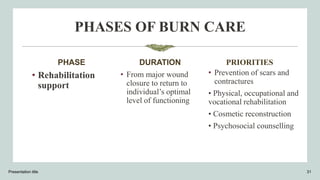
Burn
By, Kranti Jadhav ​
Content
1.Review of anatomy and physiology of skin
2.Introduction
3.Incidence
4.Definition
5.Etiology
6.Classification
7.Jackson’s thermal wound injury
8.Pathophysiology
9.Clinical manifestations
10.Diagnostic evaluation
11.Management
12.Complications
13.Prevention
14.summary
15.conclusion
Presentation title 2
BURN
REVIEW OF ANATOMY AND physiology of
skin
Presentation title 3
INTRODUCTION
Burns is one of the most devastating conditions
encountered in medicine. The injury represents an
assault on all aspects of the patient from the
physical to the psychological. The visible physical
and invisible psychological scars are long-lasting.
Presentation title 4
DEFINITION OF BURN
–Burn can be defined as any injury that results from direct contact or
exposure to any thermal, chemical, electrical, or radiation source.
INCIDENCE OF BURN
•India records 70 lacs burn injury annually of which 1.4
lacs people die and 2.4 suffers from a disability.
•70% of cases are in the 15-35 years age group.
Presentation title 6
INCIDENCE OF BURN
• 4 out of 5 cases are either women or children.
• 80% cases with women are related to kitchen-related accidents.
• 1th leading cause of death/injury of children age 1-9 years.
• 250 to 300 acid attacks are reported in India every year.
• 80% to 90% of burns occur at home
Presentation title 7
ETIOLOGY OF BURN
1. Thermal burns
2. Chemical burns
3. Electrical burns
4. Radiation burns
5. Inhalation burns
Presentation title 8
CLASSIFICATION OF BURN INJURY
1. According to burn depth
2. According to extent of the burn
Presentation title 9
•ACCORDING TO BURN DEPTH
a) Based on skin layers involvement
b) Based on the degree of burn
Presentation title 10
Based on skin layers involvement
11
Based on degree of burn
12
ACCORDING TO EXTENT OF BURN
Presentation title 13
•Rule of nine
•Lund and Browder chart
•Palmer method
Rule of nine (adult)
Presentation title 14
Rule of nine (child)
Presentation title 15
Lund and Browder Chart
Presentation title 16
Palmer method
Presentation title 17
Pathophysiology
Presentation title 18
CLINICAL FEATURES
First degree burns:
• Reddened skin
• Pain at burn site
• Involves only epidermis
• Have an in-tact epidermal barrier
• Do not result in scarring
• Examples : Sun-burn, minor scald from a kitchen accident
Presentation title 19
CLINICAL FEATURES
Superficial 2nd degree burns :
• Intense pain
• White to red skin
• Blisters
• Involves epidermis & papillary layer of dermis
• Spares hair follicles, sweat glands etc.
• Erythematous & blanch to touch
• Very painful/sensitive .
• No or minimal scarring.
• Spontaneously re-epithelialization from retained epidermal structures in
7-14 days Second Degree Burns
Presentation title 20
CLINICAL FEATURES
Deep second degree burns:
• Injury to deeper layers of dermis, i.e, reticular dermis.
• Appears pale & mottled.
• Do not blanch to touch.
• Capillary return sluggish or absent.
• Less painful, remain painful to pinprick.
• Takes 14 to 35 days to heal by re-epithelialization from
hair follicles & sweat gland, keratinocytes often with
severe scarring.
• Contractures possible.
Presentation title 21
CLINICAL FEATURES
3rd Degree Burn:
• Dry, leathery skin (white, dark brown, or charred).
• Loss of sensation (little pain).
• All dermal layers/tissue maybe involved.
Fourth degree burn:
• Involves structures beneath the skin- muscle, bone.
Presentation title 22
SYSTEMIC CHANGES
Cardiac: Decreased cardiac output.
Pulmonary: Respiratory insufficiency as a secondary
process. Can progress to respiratory failure.
Gastrointestinal: Decreased or absent GI motility.
Curling’s ulcer formation.
Metabolic: Hypermetabolic state. Increased oxygen and
calorie requirements. Increase in core body temperature.
Immunologic: Loss of protective barrier. Increased risk of
infection. Suppression of humoral and cell-mediated
immune responses.
Presentation title 23
DIAGNOSTIC EVALUATION OF BURN
1. History taking
2. Physical examination
3. Diagnostic tests-
• Complete blood count
• ABG analysis
• Human chorionic gonadotropin testing
• Serum urea, electrolytes
• Chest X-ray
• CT scan
• Laser Doppler imaging
Presentation title 24
History Taking
1. Time of injury
2. Place of injury (open/closed)
3. unconsciousness during the incidence
4. Mechanism of burn injury/agent
5. Duration of exposure to agent
6. Intentional burn injury
7. Last Tetanus shot
8. Any known Allergies
Presentation title 25
PHYSICAL EXAMINATION
1. Complete head-to-toe assessment
2. Systematic assessment
3. Burn estimation/severity assessment
Presentation title 26
Burn severity assessment
MINOR
• Adult <10% TBSA
• Young or old <5% TBSA
• <2% Full thickness Burn
MODERATE
• Adult 10-20% TBSA
• Young or old 5- 10%
TBSA
• 2-5% Full thickness burn
• High voltage injury
• Possible inhalation
injury
• Circumferential burn
• Other health problems
Presentation title 27
MAJOR
• Adult >20% TBSA
• Young or old >10%
TBSA
• >5% Full thickness burn
• Known inhalation injury
• Significant burn to face,
joints, hands or feet
• Associated injuries
DIAGNOSTIC TESTS
1. Complete blood count
2. ABG analysis
3. Serum biochemistry
4. Chest X-ray
5. CT scan
6. Laser Doppler imaging
Presentation title 28
PHASES OF BURN CARE
PHASE
• Emergent /
resuscitative phase=
DURATION
- From onset of injury to
completion of fluid
resuscitation.
Presentation title 29
PRIORITIES
• First aid
• Prevention of shock
•Prevention of
respiratory distress
•Detection and treatment
of concomitant injuries
PHASES OF BURN CARE
PHASE
• Acute /
intermediate
phase
DURATION
• From beginning of
diuresis to near
completion of wound
closure.
Presentation title 30
PRIORITIES
• Wound assessment
and care
• Wound closure
• Prevention and
treatment of
complications, including
infection
• Nutritional support
PHASES OF BURN CARE
PHASE
• Rehabilitation
support
DURATION
• From major wound
closure to return to
individual’s optimal
level of functioning
Presentation title 31
PRIORITIES
• Prevention of scars and
contractures
• Physical, occupational and
vocational rehabilitation
• Cosmetic reconstruction
• Psychosocial counselling
Emergent / resuscitative phase
Medical management
1. Assess burn severity
a) Burn depth
b) Burn size
c) Burn location
d) Age
e) General health
f) Mechanism of injury
Presentation title 32
Emergent / resuscitative phase
• Medical management
2. Treat minor burns
3. Major burns
a) Monitor airway and breathing
b) Prevent burn (hypovolemic) shock (see formula)
c) Prevent aspiration
d) Minimizing pain and anxiety
e) Wound care
f) Prevent tetanus
g) Prevent tissue ischemia
h) Transport to burn facility
Presentation title 33
Emergent / resuscitative phase
• Calculation of fluids:
1. Consensus formula:
Ringer's lactate solution= 2-4 ml X body weight kg X TBSA ½
solution in first 8 hours and rest half in next 16 hours
2. Parkland formula:
Volume of Ringer’s lactate= 4 ml X % BSA x weight (kg) ½
solution in first 8 hours and rest half in next 16 hours
Presentation title 34
Nursing management of patient in
Emergent / resuscitative phase
1. Maintaining proper oxygenation and tissues perfusion
2. Maintaining fluid and electrolyte balance
3. Relieving pain
4. Preventing hypothermia
5. Providing initial wound care
6. Preventing infection
7. Promoting comfort
8. Relieving anxiety and proving psychological support
Presentation title 35
ACUTE / INTERMEDIATE PHASE
Medical management
1. Prevent infection
• Asepsis
• Prophylactic antibiotics
• Immunization
• Environmental control
Presentation title 36
ACUTE / INTERMEDIATE PHASE
•Medical management
3. Minimizing pain
• Patient controlled analgesia devices
• Inhalation analgesic (nitrous oxide)
• Oral analgesics; opioid analgesics, NSAID’s
• Hypnosis, art and play therapy
• Guided imaginary, relaxation techniques
• Distraction therapy, biofeedback
• Music therapy
Presentation title 37
ACUTE / INTERMEDIATE PHASE
• Medical management
4. Provide wound care
a) Wound cleansing
b) Wound debridement
i. Natural debridement
ii. Mechanical debridement
iii. Chemical debridement
iv. Surgical debridement
Presentation title 38
ACUTE / INTERMEDIATE PHASE
• Medical management
c) Topical antimicrobial treatment
• Silver sulfadiazine 1%
• Mafenide acetate 5%
• Silver nitrate 0.5%
• acticoat
Presentation title 39
ACUTE / INTERMEDIATE PHASE
Medical management
d) Wound dressing
• Moist dressing
• Occlusive dressing for new grafts
• Non-adhesive dressings covers
Presentation title 40
ACUTE / INTERMEDIATE PHASE
•Medical management
5. Maximize function
• Splinting
• Positioning
• Exercise
• Ambulation performance of ADI
• Pressure therapy
Presentation title 41
ACUTE / INTERMEDIATE PHASE
•Medical management
6. Provide psychological support
• Meeting the psychological needs
• Involvement in physical therapy
• Encouragement in wound care
• Ventilation of feeling, emotions, fear
• Promoting self image
Presentation title 42
ACUTE / INTERMEDIATE PHASE
•Surgical management
1. Escharotomy: An escharotomy is a surgical procedure used to
treat full-thickness circumferential burns. In full-thickness burns,
both the epidermis and the dermis are destroyed along with
sensory nerves in the dermis.
Presentation title 43
ACUTE / INTERMEDIATE PHASE
Surgical management
2. Fasciotomy or fasciectomy
Fasciotomy or fasciectomy is a surgical procedure where
the fascia is cut to relieve tension or pressure in order to
treat the resulting loss of circulation to an area of tissue or
muscle.
Presentation title 44
ACUTE / INTERMEDIATE PHASE
• Surgical management
2. Wound grafting
# Biologic dressing / graft
# biosynthetic and synthetic grafts
Presentation title 45
ACUTE / INTERMEDIATE PHASE
Surgical management
2. Wound grafting
# Biologic dressing / graft types
• Autograft
• Isograft
• Allograft
• Xenograft
Presentation title 46
ACUTE / INTERMEDIATE PHASE
Surgical management
2. Wound grafting
# Biosynthetic and synthetic graft types
• Biobrane
• Integra
• Calcium alginate
• Non-adhering fine mesh gauze
Presentation title 47
• INTEGRA
Integra is a product that is used to help re-grow skin on body
parts where the skin has been removed or badly damaged. It
was initially used to safely cover large areas of burned tissue
where skin needed to be regrown. However, Integra is now used
far more widely as part of skin grafts in reconstructive surgery.
Presentation title 48
1.CALCIUM ALGINATE: Calcium alginate dressings are used
primarily for the granulating phase of wound repair. They are
made from alginate, a derivative of seaweed. The calcium in the
dressing interacts with sodium in the wound, providing a wound
exudate that stimulates myofibroblasts and epithelial cells and
speeds wound homeostasis.
2.NON-ADHERING FINE MESH GAUZE: Nonadherent dressings are
basically low adherent wound pad for pain-free removal of
the dressing and it is mostly used for minor wounds
Presentation title 49
Nursing management of patient in acute /
intermediate phase
1. Maintaining proper oxygenation and tissues perfusion
2. Maintaining fluid and electrolyte balance
3. Relieving pain
4. Preventing hypothermia
5. Providing wound care
6. Preventing infection
Presentation title 50
8. Relieving anxiety and proving psychological support
9. Graft care
10.Nutritional support
11.Improving mobility
12.Promoting comfort
Presentation title 51
REHABILIATION PHASE
Medical management
1. Minimizing functional loss
• Exercise
• Splinting
• positioning
Presentation title 52
REHABILIATION PHASE
3. Medical management
2. Provide psychological support
• Self image issues
• Physical limitations
• Reintegration into society
• Fear of rejection
• Good communication
• Encourage independence
Presentation title 53
REHABILIATION PHASE
3. Abnormal wound healing
Presentation title 54
REHABILIATION PHASE
3. Medical management
4. Prevention and treatment of scars
• Pressure use of topical silicon
• Scar massage
• Steroid injections
• Application of elastic pressure garments
• Cosmetic interventions
Presentation title 55
Nursing management of patient in
Rehabilitation
1. Improving mobility
2. Improving self esteem
3. Promoting independence
4. Cosmetic counselling
5. Vocational training
6. Improving body image
Presentation title 56
COMPLICATIONS OF BURN
1. Burn shock
2. Pulmonary complications due to inhalation burn
3. Acute renal failure
4. Infections and sepsis
Presentation title 57
5. Curling’s ulcers/stress ulcer
6. Extensive and disabling scarring
7. Psychological trauma
8. Marjolin’s ulcer: A Marjolin ulcer is a cutaneous malignancy that
arises in the setting of previously injured skin, longstanding scars, and
chronic wounds
9. Multiple organ failure
Presentation title 58
Summary
Presentation title 59
Thank
you

Burn.pptx

  • 1.
  • 2.
    Content 1.Review of anatomyand physiology of skin 2.Introduction 3.Incidence 4.Definition 5.Etiology 6.Classification 7.Jackson’s thermal wound injury 8.Pathophysiology 9.Clinical manifestations 10.Diagnostic evaluation 11.Management 12.Complications 13.Prevention 14.summary 15.conclusion Presentation title 2 BURN
  • 3.
    REVIEW OF ANATOMYAND physiology of skin Presentation title 3
  • 4.
    INTRODUCTION Burns is oneof the most devastating conditions encountered in medicine. The injury represents an assault on all aspects of the patient from the physical to the psychological. The visible physical and invisible psychological scars are long-lasting. Presentation title 4
  • 5.
    DEFINITION OF BURN –Burncan be defined as any injury that results from direct contact or exposure to any thermal, chemical, electrical, or radiation source.
  • 6.
    INCIDENCE OF BURN •Indiarecords 70 lacs burn injury annually of which 1.4 lacs people die and 2.4 suffers from a disability. •70% of cases are in the 15-35 years age group. Presentation title 6
  • 7.
    INCIDENCE OF BURN •4 out of 5 cases are either women or children. • 80% cases with women are related to kitchen-related accidents. • 1th leading cause of death/injury of children age 1-9 years. • 250 to 300 acid attacks are reported in India every year. • 80% to 90% of burns occur at home Presentation title 7
  • 8.
    ETIOLOGY OF BURN 1.Thermal burns 2. Chemical burns 3. Electrical burns 4. Radiation burns 5. Inhalation burns Presentation title 8
  • 9.
    CLASSIFICATION OF BURNINJURY 1. According to burn depth 2. According to extent of the burn Presentation title 9
  • 10.
    •ACCORDING TO BURNDEPTH a) Based on skin layers involvement b) Based on the degree of burn Presentation title 10
  • 11.
    Based on skinlayers involvement 11
  • 12.
    Based on degreeof burn 12
  • 13.
    ACCORDING TO EXTENTOF BURN Presentation title 13 •Rule of nine •Lund and Browder chart •Palmer method
  • 14.
    Rule of nine(adult) Presentation title 14
  • 15.
    Rule of nine(child) Presentation title 15
  • 16.
    Lund and BrowderChart Presentation title 16
  • 17.
  • 18.
  • 19.
    CLINICAL FEATURES First degreeburns: • Reddened skin • Pain at burn site • Involves only epidermis • Have an in-tact epidermal barrier • Do not result in scarring • Examples : Sun-burn, minor scald from a kitchen accident Presentation title 19
  • 20.
    CLINICAL FEATURES Superficial 2nddegree burns : • Intense pain • White to red skin • Blisters • Involves epidermis & papillary layer of dermis • Spares hair follicles, sweat glands etc. • Erythematous & blanch to touch • Very painful/sensitive . • No or minimal scarring. • Spontaneously re-epithelialization from retained epidermal structures in 7-14 days Second Degree Burns Presentation title 20
  • 21.
    CLINICAL FEATURES Deep seconddegree burns: • Injury to deeper layers of dermis, i.e, reticular dermis. • Appears pale & mottled. • Do not blanch to touch. • Capillary return sluggish or absent. • Less painful, remain painful to pinprick. • Takes 14 to 35 days to heal by re-epithelialization from hair follicles & sweat gland, keratinocytes often with severe scarring. • Contractures possible. Presentation title 21
  • 22.
    CLINICAL FEATURES 3rd DegreeBurn: • Dry, leathery skin (white, dark brown, or charred). • Loss of sensation (little pain). • All dermal layers/tissue maybe involved. Fourth degree burn: • Involves structures beneath the skin- muscle, bone. Presentation title 22
  • 23.
    SYSTEMIC CHANGES Cardiac: Decreasedcardiac output. Pulmonary: Respiratory insufficiency as a secondary process. Can progress to respiratory failure. Gastrointestinal: Decreased or absent GI motility. Curling’s ulcer formation. Metabolic: Hypermetabolic state. Increased oxygen and calorie requirements. Increase in core body temperature. Immunologic: Loss of protective barrier. Increased risk of infection. Suppression of humoral and cell-mediated immune responses. Presentation title 23
  • 24.
    DIAGNOSTIC EVALUATION OFBURN 1. History taking 2. Physical examination 3. Diagnostic tests- • Complete blood count • ABG analysis • Human chorionic gonadotropin testing • Serum urea, electrolytes • Chest X-ray • CT scan • Laser Doppler imaging Presentation title 24
  • 25.
    History Taking 1. Timeof injury 2. Place of injury (open/closed) 3. unconsciousness during the incidence 4. Mechanism of burn injury/agent 5. Duration of exposure to agent 6. Intentional burn injury 7. Last Tetanus shot 8. Any known Allergies Presentation title 25
  • 26.
    PHYSICAL EXAMINATION 1. Completehead-to-toe assessment 2. Systematic assessment 3. Burn estimation/severity assessment Presentation title 26
  • 27.
    Burn severity assessment MINOR •Adult <10% TBSA • Young or old <5% TBSA • <2% Full thickness Burn MODERATE • Adult 10-20% TBSA • Young or old 5- 10% TBSA • 2-5% Full thickness burn • High voltage injury • Possible inhalation injury • Circumferential burn • Other health problems Presentation title 27 MAJOR • Adult >20% TBSA • Young or old >10% TBSA • >5% Full thickness burn • Known inhalation injury • Significant burn to face, joints, hands or feet • Associated injuries
  • 28.
    DIAGNOSTIC TESTS 1. Completeblood count 2. ABG analysis 3. Serum biochemistry 4. Chest X-ray 5. CT scan 6. Laser Doppler imaging Presentation title 28
  • 29.
    PHASES OF BURNCARE PHASE • Emergent / resuscitative phase= DURATION - From onset of injury to completion of fluid resuscitation. Presentation title 29 PRIORITIES • First aid • Prevention of shock •Prevention of respiratory distress •Detection and treatment of concomitant injuries
  • 30.
    PHASES OF BURNCARE PHASE • Acute / intermediate phase DURATION • From beginning of diuresis to near completion of wound closure. Presentation title 30 PRIORITIES • Wound assessment and care • Wound closure • Prevention and treatment of complications, including infection • Nutritional support
  • 31.
    PHASES OF BURNCARE PHASE • Rehabilitation support DURATION • From major wound closure to return to individual’s optimal level of functioning Presentation title 31 PRIORITIES • Prevention of scars and contractures • Physical, occupational and vocational rehabilitation • Cosmetic reconstruction • Psychosocial counselling
  • 32.
    Emergent / resuscitativephase Medical management 1. Assess burn severity a) Burn depth b) Burn size c) Burn location d) Age e) General health f) Mechanism of injury Presentation title 32
  • 33.
    Emergent / resuscitativephase • Medical management 2. Treat minor burns 3. Major burns a) Monitor airway and breathing b) Prevent burn (hypovolemic) shock (see formula) c) Prevent aspiration d) Minimizing pain and anxiety e) Wound care f) Prevent tetanus g) Prevent tissue ischemia h) Transport to burn facility Presentation title 33
  • 34.
    Emergent / resuscitativephase • Calculation of fluids: 1. Consensus formula: Ringer's lactate solution= 2-4 ml X body weight kg X TBSA ½ solution in first 8 hours and rest half in next 16 hours 2. Parkland formula: Volume of Ringer’s lactate= 4 ml X % BSA x weight (kg) ½ solution in first 8 hours and rest half in next 16 hours Presentation title 34
  • 35.
    Nursing management ofpatient in Emergent / resuscitative phase 1. Maintaining proper oxygenation and tissues perfusion 2. Maintaining fluid and electrolyte balance 3. Relieving pain 4. Preventing hypothermia 5. Providing initial wound care 6. Preventing infection 7. Promoting comfort 8. Relieving anxiety and proving psychological support Presentation title 35
  • 36.
    ACUTE / INTERMEDIATEPHASE Medical management 1. Prevent infection • Asepsis • Prophylactic antibiotics • Immunization • Environmental control Presentation title 36
  • 37.
    ACUTE / INTERMEDIATEPHASE •Medical management 3. Minimizing pain • Patient controlled analgesia devices • Inhalation analgesic (nitrous oxide) • Oral analgesics; opioid analgesics, NSAID’s • Hypnosis, art and play therapy • Guided imaginary, relaxation techniques • Distraction therapy, biofeedback • Music therapy Presentation title 37
  • 38.
    ACUTE / INTERMEDIATEPHASE • Medical management 4. Provide wound care a) Wound cleansing b) Wound debridement i. Natural debridement ii. Mechanical debridement iii. Chemical debridement iv. Surgical debridement Presentation title 38
  • 39.
    ACUTE / INTERMEDIATEPHASE • Medical management c) Topical antimicrobial treatment • Silver sulfadiazine 1% • Mafenide acetate 5% • Silver nitrate 0.5% • acticoat Presentation title 39
  • 40.
    ACUTE / INTERMEDIATEPHASE Medical management d) Wound dressing • Moist dressing • Occlusive dressing for new grafts • Non-adhesive dressings covers Presentation title 40
  • 41.
    ACUTE / INTERMEDIATEPHASE •Medical management 5. Maximize function • Splinting • Positioning • Exercise • Ambulation performance of ADI • Pressure therapy Presentation title 41
  • 42.
    ACUTE / INTERMEDIATEPHASE •Medical management 6. Provide psychological support • Meeting the psychological needs • Involvement in physical therapy • Encouragement in wound care • Ventilation of feeling, emotions, fear • Promoting self image Presentation title 42
  • 43.
    ACUTE / INTERMEDIATEPHASE •Surgical management 1. Escharotomy: An escharotomy is a surgical procedure used to treat full-thickness circumferential burns. In full-thickness burns, both the epidermis and the dermis are destroyed along with sensory nerves in the dermis. Presentation title 43
  • 44.
    ACUTE / INTERMEDIATEPHASE Surgical management 2. Fasciotomy or fasciectomy Fasciotomy or fasciectomy is a surgical procedure where the fascia is cut to relieve tension or pressure in order to treat the resulting loss of circulation to an area of tissue or muscle. Presentation title 44
  • 45.
    ACUTE / INTERMEDIATEPHASE • Surgical management 2. Wound grafting # Biologic dressing / graft # biosynthetic and synthetic grafts Presentation title 45
  • 46.
    ACUTE / INTERMEDIATEPHASE Surgical management 2. Wound grafting # Biologic dressing / graft types • Autograft • Isograft • Allograft • Xenograft Presentation title 46
  • 47.
    ACUTE / INTERMEDIATEPHASE Surgical management 2. Wound grafting # Biosynthetic and synthetic graft types • Biobrane • Integra • Calcium alginate • Non-adhering fine mesh gauze Presentation title 47
  • 48.
    • INTEGRA Integra isa product that is used to help re-grow skin on body parts where the skin has been removed or badly damaged. It was initially used to safely cover large areas of burned tissue where skin needed to be regrown. However, Integra is now used far more widely as part of skin grafts in reconstructive surgery. Presentation title 48
  • 49.
    1.CALCIUM ALGINATE: Calciumalginate dressings are used primarily for the granulating phase of wound repair. They are made from alginate, a derivative of seaweed. The calcium in the dressing interacts with sodium in the wound, providing a wound exudate that stimulates myofibroblasts and epithelial cells and speeds wound homeostasis. 2.NON-ADHERING FINE MESH GAUZE: Nonadherent dressings are basically low adherent wound pad for pain-free removal of the dressing and it is mostly used for minor wounds Presentation title 49
  • 50.
    Nursing management ofpatient in acute / intermediate phase 1. Maintaining proper oxygenation and tissues perfusion 2. Maintaining fluid and electrolyte balance 3. Relieving pain 4. Preventing hypothermia 5. Providing wound care 6. Preventing infection Presentation title 50
  • 51.
    8. Relieving anxietyand proving psychological support 9. Graft care 10.Nutritional support 11.Improving mobility 12.Promoting comfort Presentation title 51
  • 52.
    REHABILIATION PHASE Medical management 1.Minimizing functional loss • Exercise • Splinting • positioning Presentation title 52
  • 53.
    REHABILIATION PHASE 3. Medicalmanagement 2. Provide psychological support • Self image issues • Physical limitations • Reintegration into society • Fear of rejection • Good communication • Encourage independence Presentation title 53
  • 54.
    REHABILIATION PHASE 3. Abnormalwound healing Presentation title 54
  • 55.
    REHABILIATION PHASE 3. Medicalmanagement 4. Prevention and treatment of scars • Pressure use of topical silicon • Scar massage • Steroid injections • Application of elastic pressure garments • Cosmetic interventions Presentation title 55
  • 56.
    Nursing management ofpatient in Rehabilitation 1. Improving mobility 2. Improving self esteem 3. Promoting independence 4. Cosmetic counselling 5. Vocational training 6. Improving body image Presentation title 56
  • 57.
    COMPLICATIONS OF BURN 1.Burn shock 2. Pulmonary complications due to inhalation burn 3. Acute renal failure 4. Infections and sepsis Presentation title 57
  • 58.
    5. Curling’s ulcers/stressulcer 6. Extensive and disabling scarring 7. Psychological trauma 8. Marjolin’s ulcer: A Marjolin ulcer is a cutaneous malignancy that arises in the setting of previously injured skin, longstanding scars, and chronic wounds 9. Multiple organ failure Presentation title 58
  • 59.
  • 60.