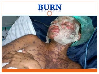
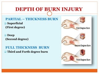
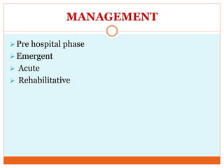
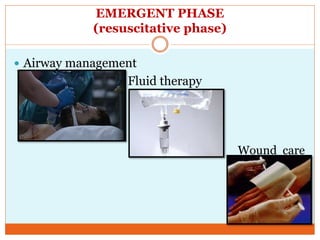
MRS. S.KAMALI KIRUBA
MSC (N),MEDICAL SURGICAL NURSING
ASSOCIATE PROFESSOR
GANGA COLLEGE OF NURSING
COIMBATORE
ANATONY OF SKIN
INTRODUCTION
Burns are one of the most common household
injuries, especially among children. The term
“burn” means more than the burning sensation
associated with this injury. Burns are
characterized by severe skin damage that
causes the affected skin cells to die.
DEFINITION
A burn is a type of injury to skin, or other tissues,
caused by heat, cold, electricity, chemicals, friction,
or radiation. Most burns are due to heat from hot
liquids (called scalding), solids, or fire.
CAUSES OF BURN
Thermal burn
Chemical burn
Electrical burn
Radiation burn Cold thermal injury
Smoke & inhalation
burn
CLASSIFICATION OF BURN INJURY
DEPTH
EXTENT
LOCATION
PATIENT
RISK
FACTOR
DEPTH OF BURN INJURY
PARTIAL – THICKNESS BURN
 Superficial
(First degree)
 Deep
(Second degree)
FULL THICKNESS BURN
 Third and Forth degree burn
DEPTH OF BURN INJURY
CLASSIFICATION CLINICAL
APPEARANCE
CAUSES STRUCTURES
INVOLVED
PARTIAL –THICKNESS BURN
Superficial
(first degree)
Erythema,
blanching on
pressure,
pain and swelling,
no vesicles or
blisters (although
after 24 hr skin
may blister and
peel)
Superficial
sunburn
Quick heat flash
Superficial
epidermal damage
with hyperemia.
Tactile and pain
sensation intact.
DEPTH OF BURN INJURY
CLASSIFICATION CLINICAL
APPEARANCE
CAUSES STRUCTURE
INVOLVED
Deep (Second
degree)
Fluid filled
vesicles that are
red, shiny, wet (if
vesicles have
ruptured);
severe pain
caused by nerve
injury;
mild to moderate
edema
Flame
Flash
Scald
Contact burns
Chemicals
Tar
Epidermis and
dermis involved
to varying depth.
Skin elements,
from which
epithelial
regeneration
occurs, remain
viable.
DEPTH OF BURN INJURY
CLASSIFICATION CLINICAL
APPEARANCE
CAUSE STRUCTURES
INVOLVED
FULL THICKNESS BURN
FULL THICKNESS
IN DESTRUCTION
(Third and Fourth
degree)
Dry, waxy white,
leathery, or hard
skin, visible
thrombosed
vessels;
insensitivity to
pain because of
nerve destruction;
possible
involvement of
muscles, tendons,
and bones.
Flame
Scald
Chemical
Tar
Electric current
All skin elements
and local nerve
endings destroyed.
Coagulation
necrosis present.
Surgical
intervention
required for
healing.
EXTENT OF INJURY
The extent of a burn wound is defined as the percentage
of total body surface damaged and may be determined by
the Rule of Nines. This divides the body into areas of 9%
or multiples of nine.
EXTENT OF INJURY
EXTENT OF INJURY
www.sagediagram.com
LOCATION OF BURN INJURY
Burn location is an important consideration.
 If the burn involves the face, nose, mouth, or neck, there is a
risk that there will be inhalation injury and enough
inflammation and swelling to obstruct the airway and
cause breathing problems.
 burns to the chest, as the burn progresses, the tissue involved
may not allow enough motion of the chest wall to allow
adequate breathing to occur.
 burns occur to arms, legs, fingers, or toes, the same
constriction may not allow blood flow.
 Burns to areas of the body with flexion creases, like the palm
of the hand, the back of the knee, the face, and the groin may
need specialized care.
PATHOPHYSIOLOGY OF BURN
BURN
↑Vascular permeability
Edema ↓intravascular
volume
↓Blood volume
↑Hematocrit
↑Peripheral Resistance
↑Viscosity
Burn shock
DIAGNOSTIC EVALUATION
 SODIUM
Hyponatremia- dilutional Hyponatremia
Water intoxication
 POTASSIUM
Hyperkalemia-renal failure
Adrenal insufficiency
Massive deep muscle injury
Hypokalemia-dilution/GI wash…
MANAGEMENT
Pre hospital phase
Emergent
 Acute
 Rehabilitative
PRE HOSPITAL PHASE
 Remove person from the source of burn
 Self shield- by rescuers
 Minor burn-<10% TBSA- cover with clean, cool, tap
water- dampened towel.
 Assessment and management of ABC
A- Airway
B-Breathing
C-Circulation
EMERGENT PHASE
(resuscitative phase)
 Airway management
Fluid therapy
Wound care
EMERGENT PHASE
(Resuscitative phase)
Airway management
Early endotracheal intubation
Ventilator assistance- with PEEP
Assess ABG values
Extubation-when edema resolves
Assess lower respiratory tract by – fiberotic bronchoscopy
Humidified oxygen
Position-high fowler’s position(not for patients with spinal
injury)
If spinal injury- reserve tendelberg position
Deep breathing and coughing exercise
Reposition every 2hrs
Bronchodialators
O2 therapy until carboxyhemoglobin become normal.
EMERGENT PHASE
(Resuscitative phase)
Easing blood flow around the wound
If a burn scab (eschar) goes completely around a limb, it can
tighten and cut off the blood circulation. Escharotomies -
to relieve respiratory distress secondary to circumferential,
full thickness burns to the neck and trunk.
FLUID THERAPY
 Patient >15% TBSA-LARGE BORE I/V access
 > 30% TBSA-central and arterial line
FORMULAS FOR ESTIMETING FLUID
REPLACEMENT
FIRST 24 HOURS SECOND 24 HOURS
FORMULA CRYSTALLOIDS COLLOIDS GLUCOSE IN
WATER
Brooke
(Modified)
Lactated Ringer’s
solution:2.0ml/kg/%TBSA
burn;
½ given during first 8 hr;
½ given during next 16 hr.
0.3-0.5ml /kg/%
TBSA burn
Amount to
replace
estimated
evaporate
losses
Parkland
(Baxter)
Lactated Ringer’s
solution:4ml/kg/%TBSA
burn;
½ given first 8hr;
¼ given each next8 hr.
20-60% of
calculated plasma
volume.
Amount to
replace
estimated
evaporate
losses
EMERGENT PHASE
(Resuscitative phase)
Wound care
Cleansing and gentle debridement- hydrotherapy
/cart shower/shower/or pattient bed /strecter
Debridement- necrotic skin remove
Escharotomies
Fasciotomies
Once daily shower and dressing
Con…
Wound care
Control infection _dressing
1. open method
Burn covered with topical antimicrobial solution without dressing
2. multiple dressing change
Sterile dressing impregnated with topical antimicrobial medication changed
every 12/24 hrs or once in every 3 days.
Moist wound healing method.
Types of dressing
 Silicone Dressings
 Foam Dressings
 Alginate Dressings
 Hydrogel Dressings
 Gel Dressings with Melaleuca
 Hydrocolloid Dressings
 Low Adherence dressings
Con..
 Water-based treatments: ultrasound mist
therapy to clean and stimulate the wound tissue.
EXCISION AND GRAFTING
 Autograft or autologous graft: skin obtained from
the patient’s own donor site.
 Allograft or heterologous graft: skin obtained from
another person
 Xenograft or heterograft: skin from other species,
such as pigs.
 Synthetic skin substitutes: manufactured products
that work as skin equivalents. They may be epidermal
(keratinocyte cultures), dermal or dermoepidermal
(artificial skin).
EXCISION AND GRAFTING
CULTURED EPITHELIAL SKINGRAFT
EXCISION AND GRAFTING
CULTURED EPITHELIAL SKINGRAFT
DRUG THERAPY
 Analgesics and sedatives
 Tetanus immunization
 Antimicrobial agents
 Nutritional support
 Antibiotics
 Tetanus toxoid
DRUG THERAPY
 NUTRITIONAL SUPPORT
VitaminsA,C,E and multivitamins
Minerals: zinc, iron (Ferrous sulfate)
Oxandrolone
ANALGESIA
Morphine
Nonsteroidal Antiinflammatory
(eg.ketaprofen)
SEDATION
Haloperidol
Lorazepam
GASTROINTESTINAL SUPPORT
Ranitidine
antacids
ACUTE PHASE
 Wound care
 Skin grafting
 Pain management
 Other pain management technique
Guided imaginary
Relaxation therapy
Hypnosis
Physical and occupational therapy
Nutritional care
Psychosocial care
ACUTE PHASE
Wound care
Debridment of necrotic tissue
Use meshed dressing with paraffin oil
Moist dressing for donor site
Enzymatic debridement
ACUTE PHASE
Nutritional care
 Provide adequate calories and protein to promote
healing.
 TPN
REHABILITATION PHASE
Manage Emotions
 Fear
 Anxiety
 Anger
 Guilt
 depression
PLASTIC SURGERY
Plastic surgery (reconstruction) can improve the
appearance of burn scars and increase the flexibility
of joints affected by scarring.
CHEMICAL BURN
 Acids
Protein injury by hydrolysis
Thermal injury is made with skin contact
 Alkali
Saponification of fat.
Hygroscopic effect-dehydrate cells.
Dissolves proteins by creation of alkaline proteinases
(hydroxide ions)
 Treatment
Late neutralization with antidote done by 0.2% acetic acid
in alkali burs, sodium bicarbonate or calcium gluconate for
acid burns.
ELECTRICAL BURN
 Low tension injury: Less than 1000 volts
 High tension injury: More than 1000 volts
 It is always deep burn
 There is a wound of entry and wound exit.
 Patient may also have major internal
organ injuries. GIT, Thoracic injuries.
 Often convulsion develop.
 Death may occur due to cardiac arrhythmias.
COMPLICATION OF BURN
 Burn shock
 Pulmonary complications due to inhalation injury
 Acute renal failure
 Infections and sepsis
 Curlin’s ulcer in large burns over 30% usually after 9th day
 Extensive and disabling scarring
 Psychological trauma
 Cancer called Marjolin’s ulcer, may take 21 years to
develop.
NURSING DIAGNOSIS
 Impaired gas exchange related to carbon monoxide
poisoning as evidenced by labored breathing.
 Ineffective airway clearance related to edema and
effects of smoke inhalation and evidenced by
ventilator support.
 Fluid volume deficit related to fluid loss as
manifested by decreased serum electrolyte level and
dry skin.
 Acute pain related to impaired skin integrity as
manifested by facial expression.
NURSING DIAGNOSIS
 Impaired skin integrity related to thermal injury as
manifested by blisters and lesions.
 Imbalanced nutrition less than body requirement
related to inability to intake as evidence by weight
loss.
 Activity intolerance related to weakness as evidenced
by verbalization.
 Risk for infection related to impaired skin integrity
and suppressed immune response.
 Risk for contracture related to the burn injury.
LIFESTYLE AND HOME REMEDIES TO TREAT
MINOR BURNS
 Cool the burn. Hold the burned area under cool (not
cold) running water or apply a cool, wet compress until the
pain eases. Don't use ice. Putting ice directly on a burn can
cause further damage to the tissue.
 Remove rings or other tight items. Try to do this
quickly and gently, before the burned area swells.
 Don't break blisters. Fluid-filled blisters protect against
infection. If a blister breaks, clean the area with water (mild
soap is optional). Apply an antibiotic ointment. But if a rash
appears, stop using the ointment.
LIFESTYLE AND HOME REMEDIES TO
TREAT MINOR BURNS
 Apply lotion. Once a burn is completely cooled, apply a
lotion, such as one that contains aloe vera or a moisturizer.
This helps prevent drying and provides relief.
 Bandage the burn. Cover the burn with a sterile gauze
bandage (not fluffy cotton). Wrap it loosely to avoid putting
pressure on burned skin. Bandaging keeps air off the area,
reduces pain and protects blistered skin.
 Take a pain reliever. Over-the-counter medications, such
as ibuprofen (Advil, Motrin IB, others), naproxen sodium
(Aleve) or acetaminophen (Tylenol, others), can help relieve
pain.
 Consider a tetanus shot. Make sure that your tetanus
booster is up to date. Doctors recommend that people get a
tetanus shot at least every 10 years.
BURN
BURN

BURN

  • 1.
    MRS. S.KAMALI KIRUBA MSC(N),MEDICAL SURGICAL NURSING ASSOCIATE PROFESSOR GANGA COLLEGE OF NURSING COIMBATORE
  • 3.
  • 4.
    INTRODUCTION Burns are oneof the most common household injuries, especially among children. The term “burn” means more than the burning sensation associated with this injury. Burns are characterized by severe skin damage that causes the affected skin cells to die.
  • 5.
    DEFINITION A burn isa type of injury to skin, or other tissues, caused by heat, cold, electricity, chemicals, friction, or radiation. Most burns are due to heat from hot liquids (called scalding), solids, or fire.
  • 6.
    CAUSES OF BURN Thermalburn Chemical burn Electrical burn Radiation burn Cold thermal injury Smoke & inhalation burn
  • 7.
    CLASSIFICATION OF BURNINJURY DEPTH EXTENT LOCATION PATIENT RISK FACTOR
  • 8.
    DEPTH OF BURNINJURY PARTIAL – THICKNESS BURN  Superficial (First degree)  Deep (Second degree) FULL THICKNESS BURN  Third and Forth degree burn
  • 9.
    DEPTH OF BURNINJURY CLASSIFICATION CLINICAL APPEARANCE CAUSES STRUCTURES INVOLVED PARTIAL –THICKNESS BURN Superficial (first degree) Erythema, blanching on pressure, pain and swelling, no vesicles or blisters (although after 24 hr skin may blister and peel) Superficial sunburn Quick heat flash Superficial epidermal damage with hyperemia. Tactile and pain sensation intact.
  • 10.
    DEPTH OF BURNINJURY CLASSIFICATION CLINICAL APPEARANCE CAUSES STRUCTURE INVOLVED Deep (Second degree) Fluid filled vesicles that are red, shiny, wet (if vesicles have ruptured); severe pain caused by nerve injury; mild to moderate edema Flame Flash Scald Contact burns Chemicals Tar Epidermis and dermis involved to varying depth. Skin elements, from which epithelial regeneration occurs, remain viable.
  • 11.
    DEPTH OF BURNINJURY CLASSIFICATION CLINICAL APPEARANCE CAUSE STRUCTURES INVOLVED FULL THICKNESS BURN FULL THICKNESS IN DESTRUCTION (Third and Fourth degree) Dry, waxy white, leathery, or hard skin, visible thrombosed vessels; insensitivity to pain because of nerve destruction; possible involvement of muscles, tendons, and bones. Flame Scald Chemical Tar Electric current All skin elements and local nerve endings destroyed. Coagulation necrosis present. Surgical intervention required for healing.
  • 12.
    EXTENT OF INJURY Theextent of a burn wound is defined as the percentage of total body surface damaged and may be determined by the Rule of Nines. This divides the body into areas of 9% or multiples of nine.
  • 13.
  • 14.
  • 15.
    LOCATION OF BURNINJURY Burn location is an important consideration.  If the burn involves the face, nose, mouth, or neck, there is a risk that there will be inhalation injury and enough inflammation and swelling to obstruct the airway and cause breathing problems.  burns to the chest, as the burn progresses, the tissue involved may not allow enough motion of the chest wall to allow adequate breathing to occur.  burns occur to arms, legs, fingers, or toes, the same constriction may not allow blood flow.  Burns to areas of the body with flexion creases, like the palm of the hand, the back of the knee, the face, and the groin may need specialized care.
  • 16.
    PATHOPHYSIOLOGY OF BURN BURN ↑Vascularpermeability Edema ↓intravascular volume ↓Blood volume ↑Hematocrit ↑Peripheral Resistance ↑Viscosity Burn shock
  • 17.
    DIAGNOSTIC EVALUATION  SODIUM Hyponatremia-dilutional Hyponatremia Water intoxication  POTASSIUM Hyperkalemia-renal failure Adrenal insufficiency Massive deep muscle injury Hypokalemia-dilution/GI wash…
  • 18.
  • 19.
    PRE HOSPITAL PHASE Remove person from the source of burn  Self shield- by rescuers  Minor burn-<10% TBSA- cover with clean, cool, tap water- dampened towel.  Assessment and management of ABC A- Airway B-Breathing C-Circulation
  • 20.
    EMERGENT PHASE (resuscitative phase) Airway management Fluid therapy Wound care
  • 21.
    EMERGENT PHASE (Resuscitative phase) Airwaymanagement Early endotracheal intubation Ventilator assistance- with PEEP Assess ABG values Extubation-when edema resolves Assess lower respiratory tract by – fiberotic bronchoscopy Humidified oxygen Position-high fowler’s position(not for patients with spinal injury) If spinal injury- reserve tendelberg position Deep breathing and coughing exercise Reposition every 2hrs Bronchodialators O2 therapy until carboxyhemoglobin become normal.
  • 22.
    EMERGENT PHASE (Resuscitative phase) Easingblood flow around the wound If a burn scab (eschar) goes completely around a limb, it can tighten and cut off the blood circulation. Escharotomies - to relieve respiratory distress secondary to circumferential, full thickness burns to the neck and trunk.
  • 23.
    FLUID THERAPY  Patient>15% TBSA-LARGE BORE I/V access  > 30% TBSA-central and arterial line
  • 24.
    FORMULAS FOR ESTIMETINGFLUID REPLACEMENT FIRST 24 HOURS SECOND 24 HOURS FORMULA CRYSTALLOIDS COLLOIDS GLUCOSE IN WATER Brooke (Modified) Lactated Ringer’s solution:2.0ml/kg/%TBSA burn; ½ given during first 8 hr; ½ given during next 16 hr. 0.3-0.5ml /kg/% TBSA burn Amount to replace estimated evaporate losses Parkland (Baxter) Lactated Ringer’s solution:4ml/kg/%TBSA burn; ½ given first 8hr; ¼ given each next8 hr. 20-60% of calculated plasma volume. Amount to replace estimated evaporate losses
  • 25.
    EMERGENT PHASE (Resuscitative phase) Woundcare Cleansing and gentle debridement- hydrotherapy /cart shower/shower/or pattient bed /strecter Debridement- necrotic skin remove Escharotomies Fasciotomies Once daily shower and dressing
  • 26.
    Con… Wound care Control infection_dressing 1. open method Burn covered with topical antimicrobial solution without dressing 2. multiple dressing change Sterile dressing impregnated with topical antimicrobial medication changed every 12/24 hrs or once in every 3 days. Moist wound healing method. Types of dressing  Silicone Dressings  Foam Dressings  Alginate Dressings  Hydrogel Dressings  Gel Dressings with Melaleuca  Hydrocolloid Dressings  Low Adherence dressings
  • 27.
    Con..  Water-based treatments:ultrasound mist therapy to clean and stimulate the wound tissue.
  • 28.
    EXCISION AND GRAFTING Autograft or autologous graft: skin obtained from the patient’s own donor site.  Allograft or heterologous graft: skin obtained from another person  Xenograft or heterograft: skin from other species, such as pigs.  Synthetic skin substitutes: manufactured products that work as skin equivalents. They may be epidermal (keratinocyte cultures), dermal or dermoepidermal (artificial skin).
  • 29.
  • 30.
  • 31.
    EXCISION AND GRAFTING CULTUREDEPITHELIAL SKINGRAFT
  • 32.
    DRUG THERAPY  Analgesicsand sedatives  Tetanus immunization  Antimicrobial agents  Nutritional support  Antibiotics  Tetanus toxoid
  • 33.
    DRUG THERAPY  NUTRITIONALSUPPORT VitaminsA,C,E and multivitamins Minerals: zinc, iron (Ferrous sulfate) Oxandrolone ANALGESIA Morphine Nonsteroidal Antiinflammatory (eg.ketaprofen) SEDATION Haloperidol Lorazepam GASTROINTESTINAL SUPPORT Ranitidine antacids
  • 34.
    ACUTE PHASE  Woundcare  Skin grafting  Pain management  Other pain management technique Guided imaginary Relaxation therapy Hypnosis Physical and occupational therapy Nutritional care Psychosocial care
  • 35.
    ACUTE PHASE Wound care Debridmentof necrotic tissue Use meshed dressing with paraffin oil Moist dressing for donor site Enzymatic debridement
  • 36.
    ACUTE PHASE Nutritional care Provide adequate calories and protein to promote healing.  TPN
  • 37.
    REHABILITATION PHASE Manage Emotions Fear  Anxiety  Anger  Guilt  depression
  • 38.
    PLASTIC SURGERY Plastic surgery(reconstruction) can improve the appearance of burn scars and increase the flexibility of joints affected by scarring.
  • 39.
    CHEMICAL BURN  Acids Proteininjury by hydrolysis Thermal injury is made with skin contact  Alkali Saponification of fat. Hygroscopic effect-dehydrate cells. Dissolves proteins by creation of alkaline proteinases (hydroxide ions)  Treatment Late neutralization with antidote done by 0.2% acetic acid in alkali burs, sodium bicarbonate or calcium gluconate for acid burns.
  • 40.
    ELECTRICAL BURN  Lowtension injury: Less than 1000 volts  High tension injury: More than 1000 volts  It is always deep burn  There is a wound of entry and wound exit.  Patient may also have major internal organ injuries. GIT, Thoracic injuries.  Often convulsion develop.  Death may occur due to cardiac arrhythmias.
  • 41.
    COMPLICATION OF BURN Burn shock  Pulmonary complications due to inhalation injury  Acute renal failure  Infections and sepsis  Curlin’s ulcer in large burns over 30% usually after 9th day  Extensive and disabling scarring  Psychological trauma  Cancer called Marjolin’s ulcer, may take 21 years to develop.
  • 42.
    NURSING DIAGNOSIS  Impairedgas exchange related to carbon monoxide poisoning as evidenced by labored breathing.  Ineffective airway clearance related to edema and effects of smoke inhalation and evidenced by ventilator support.  Fluid volume deficit related to fluid loss as manifested by decreased serum electrolyte level and dry skin.  Acute pain related to impaired skin integrity as manifested by facial expression.
  • 43.
    NURSING DIAGNOSIS  Impairedskin integrity related to thermal injury as manifested by blisters and lesions.  Imbalanced nutrition less than body requirement related to inability to intake as evidence by weight loss.  Activity intolerance related to weakness as evidenced by verbalization.  Risk for infection related to impaired skin integrity and suppressed immune response.  Risk for contracture related to the burn injury.
  • 44.
    LIFESTYLE AND HOMEREMEDIES TO TREAT MINOR BURNS  Cool the burn. Hold the burned area under cool (not cold) running water or apply a cool, wet compress until the pain eases. Don't use ice. Putting ice directly on a burn can cause further damage to the tissue.  Remove rings or other tight items. Try to do this quickly and gently, before the burned area swells.  Don't break blisters. Fluid-filled blisters protect against infection. If a blister breaks, clean the area with water (mild soap is optional). Apply an antibiotic ointment. But if a rash appears, stop using the ointment.
  • 45.
    LIFESTYLE AND HOMEREMEDIES TO TREAT MINOR BURNS  Apply lotion. Once a burn is completely cooled, apply a lotion, such as one that contains aloe vera or a moisturizer. This helps prevent drying and provides relief.  Bandage the burn. Cover the burn with a sterile gauze bandage (not fluffy cotton). Wrap it loosely to avoid putting pressure on burned skin. Bandaging keeps air off the area, reduces pain and protects blistered skin.  Take a pain reliever. Over-the-counter medications, such as ibuprofen (Advil, Motrin IB, others), naproxen sodium (Aleve) or acetaminophen (Tylenol, others), can help relieve pain.  Consider a tetanus shot. Make sure that your tetanus booster is up to date. Doctors recommend that people get a tetanus shot at least every 10 years.