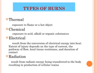
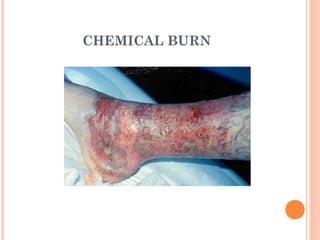
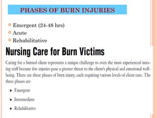
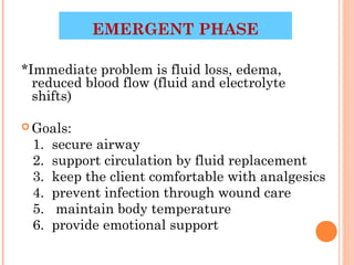
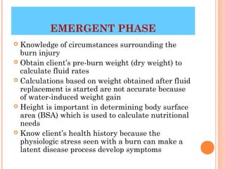
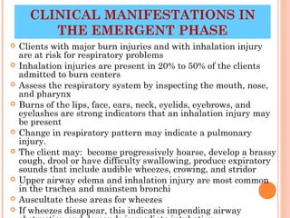
1
BURNS
Wounds caused by exposure to:
 1. excessive heat
 2. Chemicals
 3. fire/steam
 4. radiation
 5. electricity


                                2
TYPES OF BURNS

 Thermal

   exposure to flame or a hot object
 Chemical
    exposure to acid, alkali or organic substances
 Electrical

    result from the conversion of electrical energy into heat.
 Extent of injury depends on the type of current, the
 pathway of flow, local tissue resistance, and duration of
 contact
 Radiation

   result from radiant energy being transferred to the body
 resulting in production of cellular toxins
                                                     3
CHEMICAL BURN


                4
ELECTRICAL BURN


                  5
BURN WOUND
           ASSESSMENT


 Classified  according to depth of injury and
  extent of body surface area involved
 Burn wounds differentiated depending on
  the level of dermis and subcutaneous
  tissue involved
   1. superficial (first-degree)
   2. deep (second-degree)
   3. full thickness (third and fourth
  degree)

     4/1/2011                          6
SUPERFICIAL BURNS
           (FIRST DEGREE)
 Epidermal tissue only affected
 Erythema, blanching on pressure, mild swelling

   no vesicles or blister initially
 Not serious unless large areas involved

 i.e. sunburn


     4/1/2011                            8
4/1/2011
           9
10
DEEP (SECOND DEGREE)

*Involves the epidermis and deep layer of the
   dermis
Fluid-filled vesicles –red, shiny, wet, severe pain
Hospitalization required if over 25% of body
   surface involved
i.e. tar burn, flame


      4/1/2011                               11
12
13
FULL THICKNESS
    (THIRD/FOURTH DEGREE)
 Destruction of all skin layers
 Requires immediate hospitalization

 Dry, waxy white, leathery, or hard skin, no pain

 Exposure to flames, electricity or chemicals can
  cause 3rd degree burns


                                           14
15
CALCULATION OF BURNED BODY
SURFACE AREA


 Calculation  of Burned
      Body Surface Area


   4/1/2011           16
TOTAL BODY SURFACE AREA
             (TBSA)
 Superficial burns are not involved in the
  calculation
 Lund and Browder Chart is the most accurate
  because it adjusts for age
 Rule of nines divides the body – adequate for
  initial assessment for adult burns


     4/1/2011                             17
LUND BROWDER CHART USED FOR
      DETERMINING BSA


   4/1/2011      Evans, 18.1, 2007)
                                 18
RULES OF NINES

 Head & Neck = 9%
 Each upper extremity (Arms) =
  9%
 Each lower extremity (Legs) =
  18%
 Anterior trunk= 18%

 Posterior trunk = 18%

 Genitalia (perineum) = 1%


        4/1/2011                  19
FLUID IMBALANCES
 Occur as a result of fluid shift and cell
  damage
 Hypovolemia

 Metabolic acidosis

 Hyperkalemia

 Hyponatremia

 Hemoconcentration (elevated blood
  osmolarity, hematocrit/hemoglobin) due to
  dehydration

     4/1/2011                       20
PHASES OF BURN INJURIES

 Emergent (24-48 hrs)
 Acute

 Rehabilitative


                          21
EMERGENT PHASE

*Immediate problem is fluid loss, edema,
  reduced blood flow (fluid and electrolyte
  shifts)

 Goals:
 1.   secure airway
 2.   support circulation by fluid replacement
 3.   keep the client comfortable with analgesics
 4.   prevent infection through wound care
 5.    maintain body temperature
 6.   provide emotional support
                                      22
EMERGENT PHASE
 Knowledge of circumstances surrounding the
  burn injury
 Obtain client’s pre-burn weight (dry weight) to
  calculate fluid rates
 Calculations based on weight obtained after fluid
  replacement is started are not accurate because
  of water-induced weight gain
 Height is important in determining body surface
  area (BSA) which is used to calculate nutritional
  needs
 Know client’s health history because the
  physiologic stress seen with a burn can make a
  latent disease process develop symptoms
                                           23
CLINICAL MANIFESTATIONS IN
         THE EMERGENT PHASE
   Clients with major burn injuries and with inhalation injury
    are at risk for respiratory problems
   Inhalation injuries are present in 20% to 50% of the clients
    admitted to burn centers
   Assess the respiratory system by inspecting the mouth, nose,
    and pharynx
   Burns of the lips, face, ears, neck, eyelids, eyebrows, and
    eyelashes are strong indicators that an inhalation injury may
    be present
   Change in respiratory pattern may indicate a pulmonary
    injury.
   The client may: become progressively hoarse, develop a brassy
    cough, drool or have difficulty swallowing, produce expiratory
    sounds that include audible wheezes, crowing, and stridor
   Upper airway edema and inhalation injury are most common
    in the trachea and mainstem bronchi
   Auscultate these areas for wheezes
                                                      24
   If wheezes disappear, this indicates impending airway
CLINICAL
      MANIFESTATIONS


 Cardiovascular    will begin immediately
  which can include shock (Shock is a
  common cause of death in the emergent
  phase in clients with serious injuries)
 Obtain a baseline EKG

 Monitor for edema, measure central and
  peripheral pulses, blood pressure,
  capillary refill and pulse oximetry


       4/1/2011                        25
CLINICAL
        MANIFESTATIONS

 Changes   in renal function are related to
  decreased renal blood flow
 Urine is usually highly concentrated and
  has a high specific gravity
 Urine output is decreased during the first
  24 hours of the emergent phase
 Fluid resuscitation is provided at the rate
  needed to maintain adult urine output at
  30 to 50- mL/hr.
 Measure BUN, creat and NA levels


     4/1/2011                         26
CLINICAL MANIFESTATIONS
 Sympathetic   stimulation during the
  emergent phase causes reduced GI
  motility and paralytic ileus
 Auscultate the abdomen to assess bowel
  sounds which may be reduced
 Monitor for n/v and abdominal distention
 Clients with burns of 25% TBSA or who
  are intubated generally require a NG tube
  inserted to prevent aspiration and
  removal of gastric secretions


     4/1/2011                       27
SKIN ASSESSMENT

 Assess the skin to determine the size and
  depth of burn injury
 The size of the injury is first estimated in
  comparison to the total body surface area
  (TBSA). For example, a burn that
  involves 40% of the TBSA is a 40% burn
 Use the rule of nines for clients whose
  weights are in normal proportion to their
  heights

     4/1/2011                          28
IV FLUID THERAPY

 Infusion   of IV fluids is needed to maintain sufficient blood
  volume.
 Clients with burns involving 15% to 20% of the TBSA
  require IV fluid
 Severeburn requires large fluid loads in a short time to
 maintain blood flow to vital organs.
 Fluidreplacement formulas are calculated from the time of
 injury and not from the time of arrival at the hospital


                                                 29
COMMON FLUIDS

 Protenate or 5% albumin in isotonic saline (1/2
  given in first 8 hr; ½ given in next 16 hr)
 LR (Lactate Ringer) without dextrose (1/2 given
  in first 8 hr; ½ given in next 16 hr)
 Crystalloid (hypertonic saline) adjust to maintain
  urine output at 30 mL/hr
 Crystalloid only (lactated ringers)


                                           30
NURSING DIAGNOSIS IN THE
        EMERGENT PHASE
 Decreased CO
 Deficient fluid volume r/t active fluid volume loss

 Ineffective Tissue perfusion

 Ineffective breathing pattern


      4/1/2011                              31
ACUTE PHASE OF BURN
                  INJURY
•   Lasts until wound closure is complete
•   Care is directed toward continued assessment and
    maintenance of the cardiovascular and respiratory
    system
•   Pneumonia is a concern which can result in
    respiratory failure requiring mechanical ventilation
•   Combating Infection with (Topical antibiotics –
    Silvadene)
•   Tetanus toxoid
•   Weight daily without dressings or splints and compare
    to pre-burn weight
•   A 2% loss of body weight indicates a mild deficit
•   A 10% or greater weight loss requires modification of
                                             32
    calorie intake
NURSING DIAGOSIS IN THE ACUTE
               PHASE
 Impaired skin integrity
 Risk for infection

 Imbalanced nutrition

 Impaired physical mobility

 Disturbed body image


                               33
PLANNING AND IMPLEMENTATION

   Nonsurgical management: removal of exudates
    and necrotic tissue, cleaning the area,
    stimulating granulation and revascularization
    and applying dressings. Debridement may be
    needed


       4/1/2011                            34
DRESSING THE BURN
                   WOUND

 After burn wounds are cleaned and debrided,
  topical antibiotics are reapplied to prevent
  infection
 Standard wound dressings are multiple layers of
  gauze applied over the topical agents on the burn
  wound


                                           35
REHABILITATIVE PHASE OF BURN
            INJURY
 Started  at the time of admission
 Technically begins with wound closure
  and ends when the client returns to the
  highest possible level of functioning
 Provide psychosocial support
 Assess home environment, financial
  resources, medical equipment, prosthetic
  rehab
 Health teaching should include symptoms
  of infection, drugs regimens, f/u
  appointments, comfort measures to reduce
  pruritus
                                   38
DIET

 Initially NPO
 Begin oral fluids after bowel sounds return

 Do not give ice chips or free water lead to
  electrolyte imbalance
 High protein, high calorie


                                           39
GOALS

 Prevent  complications (contractures)
 Vital signs hourly
 Assess respiratory function
 Tetanus booster
 Anti-infective
 Analgesics
 No aspirin
 Strict surgical asepsis
 Turn q2h to prevent contractures
 Emotional support


                                     40
DEBRIDEMENT

 Done with forceps and curved scissor or through
  hydrotherapy (application of water for treatment)
 Only loose eschar removed

 Blisters are left alone to serve as a protector –
  controversial


                                          41
SKIN GRAFTS
 Done during the acute phase
 Used for full-thickness and deep partial-
  thickness wounds


                                          42
POST CARE OF SKIN GRAFTS

 Maintain dressing
 Use aseptic technique
 Graft should look pink if it has taken after 5 days
 Skeletal traction may be used to prevent
  contractures
 Elastic bandages may be applied for 6 mo to 1
  year to prevent hypertrophic scarring


                                            44
Burn lecture
Burn lecture

Burn lecture

  • 1.
  • 2.
    BURNS Wounds caused byexposure to: 1. excessive heat 2. Chemicals 3. fire/steam 4. radiation 5. electricity 2
  • 3.
    TYPES OF BURNS Thermal exposure to flame or a hot object  Chemical exposure to acid, alkali or organic substances  Electrical result from the conversion of electrical energy into heat. Extent of injury depends on the type of current, the pathway of flow, local tissue resistance, and duration of contact  Radiation result from radiant energy being transferred to the body resulting in production of cellular toxins 3
  • 4.
  • 5.
  • 6.
    BURN WOUND ASSESSMENT  Classified according to depth of injury and extent of body surface area involved  Burn wounds differentiated depending on the level of dermis and subcutaneous tissue involved 1. superficial (first-degree) 2. deep (second-degree) 3. full thickness (third and fourth degree) 4/1/2011 6
  • 8.
    SUPERFICIAL BURNS (FIRST DEGREE)  Epidermal tissue only affected  Erythema, blanching on pressure, mild swelling no vesicles or blister initially  Not serious unless large areas involved  i.e. sunburn 4/1/2011 8
  • 9.
  • 10.
  • 11.
    DEEP (SECOND DEGREE) *Involvesthe epidermis and deep layer of the dermis Fluid-filled vesicles –red, shiny, wet, severe pain Hospitalization required if over 25% of body surface involved i.e. tar burn, flame 4/1/2011 11
  • 12.
  • 13.
  • 14.
    FULL THICKNESS (THIRD/FOURTH DEGREE)  Destruction of all skin layers  Requires immediate hospitalization  Dry, waxy white, leathery, or hard skin, no pain  Exposure to flames, electricity or chemicals can cause 3rd degree burns 14
  • 15.
  • 16.
    CALCULATION OF BURNEDBODY SURFACE AREA Calculation of Burned Body Surface Area 4/1/2011 16
  • 17.
    TOTAL BODY SURFACEAREA (TBSA)  Superficial burns are not involved in the calculation  Lund and Browder Chart is the most accurate because it adjusts for age  Rule of nines divides the body – adequate for initial assessment for adult burns 4/1/2011 17
  • 18.
    LUND BROWDER CHARTUSED FOR DETERMINING BSA 4/1/2011 Evans, 18.1, 2007) 18
  • 19.
    RULES OF NINES Head & Neck = 9%  Each upper extremity (Arms) = 9%  Each lower extremity (Legs) = 18%  Anterior trunk= 18%  Posterior trunk = 18%  Genitalia (perineum) = 1% 4/1/2011 19
  • 20.
    FLUID IMBALANCES  Occuras a result of fluid shift and cell damage  Hypovolemia  Metabolic acidosis  Hyperkalemia  Hyponatremia  Hemoconcentration (elevated blood osmolarity, hematocrit/hemoglobin) due to dehydration 4/1/2011 20
  • 21.
    PHASES OF BURNINJURIES  Emergent (24-48 hrs)  Acute  Rehabilitative 21
  • 22.
    EMERGENT PHASE *Immediate problemis fluid loss, edema, reduced blood flow (fluid and electrolyte shifts)  Goals: 1. secure airway 2. support circulation by fluid replacement 3. keep the client comfortable with analgesics 4. prevent infection through wound care 5. maintain body temperature 6. provide emotional support 22
  • 23.
    EMERGENT PHASE  Knowledgeof circumstances surrounding the burn injury  Obtain client’s pre-burn weight (dry weight) to calculate fluid rates  Calculations based on weight obtained after fluid replacement is started are not accurate because of water-induced weight gain  Height is important in determining body surface area (BSA) which is used to calculate nutritional needs  Know client’s health history because the physiologic stress seen with a burn can make a latent disease process develop symptoms 23
  • 24.
    CLINICAL MANIFESTATIONS IN THE EMERGENT PHASE  Clients with major burn injuries and with inhalation injury are at risk for respiratory problems  Inhalation injuries are present in 20% to 50% of the clients admitted to burn centers  Assess the respiratory system by inspecting the mouth, nose, and pharynx  Burns of the lips, face, ears, neck, eyelids, eyebrows, and eyelashes are strong indicators that an inhalation injury may be present  Change in respiratory pattern may indicate a pulmonary injury.  The client may: become progressively hoarse, develop a brassy cough, drool or have difficulty swallowing, produce expiratory sounds that include audible wheezes, crowing, and stridor  Upper airway edema and inhalation injury are most common in the trachea and mainstem bronchi  Auscultate these areas for wheezes 24  If wheezes disappear, this indicates impending airway
  • 25.
    CLINICAL MANIFESTATIONS  Cardiovascular will begin immediately which can include shock (Shock is a common cause of death in the emergent phase in clients with serious injuries)  Obtain a baseline EKG  Monitor for edema, measure central and peripheral pulses, blood pressure, capillary refill and pulse oximetry 4/1/2011 25
  • 26.
    CLINICAL MANIFESTATIONS  Changes in renal function are related to decreased renal blood flow  Urine is usually highly concentrated and has a high specific gravity  Urine output is decreased during the first 24 hours of the emergent phase  Fluid resuscitation is provided at the rate needed to maintain adult urine output at 30 to 50- mL/hr.  Measure BUN, creat and NA levels 4/1/2011 26
  • 27.
    CLINICAL MANIFESTATIONS  Sympathetic stimulation during the emergent phase causes reduced GI motility and paralytic ileus  Auscultate the abdomen to assess bowel sounds which may be reduced  Monitor for n/v and abdominal distention  Clients with burns of 25% TBSA or who are intubated generally require a NG tube inserted to prevent aspiration and removal of gastric secretions 4/1/2011 27
  • 28.
    SKIN ASSESSMENT  Assessthe skin to determine the size and depth of burn injury  The size of the injury is first estimated in comparison to the total body surface area (TBSA). For example, a burn that involves 40% of the TBSA is a 40% burn  Use the rule of nines for clients whose weights are in normal proportion to their heights 4/1/2011 28
  • 29.
    IV FLUID THERAPY Infusion of IV fluids is needed to maintain sufficient blood volume.  Clients with burns involving 15% to 20% of the TBSA require IV fluid  Severeburn requires large fluid loads in a short time to maintain blood flow to vital organs.  Fluidreplacement formulas are calculated from the time of injury and not from the time of arrival at the hospital 29
  • 30.
    COMMON FLUIDS  Protenateor 5% albumin in isotonic saline (1/2 given in first 8 hr; ½ given in next 16 hr)  LR (Lactate Ringer) without dextrose (1/2 given in first 8 hr; ½ given in next 16 hr)  Crystalloid (hypertonic saline) adjust to maintain urine output at 30 mL/hr  Crystalloid only (lactated ringers) 30
  • 31.
    NURSING DIAGNOSIS INTHE EMERGENT PHASE  Decreased CO  Deficient fluid volume r/t active fluid volume loss  Ineffective Tissue perfusion  Ineffective breathing pattern 4/1/2011 31
  • 32.
    ACUTE PHASE OFBURN INJURY • Lasts until wound closure is complete • Care is directed toward continued assessment and maintenance of the cardiovascular and respiratory system • Pneumonia is a concern which can result in respiratory failure requiring mechanical ventilation • Combating Infection with (Topical antibiotics – Silvadene) • Tetanus toxoid • Weight daily without dressings or splints and compare to pre-burn weight • A 2% loss of body weight indicates a mild deficit • A 10% or greater weight loss requires modification of 32 calorie intake
  • 33.
    NURSING DIAGOSIS INTHE ACUTE PHASE  Impaired skin integrity  Risk for infection  Imbalanced nutrition  Impaired physical mobility  Disturbed body image 33
  • 34.
    PLANNING AND IMPLEMENTATION  Nonsurgical management: removal of exudates and necrotic tissue, cleaning the area, stimulating granulation and revascularization and applying dressings. Debridement may be needed 4/1/2011 34
  • 35.
    DRESSING THE BURN WOUND  After burn wounds are cleaned and debrided, topical antibiotics are reapplied to prevent infection  Standard wound dressings are multiple layers of gauze applied over the topical agents on the burn wound 35
  • 38.
    REHABILITATIVE PHASE OFBURN INJURY  Started at the time of admission  Technically begins with wound closure and ends when the client returns to the highest possible level of functioning  Provide psychosocial support  Assess home environment, financial resources, medical equipment, prosthetic rehab  Health teaching should include symptoms of infection, drugs regimens, f/u appointments, comfort measures to reduce pruritus 38
  • 39.
    DIET  Initially NPO Begin oral fluids after bowel sounds return  Do not give ice chips or free water lead to electrolyte imbalance  High protein, high calorie 39
  • 40.
    GOALS  Prevent complications (contractures)  Vital signs hourly  Assess respiratory function  Tetanus booster  Anti-infective  Analgesics  No aspirin  Strict surgical asepsis  Turn q2h to prevent contractures  Emotional support 40
  • 41.
    DEBRIDEMENT  Done withforceps and curved scissor or through hydrotherapy (application of water for treatment)  Only loose eschar removed  Blisters are left alone to serve as a protector – controversial 41
  • 42.
    SKIN GRAFTS  Doneduring the acute phase  Used for full-thickness and deep partial- thickness wounds 42
  • 44.
    POST CARE OFSKIN GRAFTS  Maintain dressing  Use aseptic technique  Graft should look pink if it has taken after 5 days  Skeletal traction may be used to prevent contractures  Elastic bandages may be applied for 6 mo to 1 year to prevent hypertrophic scarring 44