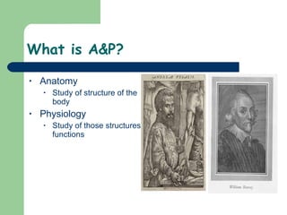
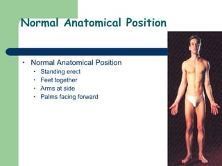
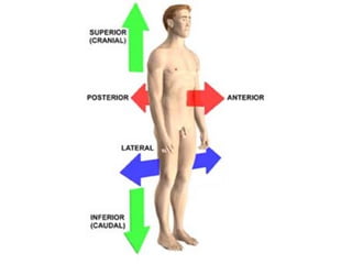
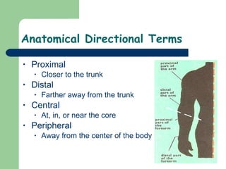
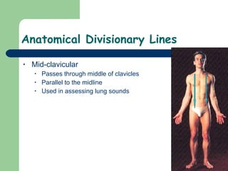
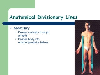
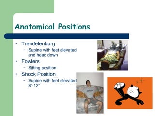
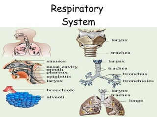
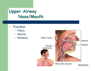
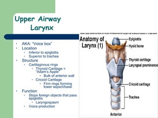
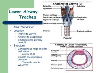
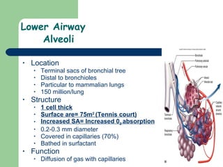
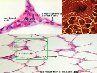
The Human Body
What is A&P? Anatomy Study of structure of the body Physiology Study of those structures functions
Normal Anatomical Position Normal Anatomical Position Standing erect Feet together Arms at side Palms facing forward
Anatomical Directional Terms Superior Further from the ground/Close to head Inferior Close to the ground/Close to feet Anterior Toward front of body (AKA. Ventral) Posterior Toward back of body (AKA. Dorsal) Medial  Close to the midline Lateral Farther away from the midline  Bilateral- Both R and L side
 
Anatomical Directional Terms Proximal Closer to the trunk Distal Farther away from the trunk Central At, in, or near the core Peripheral  Away from the center of the body
Anatomical Divisionary Lines Midline  Divides body into Right and Left halves.
Anatomical Divisionary Lines Mid-clavicular Passes through middle of clavicles Parallel to the midline  Used in assessing lung sounds
Anatomical Divisionary Lines Midaxillary Passes vertically through armpits Divides body into anterior/posterior halves
Anatomical Surfaces Palmer Surface Palm of hands Plantar Surface Sole of foot
Anatomical Positions  Supine Lying on back Prone Lying face down Recumbent Lying on side
Anatomical Positions Trendelenburg Supine with feet elevated and head down Fowlers Sitting position Shock Position Supine with feet elevated 8”-12”
Anatomical Motion Terms Abduction “ ab”=away Movement away from body Adduction “ add”= toward Movement toward to the body  Flexion Bending of a joint Extension  Straightening of a joint
Body Systems Organization Cells – Tissues – Organ - Organ System - Organism Tissue Types Muscle Contract to allow motion Smooth, Skeletal, Cardiac  Nervous Conduct impulses Epithelial Protective and semi permeable Connective  Structure/protection/attachment  Extracellular matrix, bone, cartilage, blood
 
Skeletal System 14% total body weight Function Shape, Movement, Storage  (Calcium, Phosphorus, Iron) Protection of organs Blood cell production Endocrine Regularion  Components Bones Mineralized osseous tissue Cartilage Soft connective tissue/Collagen Flexible support Tendons Attach muscle to bone Collagen, Elastin, Proteogylcans Ligaments Attach bone to bone  Collagen Axial Skeleton Skull, vertebral column, thoracic cage Appendicular Skeleton Upper limbs, pelvic girdle, lower limbs
 
Axial Skeleton Skull  Skull 22 bones Cranium + Face Encases brain Brain + CSF + Vessels Little space Facial Bones Orbits Eyes Nasal Bones Maxilla Upper Jaw Zygomatics Cheekbones  Mandible  Lower Jaw
 
Axial Skeletal Vertebral Column  Function Support Components  33 Vertebrae Intervertebral disks  Divisions of Vertebrae Cervical (C-) =  7 Neck C1 = Atlas C2 = Axis  Thoracic (T-)=  12 Chest Lumbar  (L-)=  5  Lower Back Sacral  (S-)=  5  Back of pelvis (fused)  Coccyx  (C-)=  4  Tailbone (fused)
Axial Skeletal  Thoracic Cage Function Protection of thoracic cavity Supports shoulder girdle  Components  12 pairs of ribs 1-10 attach to sternum 11-12 “float”  Sternum  Manubrium Body Xiphoid process Costal cartilage Connect ribs to sternum Thoracic vertebrae (12)
Appendicular Skeleton Upper Extremities  Clavicle Scapula Acromion (Tip of shoulder) Humerus  Olecranon (elbow) Radius Ulna Carpals (wrist) Metacarpals (hands) Phalanges (fingers)
Appendicular Skeleton Lower Extremities Greater Trochanter (head of femur) Acetabulum (socket of hip) Femur (thigh) Patella (knee) Tibia  (shin) Fibula  (lower leg) Medial/Lateral malleolus  (ankle) Tarsals and metatarsals (foot) Calcaneus (heel) Phalanges (toes)
Appendicular Skeleton Pelvis Function Support and protection Components  Ilium  Wings  Pubis Anterior portion Ischium Inferior portion Sacrum Coccyx
Joints Definition 2 or more bones connecting to bones Types Ball and socket Hip/Shoulder Wide Range of motion Hinge Elbow/knee Motion in 1 plane Flexion and extension only
Body Cavities
Body Cavities Cranial Enclosed by skull Contains: brain, eyes, ears Vertebral Enclosed by vertebrae Contains: spinal cord Thoracic Enclosed by ribcage Bounded by diaphragm  Contains: trachea, esophagus, lungs, bronchi, bronchioles, alveoli, heart, great vessels, thymus gland,  Abdominopelvic Abdominal Enclosed by ribcage and pelvis Contains: kidneys, ureters, stomach, large/small intestines, liver, gallbladder, pancreas, spleen Divided into quadrants (next slide) Pelvic Enclosed by pelvis Contains: urinary bladder, anus, reproductive organs
Abdominal Quadrants
Respiratory  System
Respiratory System  Function Gas exchange with outside environment Filtration/Humidification/Warming/Conduction of air Structures Nose Mouth Naso/Oro/Laryngopharynx Larynx Bronchi Bronchioles Lungs Diaphragm Associated muscles  Alveoli
Upper Airway  Nose/Mouth Function Filters Warms Moistens
Upper Airway  Pharynx Location Posterior to mouth Superior to esophagus, larynx, trachea Function Conducts air to bronchi 3 Divisions Nasopharynx Oropharynx Laryngopharynx
Upper Airway  Epiglottis  Location Sits posterior to larynx Attached to tongue  Structure Leaf shaped cartilage Function Prevents food/liquid from entering larynx during swallowing  Guards opening to vocal cords (glottis)
Upper Airway  Larynx AKA: “Voice box” Location Inferior to epiglottis Superior to trachea   Structure   Cartilaginous rings Thyroid Cartilage = “Adam’s Apple” Bulk of anterior wall Cricoid Cartilage Firm rings forming lower aspect/base Function Stops foreign objects that pass epiglottis Laryngospasm Voice production
Lower Airway  Trachea AKA: “Windpipe”  Location Inferior to Larynx Anterior to Esophagus Bifurcates into primary bronchi Structure  Cartilaginous rings anterior and lateral Approx 15-20  Smooth muscle tissue posterior  Trachealis muscle  Why????
Lower Airway  Bronchi Location Bifurcation of trachea  2 nd  Intercostal space Angle of Louis Right and Left main stem Structure Smooth muscle Irregular hyaline cartilage rings  Function Conducts air to lungs
Lower Airway  Bronchioles  Location Distal bifurcations of the bronchi Terminate at alveoli Function Conduct air to alveoli  Structure 1 st  airways with NO cartilage ALL muscle  Bronchoconstriction  Bronchospasm < 1 mm wide =Tiny
Lower Airway  Alveoli Location Terminal sacs of bronchial tree Distal to bronchioles Particular to mammalian lungs 150 million/lung Structure 1 cell thick Surface are= 75m 2  (Tennis court) Increased SA= Increased 0 2  absorption 0.2-0.3 mm diameter  Covered in capillaries (70%) Bathed in surfactant  Function Diffusion of gas with capillaries
 
Lower Airway  Lungs Location Bilateral of midline Structure Divided into lobes Left= 2 Right= 3 Function Houses structure for gas exchange Alteration of pH
Lower Airway Mucociliary Escalator  Location Along epithelium of primary bronchi Beat in rhythm  Structure Cilia projections “ Hair like” Function Move debris up out of lungs Cough or swallow Smokers… Prevent mucous accumulation
 
Respiratory Physiology How we breathe…  Ventilation Mechanical movement of air into/out of the body Inhalation ( Active ) Muscles Used Diaphragm & External Intercostals Physiology  Diaphragm contracts downward External intercostals pull ribs up and out Increases dimension of chest cavity Increased diameter of chest drops intra thoracic pressure Air rushes in until pressure is equalized
Respiratory Physiology How we breathe… Ventilation Mechanical movement of air into/out of the body Exhalation ( Passive ) Physiology Diaphragm relaxes as well as intercostals Chest cavity dimension decreases Decrease in dimension increases intrathoracic pressure Air rushes out  Lungs recoil
 
Respiratory Physiology Gas Exchange Respiration Process by which the body utilizes oxygen Diffusion Net movement of molecules from an area of high concentration to an area of low concentration
 
Respiratory Physiology Gas Exchange Respiration Process by which the body utilizes oxygen Alveolar/Capillary Exchange Physiology O 2  rich air enters alveoli O 2  poor blood in capillaries pass alveoli O 2  diffuses down its concentration gradient into the capillaries CO 2  diffuses down its concentration gradient into the alveoli CO 2  is exhaled and O 2  transported to tissues
Respiratory Physiology Gas Exchange Respiration Process by which the body utilizes oxygen Capillary/Cellular Exchange Physiology O 2  rich blood passes cells O 2  diffuses across its concentration gradient into the cells CO 2  diffuses across its concentration gradient into the capillary CO 2  is transported to the alveoli
Respiratory Evaluation Areas of assessment Rate. Rhythm. Depth. Quality. Rate Adult = 12-20 per minute Child = 15-30 per minute Infant -= 30-60 per minute Rhythm Regular or irregular Depth Tidal volume adequate or inadequate Amount of air breathed in/out in one ventilation Approx 500 mL
Respiratory Evaluation cont’d. Quality Breath sounds Midclavicular & Midaxillary lines Present or diminished or absent Chest expansion Unequal or symmetrical Increased effort Accessory muscles  “ Seesaw” breathing  Infants Nasal flaring  Retractions  Above clavicles, between ribs Cyanosis Shortness of breath Altered mental status
Accessory Muscle Use Nasal Flaring Retractions
Respiratory Evaluation cont’d. Cyanosis Blue/pale coloring of skin Nail beds Lips Eyelids  Why is this seen in these areas first???  Indicates poor perfusion
Pediatric Considerations Mouth/Nose Smaller and easily obstructed Pharynx Tongue is BIG  Trachea Narrower Softer and more flexible  Cricoid Cartilage Less developed/Less rigid = easily kinked Diaphragm  Chest is soft Depend on diaphragm to do most of the work of breathing Seesaw Breathing….
The Circulatory System
The Circulatory System Function/Components Function Transport system of the body Delivers O2 and nutrients to cells Returns waste to liver/kidneys/lungs Transports specialized cells to fight infection. Components Blood Blood vessels 60,000 – 100,000 miles Heart
The Circulatory System The heart Location Just left of midline Posterior to sternum Anterior to T-spine Function Pump for driving of blood flow
 
The Circulatory System Chambers of the Heart 4 Chambers Divided by a septum 2 Atria Receiving chambers Contract together Superior to ventricles 2 Ventricles  Pumping Chambers Contract together Inferior to atria
The Circulatory System Valves of The Heart Function Prevents backflow of blood Create heart sounds Atrioventricular Valvues Between each atria and its ventricle Tricuspid Valve Between Right Atria/Ventricle Bicuspid/Mitral Valve Between Left Atria/Ventricle Semilunar Valves Between each ventricle and its artery Pulmonic Valve Right Ventricle and Pulmonary Artery Aortic Valve Left Ventricle and Aorta
The Circulatory System Types of Circulation Separate Systems Pulmonary Right ventricle Blood to the lungs Oxygenation Systemic Left ventricle Blood to the body Perfusion
The Circulatory System Conductive System Automaticity Ability to create own electrical signal Pacemaker Sites Sinoatrail Node (SA) 60-100 bpm Atrioventricular Node (AV) 40-60 bpm Bundle of HIS 40-60 bpm Purkinje Fibers 20-40 bpm
 
The Circulatory System Blood Vessels Arteries  Arterioles  Capillaries  Venules  Veins
The Circulatory System Arteries Function Conduct blood away from heart High pressure Structure Endothelial lining Connective tissue  THICK Smooth muscle Connective tissue Allows for great expansion of vessels
The Circulatory System Major Arteries  Coronary Supply heart with blood Aorta Major artery from the heart to the body 1” diameter Pulmonary Carries O2 poor blood to lungs Umbilical  Carries O2 poor blood to lungs Carotid Major artery of the neck Supplies the head with blood Femoral Major artery of the thigh Supplies lower extremities with blood Bifurcation of aorta at navel  Radial Major artery of the lower arm Brachial Artery of the upper arm Posterior tibial Artery running posterior to ankle Dorsalis pedis Artery of the foot Anterior
 
The Circulatory System Arterioles Smallest branch of an artery Leads to a capillary Structure Thin smooth muscle wall (1-2 layers) Function Main site of vascular resistance  Important in blood pressure
The Circulatory System Capillaries Structure Tiny blood vessel 5-10  μ m diameter 1 endothelial cell thick Function Connect arterioles to venules Exchange of gases, nutrients, etc. Decrease pressure Usually carries no more than 50% of the volume it could
The Circulatory System Venule Smallest branch of a vein leading from a capillary Structure A vein on small scale Internal valves   Function  Conducts deoxygenated blood out of capillaries into veins
The Circulatory System Veins Function Return blood to the heart Low pressure system Structure Endothelial lining Connective tissue  Thinner Smooth muscle Connective tissue Internal Valves
The Circulatory System Major Veins Pulmonary Carries O2 rich blood from lungs to left atrium Umbilical Carries O2 rich blood from lungs to left atrium Vena Cava Superior Drains head/upper extremities Inferior Drains trunk/lower extremities
 
Path of blood through the heart Putting it all together Pulmonary Circulation:  Blood from the body enters Right atrium via venae cavae  Right Atrium contracts Blood enters Right ventricle via Tricuspid valve Right Ventricle contracts Blood enters Pulmonary artery via Pulmonic Valve Pulmonary artery carries blood to lungs for gas exchange
 
Path of blood through the heart Putting it all together Systemic Circulation: Blood enters the L atrium via Pulmonary veins Atrium contracts Blood enters the L ventricle via Mitral/Bicuspid Valve Ventricle contracts Blood enters the aorta via Aortic Valve Aorta conducts blood to body  Pumps Your Blood Song
 
The Circulatory System Blood 5-6 Liters Components Plasma Fluid that carries blood cells/nutrients 55% Formed Elements  40% Red Blood Cells Carry O2 to organs & CO2 away Give blood its color Hemoglobin 45%, 4.2-6.1 million/mL White Blood Cells Defense  4,3000-10,800 WBC/mL  Platelets   Clotting  150,000 - 350,000/mL
The Circulatory System Physiology  Pulse Pulse Palpable wave of blood sent though arteries after contraction of L ventricle  Peripheral Radial Brachial Posterior tibial Dorsalis pedis Central Carotid  Femoral
The Circulatory System  Physiology Blood Pressure Blood pressure Force exerted from blood on walls of vessels  Phases of Cardiac Cycle Systolic Pressure against the walls when the L ventricle contracts Diastolic  Pressure against the walls when the L ventricle relaxes
The Circulatory System Physiology Perfusion Perfusion In an organ system:  Delivery of O2/nutrients  Removal of waste products
The Circulatory System Pathology Shock Shock/Hypoperfusion  Failure of the circulatory system to adequately perfuse and oxygenate the tissues of the body  Signs/Symptoms Pale, cool, cyanotic, clammy skin Rapid/shallow breathing Restlessness/anxiety Nausea/vomiting  Weak pulse Low blood pressure/volume
Musculoskeletal System
Musculoskeletal System Function Body shape Protection of organs Movement Blood cell production Components   Muscle tissue Skeletal Smooth Cardiac   Skeletal  Ligaments Tendons Skeletal tissue
Musculoskeletal System Skeletal Tissue Skeletal Voluntary Attached to bone Tendons Form major muscle groups of the body
Musculoskeletal System Smooth Tissue Involuntary Location Walls of tubular structures  GI and urinary tract Blood vessels Function Control flow  Carry out automatic functions of the body
Musculoskeletal System Cardiac Tissue Involuntary Location Only in heart Function Create/conduct electrical impulses Automaticity
The Nervous System
The Nervous System Function Controls voluntary/involuntary activity Components  Central Nervous System (Computer) Brain Brainstem  Spinal Cord Peripheral Nervous System (Communicator) Associated nerves Sensory- Carry info from body to brain Motor – Carry info from the brain to the body Divided into  Somatic NS = voluntary Autonomic NS= Involuntary
 
Divisions of the Autonomic  Nervous System Sympathetic “Fight or flight” Parasympathetic “Feed or breed” OR
 
The Nervous System  The Brain Cerebrum Largest most superior portion of the brain Divided into R & L hemispheres Hemispheres divided into specialized lobes Frontal = Intellect and motor function Occipital = Eyesight Temporal = Smell/Hearing Parietal = Sensory information Brainstem Lower part of the brain Circulation, Respiration, BP Cerebellum Outpocketing of brain, posterior to brainstem Coordination and movement
 
The Nervous System  The Brain: Blood Supply Cerebral Blood Supply 15% of Cardiac output 80% of blood is supplied by the carotid arteries Vertebral arteries supply the rest Circle of Willis Each area of the brain has its own blood supply Sensitivity to Deprivation of glucose and O2 Cannot store glucose itself Deprivation = AMS   Interruption in O2 supply Unconsciousness 5-10 seconds  Blockage of O2 supply  Neural death 4-6 minutes
 
Integumentary System   Skin
Integumentary System   Skin Function Largest organ system in the body Protection from environment  Temperature regulation Senses  Heat, Cold, Touch, Pressure, Pain, etc.  Vitamin D synthesis Storage  Structure Epidermis Outermost layer No blood vessels Protection, absorption of nutrients  homeostasis Dermis Deeper layer Contains sweat/sebaceous glands, hair folicles, blood vessels, nerve endings Gives skin its flexibility  Subcutaneous layer Fat layer Insulation, protective padding, energy storage
 
Endocrine System Function Secretes chemicals (hormones) that regulate body activities i.e. Insulin & adrenalin  Structures Pituitary Pineal gland Hypothalamus  Thyroid Parathyroid Adrenals Pancreas Islets of Langerhans Ovaries Testes
That does it

6)The Human Body

  • 1.
  • 2.
    What is A&P?Anatomy Study of structure of the body Physiology Study of those structures functions
  • 3.
    Normal Anatomical PositionNormal Anatomical Position Standing erect Feet together Arms at side Palms facing forward
  • 4.
    Anatomical Directional TermsSuperior Further from the ground/Close to head Inferior Close to the ground/Close to feet Anterior Toward front of body (AKA. Ventral) Posterior Toward back of body (AKA. Dorsal) Medial Close to the midline Lateral Farther away from the midline Bilateral- Both R and L side
  • 5.
  • 6.
    Anatomical Directional TermsProximal Closer to the trunk Distal Farther away from the trunk Central At, in, or near the core Peripheral Away from the center of the body
  • 7.
    Anatomical Divisionary LinesMidline Divides body into Right and Left halves.
  • 8.
    Anatomical Divisionary LinesMid-clavicular Passes through middle of clavicles Parallel to the midline Used in assessing lung sounds
  • 9.
    Anatomical Divisionary LinesMidaxillary Passes vertically through armpits Divides body into anterior/posterior halves
  • 10.
    Anatomical Surfaces PalmerSurface Palm of hands Plantar Surface Sole of foot
  • 11.
    Anatomical Positions Supine Lying on back Prone Lying face down Recumbent Lying on side
  • 12.
    Anatomical Positions TrendelenburgSupine with feet elevated and head down Fowlers Sitting position Shock Position Supine with feet elevated 8”-12”
  • 13.
    Anatomical Motion TermsAbduction “ ab”=away Movement away from body Adduction “ add”= toward Movement toward to the body Flexion Bending of a joint Extension Straightening of a joint
  • 14.
    Body Systems OrganizationCells – Tissues – Organ - Organ System - Organism Tissue Types Muscle Contract to allow motion Smooth, Skeletal, Cardiac Nervous Conduct impulses Epithelial Protective and semi permeable Connective Structure/protection/attachment Extracellular matrix, bone, cartilage, blood
  • 15.
  • 16.
    Skeletal System 14%total body weight Function Shape, Movement, Storage (Calcium, Phosphorus, Iron) Protection of organs Blood cell production Endocrine Regularion Components Bones Mineralized osseous tissue Cartilage Soft connective tissue/Collagen Flexible support Tendons Attach muscle to bone Collagen, Elastin, Proteogylcans Ligaments Attach bone to bone Collagen Axial Skeleton Skull, vertebral column, thoracic cage Appendicular Skeleton Upper limbs, pelvic girdle, lower limbs
  • 17.
  • 18.
    Axial Skeleton Skull Skull 22 bones Cranium + Face Encases brain Brain + CSF + Vessels Little space Facial Bones Orbits Eyes Nasal Bones Maxilla Upper Jaw Zygomatics Cheekbones Mandible Lower Jaw
  • 19.
  • 20.
    Axial Skeletal VertebralColumn Function Support Components 33 Vertebrae Intervertebral disks Divisions of Vertebrae Cervical (C-) = 7 Neck C1 = Atlas C2 = Axis Thoracic (T-)= 12 Chest Lumbar (L-)= 5 Lower Back Sacral (S-)= 5 Back of pelvis (fused) Coccyx (C-)= 4 Tailbone (fused)
  • 21.
    Axial Skeletal Thoracic Cage Function Protection of thoracic cavity Supports shoulder girdle Components 12 pairs of ribs 1-10 attach to sternum 11-12 “float” Sternum Manubrium Body Xiphoid process Costal cartilage Connect ribs to sternum Thoracic vertebrae (12)
  • 22.
    Appendicular Skeleton UpperExtremities Clavicle Scapula Acromion (Tip of shoulder) Humerus Olecranon (elbow) Radius Ulna Carpals (wrist) Metacarpals (hands) Phalanges (fingers)
  • 23.
    Appendicular Skeleton LowerExtremities Greater Trochanter (head of femur) Acetabulum (socket of hip) Femur (thigh) Patella (knee) Tibia (shin) Fibula (lower leg) Medial/Lateral malleolus (ankle) Tarsals and metatarsals (foot) Calcaneus (heel) Phalanges (toes)
  • 24.
    Appendicular Skeleton PelvisFunction Support and protection Components Ilium Wings Pubis Anterior portion Ischium Inferior portion Sacrum Coccyx
  • 25.
    Joints Definition 2or more bones connecting to bones Types Ball and socket Hip/Shoulder Wide Range of motion Hinge Elbow/knee Motion in 1 plane Flexion and extension only
  • 26.
  • 27.
    Body Cavities CranialEnclosed by skull Contains: brain, eyes, ears Vertebral Enclosed by vertebrae Contains: spinal cord Thoracic Enclosed by ribcage Bounded by diaphragm Contains: trachea, esophagus, lungs, bronchi, bronchioles, alveoli, heart, great vessels, thymus gland, Abdominopelvic Abdominal Enclosed by ribcage and pelvis Contains: kidneys, ureters, stomach, large/small intestines, liver, gallbladder, pancreas, spleen Divided into quadrants (next slide) Pelvic Enclosed by pelvis Contains: urinary bladder, anus, reproductive organs
  • 28.
  • 29.
  • 30.
    Respiratory System Function Gas exchange with outside environment Filtration/Humidification/Warming/Conduction of air Structures Nose Mouth Naso/Oro/Laryngopharynx Larynx Bronchi Bronchioles Lungs Diaphragm Associated muscles Alveoli
  • 31.
    Upper Airway Nose/Mouth Function Filters Warms Moistens
  • 32.
    Upper Airway Pharynx Location Posterior to mouth Superior to esophagus, larynx, trachea Function Conducts air to bronchi 3 Divisions Nasopharynx Oropharynx Laryngopharynx
  • 33.
    Upper Airway Epiglottis Location Sits posterior to larynx Attached to tongue Structure Leaf shaped cartilage Function Prevents food/liquid from entering larynx during swallowing Guards opening to vocal cords (glottis)
  • 34.
    Upper Airway Larynx AKA: “Voice box” Location Inferior to epiglottis Superior to trachea Structure Cartilaginous rings Thyroid Cartilage = “Adam’s Apple” Bulk of anterior wall Cricoid Cartilage Firm rings forming lower aspect/base Function Stops foreign objects that pass epiglottis Laryngospasm Voice production
  • 35.
    Lower Airway Trachea AKA: “Windpipe” Location Inferior to Larynx Anterior to Esophagus Bifurcates into primary bronchi Structure Cartilaginous rings anterior and lateral Approx 15-20 Smooth muscle tissue posterior Trachealis muscle Why????
  • 36.
    Lower Airway Bronchi Location Bifurcation of trachea 2 nd Intercostal space Angle of Louis Right and Left main stem Structure Smooth muscle Irregular hyaline cartilage rings Function Conducts air to lungs
  • 37.
    Lower Airway Bronchioles Location Distal bifurcations of the bronchi Terminate at alveoli Function Conduct air to alveoli Structure 1 st airways with NO cartilage ALL muscle Bronchoconstriction Bronchospasm < 1 mm wide =Tiny
  • 38.
    Lower Airway Alveoli Location Terminal sacs of bronchial tree Distal to bronchioles Particular to mammalian lungs 150 million/lung Structure 1 cell thick Surface are= 75m 2 (Tennis court) Increased SA= Increased 0 2 absorption 0.2-0.3 mm diameter Covered in capillaries (70%) Bathed in surfactant Function Diffusion of gas with capillaries
  • 39.
  • 40.
    Lower Airway Lungs Location Bilateral of midline Structure Divided into lobes Left= 2 Right= 3 Function Houses structure for gas exchange Alteration of pH
  • 41.
    Lower Airway MucociliaryEscalator Location Along epithelium of primary bronchi Beat in rhythm Structure Cilia projections “ Hair like” Function Move debris up out of lungs Cough or swallow Smokers… Prevent mucous accumulation
  • 42.
  • 43.
    Respiratory Physiology Howwe breathe… Ventilation Mechanical movement of air into/out of the body Inhalation ( Active ) Muscles Used Diaphragm & External Intercostals Physiology Diaphragm contracts downward External intercostals pull ribs up and out Increases dimension of chest cavity Increased diameter of chest drops intra thoracic pressure Air rushes in until pressure is equalized
  • 44.
    Respiratory Physiology Howwe breathe… Ventilation Mechanical movement of air into/out of the body Exhalation ( Passive ) Physiology Diaphragm relaxes as well as intercostals Chest cavity dimension decreases Decrease in dimension increases intrathoracic pressure Air rushes out Lungs recoil
  • 45.
  • 46.
    Respiratory Physiology GasExchange Respiration Process by which the body utilizes oxygen Diffusion Net movement of molecules from an area of high concentration to an area of low concentration
  • 47.
  • 48.
    Respiratory Physiology GasExchange Respiration Process by which the body utilizes oxygen Alveolar/Capillary Exchange Physiology O 2 rich air enters alveoli O 2 poor blood in capillaries pass alveoli O 2 diffuses down its concentration gradient into the capillaries CO 2 diffuses down its concentration gradient into the alveoli CO 2 is exhaled and O 2 transported to tissues
  • 49.
    Respiratory Physiology GasExchange Respiration Process by which the body utilizes oxygen Capillary/Cellular Exchange Physiology O 2 rich blood passes cells O 2 diffuses across its concentration gradient into the cells CO 2 diffuses across its concentration gradient into the capillary CO 2 is transported to the alveoli
  • 50.
    Respiratory Evaluation Areasof assessment Rate. Rhythm. Depth. Quality. Rate Adult = 12-20 per minute Child = 15-30 per minute Infant -= 30-60 per minute Rhythm Regular or irregular Depth Tidal volume adequate or inadequate Amount of air breathed in/out in one ventilation Approx 500 mL
  • 51.
    Respiratory Evaluation cont’d.Quality Breath sounds Midclavicular & Midaxillary lines Present or diminished or absent Chest expansion Unequal or symmetrical Increased effort Accessory muscles “ Seesaw” breathing Infants Nasal flaring Retractions Above clavicles, between ribs Cyanosis Shortness of breath Altered mental status
  • 52.
    Accessory Muscle UseNasal Flaring Retractions
  • 53.
    Respiratory Evaluation cont’d.Cyanosis Blue/pale coloring of skin Nail beds Lips Eyelids Why is this seen in these areas first??? Indicates poor perfusion
  • 54.
    Pediatric Considerations Mouth/NoseSmaller and easily obstructed Pharynx Tongue is BIG Trachea Narrower Softer and more flexible Cricoid Cartilage Less developed/Less rigid = easily kinked Diaphragm Chest is soft Depend on diaphragm to do most of the work of breathing Seesaw Breathing….
  • 55.
  • 56.
    The Circulatory SystemFunction/Components Function Transport system of the body Delivers O2 and nutrients to cells Returns waste to liver/kidneys/lungs Transports specialized cells to fight infection. Components Blood Blood vessels 60,000 – 100,000 miles Heart
  • 57.
    The Circulatory SystemThe heart Location Just left of midline Posterior to sternum Anterior to T-spine Function Pump for driving of blood flow
  • 58.
  • 59.
    The Circulatory SystemChambers of the Heart 4 Chambers Divided by a septum 2 Atria Receiving chambers Contract together Superior to ventricles 2 Ventricles Pumping Chambers Contract together Inferior to atria
  • 60.
    The Circulatory SystemValves of The Heart Function Prevents backflow of blood Create heart sounds Atrioventricular Valvues Between each atria and its ventricle Tricuspid Valve Between Right Atria/Ventricle Bicuspid/Mitral Valve Between Left Atria/Ventricle Semilunar Valves Between each ventricle and its artery Pulmonic Valve Right Ventricle and Pulmonary Artery Aortic Valve Left Ventricle and Aorta
  • 61.
    The Circulatory SystemTypes of Circulation Separate Systems Pulmonary Right ventricle Blood to the lungs Oxygenation Systemic Left ventricle Blood to the body Perfusion
  • 62.
    The Circulatory SystemConductive System Automaticity Ability to create own electrical signal Pacemaker Sites Sinoatrail Node (SA) 60-100 bpm Atrioventricular Node (AV) 40-60 bpm Bundle of HIS 40-60 bpm Purkinje Fibers 20-40 bpm
  • 63.
  • 64.
    The Circulatory SystemBlood Vessels Arteries Arterioles Capillaries Venules Veins
  • 65.
    The Circulatory SystemArteries Function Conduct blood away from heart High pressure Structure Endothelial lining Connective tissue THICK Smooth muscle Connective tissue Allows for great expansion of vessels
  • 66.
    The Circulatory SystemMajor Arteries Coronary Supply heart with blood Aorta Major artery from the heart to the body 1” diameter Pulmonary Carries O2 poor blood to lungs Umbilical Carries O2 poor blood to lungs Carotid Major artery of the neck Supplies the head with blood Femoral Major artery of the thigh Supplies lower extremities with blood Bifurcation of aorta at navel Radial Major artery of the lower arm Brachial Artery of the upper arm Posterior tibial Artery running posterior to ankle Dorsalis pedis Artery of the foot Anterior
  • 67.
  • 68.
    The Circulatory SystemArterioles Smallest branch of an artery Leads to a capillary Structure Thin smooth muscle wall (1-2 layers) Function Main site of vascular resistance Important in blood pressure
  • 69.
    The Circulatory SystemCapillaries Structure Tiny blood vessel 5-10 μ m diameter 1 endothelial cell thick Function Connect arterioles to venules Exchange of gases, nutrients, etc. Decrease pressure Usually carries no more than 50% of the volume it could
  • 70.
    The Circulatory SystemVenule Smallest branch of a vein leading from a capillary Structure A vein on small scale Internal valves Function Conducts deoxygenated blood out of capillaries into veins
  • 71.
    The Circulatory SystemVeins Function Return blood to the heart Low pressure system Structure Endothelial lining Connective tissue Thinner Smooth muscle Connective tissue Internal Valves
  • 72.
    The Circulatory SystemMajor Veins Pulmonary Carries O2 rich blood from lungs to left atrium Umbilical Carries O2 rich blood from lungs to left atrium Vena Cava Superior Drains head/upper extremities Inferior Drains trunk/lower extremities
  • 73.
  • 74.
    Path of bloodthrough the heart Putting it all together Pulmonary Circulation: Blood from the body enters Right atrium via venae cavae Right Atrium contracts Blood enters Right ventricle via Tricuspid valve Right Ventricle contracts Blood enters Pulmonary artery via Pulmonic Valve Pulmonary artery carries blood to lungs for gas exchange
  • 75.
  • 76.
    Path of bloodthrough the heart Putting it all together Systemic Circulation: Blood enters the L atrium via Pulmonary veins Atrium contracts Blood enters the L ventricle via Mitral/Bicuspid Valve Ventricle contracts Blood enters the aorta via Aortic Valve Aorta conducts blood to body Pumps Your Blood Song
  • 77.
  • 78.
    The Circulatory SystemBlood 5-6 Liters Components Plasma Fluid that carries blood cells/nutrients 55% Formed Elements 40% Red Blood Cells Carry O2 to organs & CO2 away Give blood its color Hemoglobin 45%, 4.2-6.1 million/mL White Blood Cells Defense 4,3000-10,800 WBC/mL Platelets Clotting 150,000 - 350,000/mL
  • 79.
    The Circulatory SystemPhysiology Pulse Pulse Palpable wave of blood sent though arteries after contraction of L ventricle Peripheral Radial Brachial Posterior tibial Dorsalis pedis Central Carotid Femoral
  • 80.
    The Circulatory System Physiology Blood Pressure Blood pressure Force exerted from blood on walls of vessels Phases of Cardiac Cycle Systolic Pressure against the walls when the L ventricle contracts Diastolic Pressure against the walls when the L ventricle relaxes
  • 81.
    The Circulatory SystemPhysiology Perfusion Perfusion In an organ system: Delivery of O2/nutrients Removal of waste products
  • 82.
    The Circulatory SystemPathology Shock Shock/Hypoperfusion Failure of the circulatory system to adequately perfuse and oxygenate the tissues of the body Signs/Symptoms Pale, cool, cyanotic, clammy skin Rapid/shallow breathing Restlessness/anxiety Nausea/vomiting Weak pulse Low blood pressure/volume
  • 83.
  • 84.
    Musculoskeletal System FunctionBody shape Protection of organs Movement Blood cell production Components Muscle tissue Skeletal Smooth Cardiac Skeletal Ligaments Tendons Skeletal tissue
  • 85.
    Musculoskeletal System SkeletalTissue Skeletal Voluntary Attached to bone Tendons Form major muscle groups of the body
  • 86.
    Musculoskeletal System SmoothTissue Involuntary Location Walls of tubular structures GI and urinary tract Blood vessels Function Control flow Carry out automatic functions of the body
  • 87.
    Musculoskeletal System CardiacTissue Involuntary Location Only in heart Function Create/conduct electrical impulses Automaticity
  • 88.
  • 89.
    The Nervous SystemFunction Controls voluntary/involuntary activity Components Central Nervous System (Computer) Brain Brainstem Spinal Cord Peripheral Nervous System (Communicator) Associated nerves Sensory- Carry info from body to brain Motor – Carry info from the brain to the body Divided into Somatic NS = voluntary Autonomic NS= Involuntary
  • 90.
  • 91.
    Divisions of theAutonomic Nervous System Sympathetic “Fight or flight” Parasympathetic “Feed or breed” OR
  • 92.
  • 93.
    The Nervous System The Brain Cerebrum Largest most superior portion of the brain Divided into R & L hemispheres Hemispheres divided into specialized lobes Frontal = Intellect and motor function Occipital = Eyesight Temporal = Smell/Hearing Parietal = Sensory information Brainstem Lower part of the brain Circulation, Respiration, BP Cerebellum Outpocketing of brain, posterior to brainstem Coordination and movement
  • 94.
  • 95.
    The Nervous System The Brain: Blood Supply Cerebral Blood Supply 15% of Cardiac output 80% of blood is supplied by the carotid arteries Vertebral arteries supply the rest Circle of Willis Each area of the brain has its own blood supply Sensitivity to Deprivation of glucose and O2 Cannot store glucose itself Deprivation = AMS Interruption in O2 supply Unconsciousness 5-10 seconds Blockage of O2 supply Neural death 4-6 minutes
  • 96.
  • 97.
  • 98.
    Integumentary System Skin Function Largest organ system in the body Protection from environment Temperature regulation Senses Heat, Cold, Touch, Pressure, Pain, etc. Vitamin D synthesis Storage Structure Epidermis Outermost layer No blood vessels Protection, absorption of nutrients homeostasis Dermis Deeper layer Contains sweat/sebaceous glands, hair folicles, blood vessels, nerve endings Gives skin its flexibility Subcutaneous layer Fat layer Insulation, protective padding, energy storage
  • 99.
  • 100.
    Endocrine System FunctionSecretes chemicals (hormones) that regulate body activities i.e. Insulin & adrenalin Structures Pituitary Pineal gland Hypothalamus Thyroid Parathyroid Adrenals Pancreas Islets of Langerhans Ovaries Testes
  • 101.