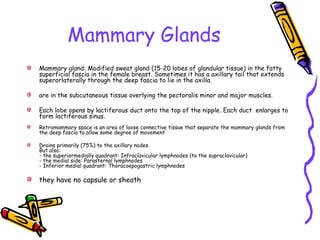
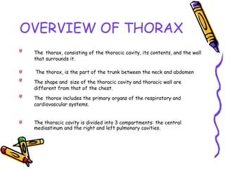
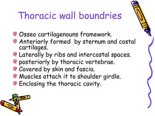
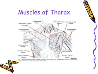
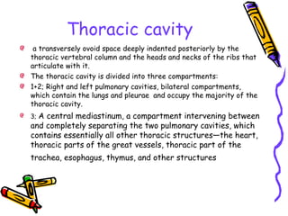
Thorax
OVERVIEW OF THORAX
The thorax, consisting of the thoracic cavity, its contents, and the wall
that surrounds it.
The thorax, is the part of the trunk between the neck and abdomen
The shape and size of the thoracic cavity and thoracic wall are
different from that of the chest.
The thorax includes the primary organs of the respiratory and
cardiovascular systems.
The thoracic cavity is divided into 3 compartments: the central
mediastinum and the right and left pulmonary cavities.
Boundries
Sternum:
manibrum.
sternal angle.
body.
oxphoid process.
12 pairs of ribs:
6 or 7 pairs of true ribs.
3 or 4 pairs of false ribs.
2 pairs of floating ribs.
Superiorly: Thoracic Inlet.
Inferiorly: Thoracic Outlet.
Posterior: Thoracic Vertebrae.
Landmarks of Thorax
Anterior Landmarks
Suprasternal notch
Sternum.
Manibrusternal
angle.
Costal angle.
Posterior Landmarks:
Vertebrae
prominens.
Spinous process.
Inferior border of
scapula.
12th
Rib.
Location finding in the
lung
Anterior Landmarks:
• Sternal angel best guide
anteriorly .
• First 7 ribs of costal
cartilage articulate with
sternum.
• 8th, 9th,10th, ribs
articulate with costal
cartilage just above them.
• 11th, and 12th
ribs are
flouting ribs.
Posterior Landmarks:
• 12th
rib.
• inferior angel of the
scapula lies at the level of
the 7th
rib.
• Spinous process C7 is the
most prominent process.
• Second ICS at the level of
Spinous process T1.
Lines of Orientation
Anterior:
midclavicular line.
midsternal line.
anterior axillary line.
Posterior:
scapular line.
vertebral line.
Laterl:
anterior axillary line.
posterior axillary line.
mid-axillary line.
Lines of Orientation
Thoracic wall boundries
Osseo cartilagenouns framework.
Anteriorly formed by sternum and costal
cartilages.
Laterally by ribs and intercostal spaces.
posteriorly by thoracic vertebrae.
Covered by skin and fascia.
Muscles attach it to shoulder girdle.
Enclosing the thoracic cavity.
Layers of chest wall
Skin.
Subcutanous fatt tissue.
Superficial fascia.
Mammary glands.
Muscles.
Deep fascia; “endothoracic fascia”.
Parietal pleura.
Cutaneous innervation of
chest wall
Anteriorly:
above sternal angle; T4:
from supraclavicular nerves.
Below sternal angle:
anterior and lateral cutanous branches of intercostal nerve.
Posteriorly:
dorsal rami of spinal nerves.
Muscles of Thorax
Extrinsic Muscles:
Serratus anterior.
pectoralis minor.
pectoralis major.
Intrinsic Muscles:
intercostales externi.
intercostales interni.
transverses thoracics.
Muscles of Thorax
Muscles of Thorax
Nerve supply to thorax
Intercostal nerves:
From ventral rami of T1-11 spinal nn.
Subcostal nerves:
Ventral ramus T12mspinal n.
They travel through intercostal
space.
Azygous v.
The azygos vein is located along the right side of the thoracic
vertebral column.
it carries deoxygenated blood from areas of the chest and abdomen to
the heart.
thus explaining its name, which means "unpaired" in Greek.
Blood in the azygos vein empties into the superior vena cava, which
then carries it to the heart to be recirculated.
Hemiazygous V.
The hemiazygos vein arises from one or more of
three possible roots (lateral, intermediate and
medial).
The hemiazygos vein usually receives the ninth to
eleventh posterior intercostal veins, and may
include the eighth.
The hemiazygos vein passes behind the aorta
from the left side to join the right-sided azygos.
The communication may be doubled.
Arteries
Un-paired visceral a.a.:
Bronchial, oesophagual, pericardial and medistinal branches.
Parietal visceral a.a.:
3rd
- 11th
posterior intercostal aa. And subcostal aa.
1st
-2nd
posterior intercostal aa. From subclavian a.
Anterior intercostal a. from internal thoracic a.
Superior phrenic aa.
Veins
Un-paired visceral vv.:
Bronchial, oesophagual, pericardial and medistinal branches.
Parietal visceral vv.:
3rd
- 11th
posterior intercostal vv. And subcostal vv.
1st
-2nd
posterior intercostal vv. From subclavian v.
Anterior intercostal vv. from internal thoracic v.
Superior phrenic vv.
Intercostal Spaces
spaces between robs are 11 in numbers.
Contain 3 layers of muscles of respiration:
extenal intercostal muscle.
internal intercostal muscle.
transverses thoracics muscle.
Each intercostal space has it own neurovascular bundle.
Neurovascular bundle of intercostal muscles contain:
intercostal V.A. N.
sympathetic trunk.
Innermost intercostal muscles bridge one intercostal space; subcostal
muscles bridge two
Breast
Breast consist of: Mammary gland, fibrous and fatty tissue. Also blood, lymph and
nerves.
Location: Extends from the second to sixth rib and from the sternum to the midaxillary
line.
Nipple: lies at the level of 4th intercostal space and contain smooth muscle fibers that
contract on tactile stimulation.
Areola: is a ring of pigmented skin around the nipple.
Innervated by anterior and lateral cutaneous branches of the second to the sixth
intercostal nerves.
Blood supply: Receives blood from the medial mammary (branch of the
anterior perforating branches of the internal thoracic), lateral
mammary (branch from the lateral thoracic artery) and pectoral
branches (branches from the thoracoacromial trunk). Also the lateral
cutaneous branches of the posterior intercostal arteries
Mammary Glands
Mammary gland: Modified sweat gland (15-20 lobes of glandular tissue) in the fatty
superficial fascia in the female breast. Sometimes it has a axillary tail that extends
superorlaterally through the deep fascia to lie in the axilla.
are in the subcutaneous tissue overlying the pectoralis minor and major muscles.
Each lobe opens by lactiferous duct onto the top of the nipple. Each duct enlarges to
form lactiferous sinus.
Retromammary space is an area of loose connective tissue that separate the mammary glands from
the deep fascia to allow some degree of movement
Drains primarily (75%) to the axillary nodes.
But also:
- the superiormedially quadrant: Infraclavicular lymphnodes (to the supraclavicular)
- the medial side: Parasternal lymphnodes
- Inferior medial quadrant: Thoracoepogastric lymphnodes
they have no capsule or sheath
Mammary Glands
The rounded contour and most of the volume of the breasts are
produced by subcutaneous fat, except during pregnancy when the
mammary glands enlarge and new glandular tissue forms.
The milksecreting alveoli (L. small hollow spaces) are
arranged in grape-like clusters. In most women, the breasts
enlarge slightly during the menstrual period from increased
release of gonadotropic hormones—follicle-stimulating
hormone (FSH) and luteinizing hormone (LH)—on the
glandular tissue.
The lymphatic drainage
the lymphatic drainage of the breast is important because of
its role in the metastasis of cancer cells.
Lymph passes from the nipple, areola, and lobules of the gland
to the subareolar lymphatic plexus.
Most lymph from the breast drains to the axillary lymph
nodes; this is significant when treating breast cancer.
the mammary glands and axillary lymph nodes are superficial,
the ability to palpate primary and metastatic tumors during
routine breast examination enables early detection and
treatment.
Thoracic cavity
a transversely ovoid space deeply indented posteriorly by the
thoracic vertebral column and the heads and necks of the ribs that
articulate with it.
The thoracic cavity is divided into three compartments:
1+2; Right and left pulmonary cavities, bilateral compartments,
which contain the lungs and pleurae and occupy the majority of the
thoracic cavity.
3; A central mediastinum, a compartment intervening between
and completely separating the two pulmonary cavities, which
contains essentially all other thoracic structures—the heart,
thoracic parts of the great vessels, thoracic part of the
trachea, esophagus, thymus, and other structures
Pleural sacs
Serous membranes forming closed sacs: visceral + parietal.
Visceral pleura: serous membrane that cover the outer service of
the lung.
Parietal pleura: serous membrane that line the inner rib cage of
the upper surface of diaphragm.
Function of the pleura: smooth the opposing service and lubricant
by plural fluid to allow the ling to move easily with the rib cage.
Pleural space: potential space between visceral and parietal
pleura; and contains a small amounts of pleural fluids and
subatmospheric in it.
Pleural Recesses
Potential spaces of pleural cavity with lungs are not occupied in
quiet respiration.
“costodiaphragmatic recess”; are the slit-like intervals between
costal and diaphragmatic pleurae on each side, the lowest point of
pleural cavity.
Vertically it measures about 5cm, extends from 8th
to 10th
rib along
the midaxillary line.
“costomedistinal recess”; on the left side between the medistinal
pleura and costal pleura.
Trachea
Position:
Extends from the lower bordes of cricoid cartilage to the level of
sternal angle (between T4-T5 V.) where its divides into left and right
principal bronchi.
Structure features:
Consist of about 16-20 C-shaped incomplete tracheal cartilage for
patency connected by smooth m. and connective tissue.
“Carina of trachea”; a ridge of cartilage at Bi-furication at principal
bronchi.
Bronchi
Right Principal Bronchus:
Shorter, wider, and more vertical than the left. Is about 2.5cm longs,
and leveas the extend line of the middle line of the trachea at a
22-25 degree angle.
Forgein bodies are therefore more likely to lodge in this bronchus or
of its branches.
Left principal Bronchus:
Narrower, longer and more horizontal than the right, its about 5cm,
and leveas the extend line of the middle line of the trachea at
about 35-36 angle.
Bronchial Tree
Each principal bronchus divides into lobar
bronchi “ 2 in the left, and 3 on the right”,
each of which supplies a lobe of the lung.
Each lobar bronchus then divides into segmental
bronchi, which supply specific segment of the
lung.
Lungs
Position:
Located in the thoracic cavity by both sides of the mediastinum.
General Features:
cone shaped, the right lung is shorter and broader and the left lung is longer and
narrower.
Apex:
Rises 2-3 cm above the medial third of the clavicle into the neck.
Base:
concave;, related to diaphragm, also called: “diaphragmatic surface “.
Costal surface:
Large, convex, related to the thoracic wall.
Lungs
Medial surface:
Concave, related to mediastinum and vertebrae.
Hilum of Lung:
Area on medial surface where the structures of root enter or leaves the lung.
Root of Lung:
contents:
principal bronchus
pulomanry artery and vein
nerves and lymphatics
Surrounded by connective tissue.
Lungs
Aneriorly:
Apex of each lung rises about 2-4 cm above the inner third of the clavicle.
The lower border of the lung cross the 6thrib at the midclaviculerline and 8thrib
in the mid auxiliary line.
Posteriorly:
The lower border of the lung lies at about the level of T10 spinous process.
Rigth lung: upper, middle, lower lobes.
Left lung: upper and lower lobes.
Mediastinum
Is the regoin that located between the 2 pleural cavities.
BOundries:
Superior: superior thoracic aperture.
Inferior: Diaphragm.
Anterior: Sternum.
Posterior: bodies of T1-T12.
Lateral: medistinal pariteal pleura.
Contents of mediastinum
Anterior mediastinm:
The part that lies between the sternum and the pericardium.
In the children and adolescents, part of the thymus may be found in the anterior
mediastinum.
Middle mediastinum:
The part that contains the pericardium, the heart, and the roots of the great vessels.
Posterior mediastinum:
The part that lies posteriorly to the pericarduim and anteriorly to the bodies of the
vertebrae T5-T12.
It contains structures that pass between the neck, thorax, and the abdomen. Ex:
vagus n, oesophagous, thoracic duct and aorta, azygos system of veins.
Anterior Mediastinum
Anterior border:
Sternum.
Posterior border:
Ventral cardiac surface and
brachio-cephalic vessels.
Contents:
Thymus.
Fat.
Lymph nodes.
Sternum and anterior ribs.
Middle mediastinum
Anterior Border:
Ventral heart border.
Posterior Border:
Anterior surface of the spine.
Contents:
Heart and pericarduim.
Ascending aorta & arch of aorta.
Vena cava.
Brachio-cephalic vessels.
Main pulmonary aa.and vv.
Trachea, bronch i& oesophagous.
Lymph nodes.
Posterior Mediastinum
Anterior Border:
Anterior surface of spine.
Posterior Border:
Posterior ribs.
Contents:
Descending aorta.
Spine and posterior ribs.
Nerves, ganglia, roots and spinal
cord.
Lymph nodes.
Azygous and hemi-azygous vv.
Questions?

Presentation1

  • 1.
  • 2.
    OVERVIEW OF THORAX Thethorax, consisting of the thoracic cavity, its contents, and the wall that surrounds it. The thorax, is the part of the trunk between the neck and abdomen The shape and size of the thoracic cavity and thoracic wall are different from that of the chest. The thorax includes the primary organs of the respiratory and cardiovascular systems. The thoracic cavity is divided into 3 compartments: the central mediastinum and the right and left pulmonary cavities.
  • 3.
    Boundries Sternum: manibrum. sternal angle. body. oxphoid process. 12pairs of ribs: 6 or 7 pairs of true ribs. 3 or 4 pairs of false ribs. 2 pairs of floating ribs. Superiorly: Thoracic Inlet. Inferiorly: Thoracic Outlet. Posterior: Thoracic Vertebrae.
  • 4.
    Landmarks of Thorax AnteriorLandmarks Suprasternal notch Sternum. Manibrusternal angle. Costal angle. Posterior Landmarks: Vertebrae prominens. Spinous process. Inferior border of scapula. 12th Rib.
  • 5.
    Location finding inthe lung Anterior Landmarks: • Sternal angel best guide anteriorly . • First 7 ribs of costal cartilage articulate with sternum. • 8th, 9th,10th, ribs articulate with costal cartilage just above them. • 11th, and 12th ribs are flouting ribs. Posterior Landmarks: • 12th rib. • inferior angel of the scapula lies at the level of the 7th rib. • Spinous process C7 is the most prominent process. • Second ICS at the level of Spinous process T1.
  • 6.
    Lines of Orientation Anterior: midclavicularline. midsternal line. anterior axillary line. Posterior: scapular line. vertebral line. Laterl: anterior axillary line. posterior axillary line. mid-axillary line.
  • 7.
  • 8.
    Thoracic wall boundries Osseocartilagenouns framework. Anteriorly formed by sternum and costal cartilages. Laterally by ribs and intercostal spaces. posteriorly by thoracic vertebrae. Covered by skin and fascia. Muscles attach it to shoulder girdle. Enclosing the thoracic cavity.
  • 9.
    Layers of chestwall Skin. Subcutanous fatt tissue. Superficial fascia. Mammary glands. Muscles. Deep fascia; “endothoracic fascia”. Parietal pleura.
  • 10.
    Cutaneous innervation of chestwall Anteriorly: above sternal angle; T4: from supraclavicular nerves. Below sternal angle: anterior and lateral cutanous branches of intercostal nerve. Posteriorly: dorsal rami of spinal nerves.
  • 11.
    Muscles of Thorax ExtrinsicMuscles: Serratus anterior. pectoralis minor. pectoralis major. Intrinsic Muscles: intercostales externi. intercostales interni. transverses thoracics.
  • 12.
  • 13.
  • 14.
    Nerve supply tothorax Intercostal nerves: From ventral rami of T1-11 spinal nn. Subcostal nerves: Ventral ramus T12mspinal n. They travel through intercostal space.
  • 15.
    Azygous v. The azygosvein is located along the right side of the thoracic vertebral column. it carries deoxygenated blood from areas of the chest and abdomen to the heart. thus explaining its name, which means "unpaired" in Greek. Blood in the azygos vein empties into the superior vena cava, which then carries it to the heart to be recirculated.
  • 16.
    Hemiazygous V. The hemiazygosvein arises from one or more of three possible roots (lateral, intermediate and medial). The hemiazygos vein usually receives the ninth to eleventh posterior intercostal veins, and may include the eighth. The hemiazygos vein passes behind the aorta from the left side to join the right-sided azygos. The communication may be doubled.
  • 17.
    Arteries Un-paired visceral a.a.: Bronchial,oesophagual, pericardial and medistinal branches. Parietal visceral a.a.: 3rd - 11th posterior intercostal aa. And subcostal aa. 1st -2nd posterior intercostal aa. From subclavian a. Anterior intercostal a. from internal thoracic a. Superior phrenic aa.
  • 18.
    Veins Un-paired visceral vv.: Bronchial,oesophagual, pericardial and medistinal branches. Parietal visceral vv.: 3rd - 11th posterior intercostal vv. And subcostal vv. 1st -2nd posterior intercostal vv. From subclavian v. Anterior intercostal vv. from internal thoracic v. Superior phrenic vv.
  • 19.
    Intercostal Spaces spaces betweenrobs are 11 in numbers. Contain 3 layers of muscles of respiration: extenal intercostal muscle. internal intercostal muscle. transverses thoracics muscle. Each intercostal space has it own neurovascular bundle. Neurovascular bundle of intercostal muscles contain: intercostal V.A. N. sympathetic trunk. Innermost intercostal muscles bridge one intercostal space; subcostal muscles bridge two
  • 20.
    Breast Breast consist of:Mammary gland, fibrous and fatty tissue. Also blood, lymph and nerves. Location: Extends from the second to sixth rib and from the sternum to the midaxillary line. Nipple: lies at the level of 4th intercostal space and contain smooth muscle fibers that contract on tactile stimulation. Areola: is a ring of pigmented skin around the nipple. Innervated by anterior and lateral cutaneous branches of the second to the sixth intercostal nerves. Blood supply: Receives blood from the medial mammary (branch of the anterior perforating branches of the internal thoracic), lateral mammary (branch from the lateral thoracic artery) and pectoral branches (branches from the thoracoacromial trunk). Also the lateral cutaneous branches of the posterior intercostal arteries
  • 21.
    Mammary Glands Mammary gland:Modified sweat gland (15-20 lobes of glandular tissue) in the fatty superficial fascia in the female breast. Sometimes it has a axillary tail that extends superorlaterally through the deep fascia to lie in the axilla. are in the subcutaneous tissue overlying the pectoralis minor and major muscles. Each lobe opens by lactiferous duct onto the top of the nipple. Each duct enlarges to form lactiferous sinus. Retromammary space is an area of loose connective tissue that separate the mammary glands from the deep fascia to allow some degree of movement Drains primarily (75%) to the axillary nodes. But also: - the superiormedially quadrant: Infraclavicular lymphnodes (to the supraclavicular) - the medial side: Parasternal lymphnodes - Inferior medial quadrant: Thoracoepogastric lymphnodes they have no capsule or sheath
  • 22.
    Mammary Glands The roundedcontour and most of the volume of the breasts are produced by subcutaneous fat, except during pregnancy when the mammary glands enlarge and new glandular tissue forms. The milksecreting alveoli (L. small hollow spaces) are arranged in grape-like clusters. In most women, the breasts enlarge slightly during the menstrual period from increased release of gonadotropic hormones—follicle-stimulating hormone (FSH) and luteinizing hormone (LH)—on the glandular tissue.
  • 23.
    The lymphatic drainage thelymphatic drainage of the breast is important because of its role in the metastasis of cancer cells. Lymph passes from the nipple, areola, and lobules of the gland to the subareolar lymphatic plexus. Most lymph from the breast drains to the axillary lymph nodes; this is significant when treating breast cancer. the mammary glands and axillary lymph nodes are superficial, the ability to palpate primary and metastatic tumors during routine breast examination enables early detection and treatment.
  • 24.
    Thoracic cavity a transverselyovoid space deeply indented posteriorly by the thoracic vertebral column and the heads and necks of the ribs that articulate with it. The thoracic cavity is divided into three compartments: 1+2; Right and left pulmonary cavities, bilateral compartments, which contain the lungs and pleurae and occupy the majority of the thoracic cavity. 3; A central mediastinum, a compartment intervening between and completely separating the two pulmonary cavities, which contains essentially all other thoracic structures—the heart, thoracic parts of the great vessels, thoracic part of the trachea, esophagus, thymus, and other structures
  • 25.
    Pleural sacs Serous membranesforming closed sacs: visceral + parietal. Visceral pleura: serous membrane that cover the outer service of the lung. Parietal pleura: serous membrane that line the inner rib cage of the upper surface of diaphragm. Function of the pleura: smooth the opposing service and lubricant by plural fluid to allow the ling to move easily with the rib cage. Pleural space: potential space between visceral and parietal pleura; and contains a small amounts of pleural fluids and subatmospheric in it.
  • 26.
    Pleural Recesses Potential spacesof pleural cavity with lungs are not occupied in quiet respiration. “costodiaphragmatic recess”; are the slit-like intervals between costal and diaphragmatic pleurae on each side, the lowest point of pleural cavity. Vertically it measures about 5cm, extends from 8th to 10th rib along the midaxillary line. “costomedistinal recess”; on the left side between the medistinal pleura and costal pleura.
  • 27.
    Trachea Position: Extends from thelower bordes of cricoid cartilage to the level of sternal angle (between T4-T5 V.) where its divides into left and right principal bronchi. Structure features: Consist of about 16-20 C-shaped incomplete tracheal cartilage for patency connected by smooth m. and connective tissue. “Carina of trachea”; a ridge of cartilage at Bi-furication at principal bronchi.
  • 28.
    Bronchi Right Principal Bronchus: Shorter,wider, and more vertical than the left. Is about 2.5cm longs, and leveas the extend line of the middle line of the trachea at a 22-25 degree angle. Forgein bodies are therefore more likely to lodge in this bronchus or of its branches. Left principal Bronchus: Narrower, longer and more horizontal than the right, its about 5cm, and leveas the extend line of the middle line of the trachea at about 35-36 angle.
  • 29.
    Bronchial Tree Each principalbronchus divides into lobar bronchi “ 2 in the left, and 3 on the right”, each of which supplies a lobe of the lung. Each lobar bronchus then divides into segmental bronchi, which supply specific segment of the lung.
  • 30.
    Lungs Position: Located in thethoracic cavity by both sides of the mediastinum. General Features: cone shaped, the right lung is shorter and broader and the left lung is longer and narrower. Apex: Rises 2-3 cm above the medial third of the clavicle into the neck. Base: concave;, related to diaphragm, also called: “diaphragmatic surface “. Costal surface: Large, convex, related to the thoracic wall.
  • 31.
    Lungs Medial surface: Concave, relatedto mediastinum and vertebrae. Hilum of Lung: Area on medial surface where the structures of root enter or leaves the lung. Root of Lung: contents: principal bronchus pulomanry artery and vein nerves and lymphatics Surrounded by connective tissue.
  • 32.
    Lungs Aneriorly: Apex of eachlung rises about 2-4 cm above the inner third of the clavicle. The lower border of the lung cross the 6thrib at the midclaviculerline and 8thrib in the mid auxiliary line. Posteriorly: The lower border of the lung lies at about the level of T10 spinous process. Rigth lung: upper, middle, lower lobes. Left lung: upper and lower lobes.
  • 33.
    Mediastinum Is the regointhat located between the 2 pleural cavities. BOundries: Superior: superior thoracic aperture. Inferior: Diaphragm. Anterior: Sternum. Posterior: bodies of T1-T12. Lateral: medistinal pariteal pleura.
  • 34.
    Contents of mediastinum Anteriormediastinm: The part that lies between the sternum and the pericardium. In the children and adolescents, part of the thymus may be found in the anterior mediastinum. Middle mediastinum: The part that contains the pericardium, the heart, and the roots of the great vessels. Posterior mediastinum: The part that lies posteriorly to the pericarduim and anteriorly to the bodies of the vertebrae T5-T12. It contains structures that pass between the neck, thorax, and the abdomen. Ex: vagus n, oesophagous, thoracic duct and aorta, azygos system of veins.
  • 35.
    Anterior Mediastinum Anterior border: Sternum. Posteriorborder: Ventral cardiac surface and brachio-cephalic vessels. Contents: Thymus. Fat. Lymph nodes. Sternum and anterior ribs.
  • 36.
    Middle mediastinum Anterior Border: Ventralheart border. Posterior Border: Anterior surface of the spine. Contents: Heart and pericarduim. Ascending aorta & arch of aorta. Vena cava. Brachio-cephalic vessels. Main pulmonary aa.and vv. Trachea, bronch i& oesophagous. Lymph nodes.
  • 37.
    Posterior Mediastinum Anterior Border: Anteriorsurface of spine. Posterior Border: Posterior ribs. Contents: Descending aorta. Spine and posterior ribs. Nerves, ganglia, roots and spinal cord. Lymph nodes. Azygous and hemi-azygous vv.
  • 38.