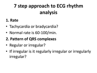
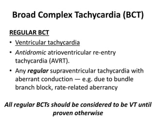
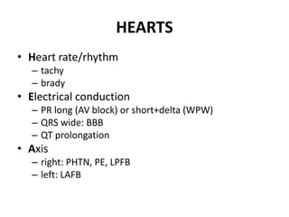
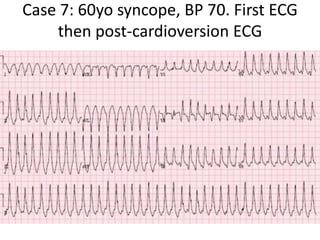
ECG
Fakhir
SIUT
22-Jan-2020
Leads
• 6 Limb Lead
• 6 Chest Leads
Looking at heart from different angles.
Rate
Rythm
7 step approach to ECG rhythm
analysis
1. Rate
• Tachycardia or bradycardia?
• Normal rate is 60-100/min.
2. Pattern of QRS complexes
• Regular or irregular?
• If irregular is it regularly irregular or irregularly
irregular?
7 step approach to ECG rhythm
analysis
3. QRS morphology
• Narrow complex: sinus, atrial or junctional origin.
• Wide complex: ventricular origin, or
supraventricular with aberrant conduction.
4. P waves
• Absent: sinus arrest, atrial fibrillation
• Present: morphology and PR interval may suggest
sinus, atrial, junctional or even retrograde from
the ventricles.
7 step approach to ECG rhythm
analysis
5. Relationship between P waves and QRS complexes
• AV association (may be difficult to distinguish from
isorhythmic dissociation)
• AV dissociation
– complete: atrial and ventricular activity is always
independent.
– incomplete: intermittent capture.
6. Onset and termination
• Abrupt: suggests re-entrant process.
• Gradual: suggests increased automaticity.
7 step approach to ECG rhythm
analysis
7. Response to vagal manoeuvres
• Sinus tachycardia, ectopic atrial
tachydysrhythmia: gradual slowing during the
vagal manoeuvre, but resumes on cessation.
• AVNRT or AVRT: abrupt termination or no
response.
• Atrial fibrillation and atrial flutter: gradual
slowing during the manoeuvre.
• VT: no response.
Narrow Complex (Supraventricular)
Tachycardia
ATRIAL – REGULAR
• Sinus tachycardia
• Atrial tachycardia
• Atrial flutter
• Inappropriate sinus tachycardia
• Sinus node re-entrant tachycardia
ATRIAL FLUTTER
Narrow Complex (Supraventricular)
Tachycardia
ATRIAL – IRREGULAR
• Atrial fibrillation
• Atrial flutter with variable block
• Multifocal atrial tachycardia
Atrial Fibrillation
Multifocal atrial tachycardia
Narrow Complex (Supraventricular)
Tachycardia
ATRIOVENTRICULAR
• Atrioventricular re-entry tachycardia (AVRT)
• AV nodal re-entry tachycardia (AVNRT)
• Automatic junctional tachycardia
Broad Complex Tachycardia (BCT)
REGULAR BCT
• Ventricular tachycardia
• Antidromic atrioventricular re-entry
tachycardia (AVRT).
• Any regular supraventricular tachycardia with
aberrant conduction — e.g. due to bundle
branch block, rate-related aberrancy
All regular BCTs should be considered to be VT until
proven otherwise
Ventricular Tachycardia
Broad Complex Tachycardia (BCT)
• IRREGULAR
• Ventricular fibrillation
• Polymorphic VT
• Torsades de Pointes
• AF with Wolff-Parkinson-White syndrome
• Any irregular supraventricular tachycardia
with aberrant conduction — e.g. due to
bundle branch block, rate-related aberrancy
Ventricular Fibrillation
Polymorphic VT
Torsades de Pointes
Bradycardia
P WAVES PRESENT
1. Every P wave is followed by a QRS complex
(= sinus node dysfunction)
• Sinus bradycardia
• Sinus node exit block
• Sinus pause / arrest
Bradycardia
P WAVES PRESENT
2. Not every P wave is followed by a QRS complex
(= AV node dysfunction)
• AV block: 2nd degree, Mobitz I (Wenckebach)
• AV block: 2nd degree, Mobitz II (Hay)
• AV block: 2nd degree, “fixed ratio blocks” (2:1,
3:1)
• AV block: 2nd degree, “high grade AV block”
• AV block: 3rd degree (complete heart block)
Mobitz Type I
Mobitz Type II
3rd Degree Heart Block
What is This
Bradycardia
• P WAVES ABSENT
• Narrow complex: Junctional escape rhythm
• Broad complex: Ventricular escape rhythm
Axis
ECG Axis Interpretation
• Normal Axis = QRS axis between -30° and +90°.
• Left Axis Deviation = QRS axis less than -30°.
• Right Axis Deviation = QRS axis greater than +90°.
• Extreme Axis Deviation = QRS axis between -90°
and 180° (AKA “Northwest Axis”)
Methods of ECG Axis Interpretation
There are several complementary approaches to
estimating QRS axis, which are summarized
below:
• The Quadrant Method – (Lead I and aVF)
• Three Lead analysis – (Lead I, Lead II and aVF)
• Isoelectric Lead analysis
• Super SAM the Axis Man
Intervels
Cases
Sycope
Case 1: 20yo previously well with half
an hour of palpitations and
presyncope, resolved
Case 2: 80yo recurring presyncope
while sitting
Case 3: 25yo with exertional syncope
Case 4: 70yo history of cancer,
presents with shortness of breath.
Shortly after ECG patient collapsed
Case 5: 70yo unwell. After ECG patient
collapsed
Case 6: 60yo chest pain then syncope
Case 7: 60yo syncope, BP 70. First ECG
then post-cardioversion ECG
Case 7: 60yo syncope, BP 70. First ECG
then post-cardioversion ECG
Syncope
The 2018 ESC guidelines for the diagnosis and
management of syncope classifies syncope as
• Reflex (vasovagal, situational or carotid sinus)
• Orthostatic (drug-induced, volume depletion
or autonomic)
• Cardiac
Cardiac syncope
• arrhythmia
– bradycardia: sinus or AV conduction disease
– tachycardia: supraventricular or ventricular
• structural
– tamponade
– ACS
– aortic stenosis
– hypertrophic cardiomyopathy (HCM)
– cardiac mass
– prosthetic valve dysfunction
• cardiopulmonary
– PE
– aortic dissection
– pulmonary hypertension
HEARTS
• Heart rate/rhythm
– tachy
– brady
• Electrical conduction
– PR long (AV block) or short+delta (WPW)
– QRS wide: BBB
– QT prolongation
• Axis
– right: PHTN, PE, LPFB
– left: LAFB
HEARTS
• R wave size/progression
– early R wave: RBBB, WPW type A, posterior MI, pulmonary
hypertension
– late R wave: cardiomyopathy
– high voltages: hypertrophy
– low voltages: pericardial effusion
• Tension (hypertrophy)
– aortic stenosis
– HCM
• ST/T wave changes
– inherited: ARVD, Brugada
– ischemic: ACS, PE, dissection
Back to the cases
Case 1: 20yo previously well with half
an hour of palpitations and
presyncope, resolved
HR/rhythm: NSR.
Electrical conduction: short PR with delta wave
in precordial leads.
Axis: normal.
R wave: early progression.
Tension: none.
ST/ T changes: mild anterior ST depression,
discordant to delta.
WPW. Referred for ablatation
Case 2: 80yo recurring presyncope
while sitting
“trifascicular block” HR/rhythm: sinus brady.
Electrical conduction: first degree AV block +
RBBB + LAFB.
Axis: left (from LAFB).
R wave: early (from RBBB)
Tension: none
ST/T wave changes: none
bifascicular block with first degree AV block.
Admitted for pacemaker
Case 3: 25yo with exertional syncope
HR/rhythm: borderline sinus tach
Electrical conduction: normal
Axis: normal
R wave: large voltages with deep inferolateral
Q waves
Tension: LVH
ST/T wave: no changes
POCUS: HCM
Case 4: 70yo history of cancer, presents with shortness
of breath. Shortly after ECG patient collapsed
HR/rhythm: sinus tach
Electrical conduction: normal
Axis: normal
R wave: small voltages
Tension: none
ST/T wave: no significance changes
POCUS: no pericardial effusion but RV>LV.
Patient arrested, achieved ROSC after tPA and
CT showed massive PE
Case 5: 70yo unwell. After ECG patient collapsed
HR/rhythm: NSR
Electrical conduction: RBBB
Axis: normal
R wave: normal size/progression
Tension: none
ST/T wave: V1-2 coved ST elevation
Diagnosis: Brugada. Patient had VF arrest
shortly after ECG, was cardioverted and
admitted for ICD
Case 6: 60yo chest pain then syncope
HR/rhytm: sinus bradycardia
Electrical conduction: normal
Axis: normal
R wave: normal size/progression
Tension: none
ST/T wave: anterior ST depression with T wave
inversion
No early R waves/right axis to suggest
pulmonary hypertension, no other features to
suggest PE.
Posterior leads showed ST elevation and cath
lab activated: occluded circumflex artery
Case 7: 60yo syncope, BP 70. First ECG
then post-cardioversion ECG
Case 7: then post-cardioversion ECG
HR/rhythm: VT cardioverted into junctional rhythm
Electrical conduction: RBBB + LAFB
Axis: left (from LAFB)
R wave: RBBB superimposed on anterior Q and delayed R
wave progression
Tension: none
ST/T wave: diffuse ST depression with reciprocal elevation in
aVR. This pattern can be seen post-cardioversion, but this
patient also had anterior Q waves and a bifasicular block
Shukria and JazakAllah for listening
‫ت‬‫ت‬‫ت‬‫ت‬‫ت‬‫ت‬‫ت‬‫ت‬‫ت‬‫ت‬‫ت‬ ‫ک‬ ‫ت‬‫ت‬‫ت‬‫ت‬‫ت‬‫ت‬‫ت‬‫ت‬‫ت‬‫ت‬‫ت‬ ‫ک‬ ‫ا‬ ‫ت‬‫ت‬‫ت‬‫ت‬‫ت‬‫ت‬‫ت‬‫ت‬‫ت‬‫ت‬‫ت‬‫س‬‫ک‬ ‫ت‬‫ت‬‫ت‬‫ت‬‫ت‬‫ت‬‫ت‬‫ت‬‫ت‬‫ت‬‫ت‬ ‫کانک‬ ‫کد‬‫تئ‬‫ت‬‫ت‬‫ت‬‫ت‬‫ت‬‫ت‬‫ت‬‫ت‬‫ت‬‫ت‬‫و‬‫ک‬‫ان‬ ‫ت‬‫ت‬‫ت‬‫ت‬‫ت‬‫ت‬‫ت‬‫ت‬‫ت‬‫ت‬‫ت‬‫س‬‫دا‬
‫ت‬‫ت‬‫ت‬ ‫ک‬ ‫ت‬‫ت‬‫ت‬ ‫ک‬ ‫تا‬‫ت‬‫ت‬ ‫کب‬ ‫ت‬‫ت‬‫ت‬ ‫ک‬ ‫تگ‬‫ت‬‫ت‬ ‫تنکای‬‫ت‬‫ت‬‫ک‬ ‫ک‬ ‫ت‬‫ت‬‫ت‬ ‫کھ‬‫ک‬ ‫ت‬‫ت‬‫ت‬ ‫تاگکب‬‫ت‬‫ت‬‫ب‬
(‫ادکآبادک‬‫ر‬‫رکم‬ ‫ج‬)

ECG electrocardigram

  • 1.
  • 2.
    Leads • 6 LimbLead • 6 Chest Leads Looking at heart from different angles.
  • 7.
  • 9.
  • 10.
    7 step approachto ECG rhythm analysis 1. Rate • Tachycardia or bradycardia? • Normal rate is 60-100/min. 2. Pattern of QRS complexes • Regular or irregular? • If irregular is it regularly irregular or irregularly irregular?
  • 11.
    7 step approachto ECG rhythm analysis 3. QRS morphology • Narrow complex: sinus, atrial or junctional origin. • Wide complex: ventricular origin, or supraventricular with aberrant conduction. 4. P waves • Absent: sinus arrest, atrial fibrillation • Present: morphology and PR interval may suggest sinus, atrial, junctional or even retrograde from the ventricles.
  • 12.
    7 step approachto ECG rhythm analysis 5. Relationship between P waves and QRS complexes • AV association (may be difficult to distinguish from isorhythmic dissociation) • AV dissociation – complete: atrial and ventricular activity is always independent. – incomplete: intermittent capture. 6. Onset and termination • Abrupt: suggests re-entrant process. • Gradual: suggests increased automaticity.
  • 13.
    7 step approachto ECG rhythm analysis 7. Response to vagal manoeuvres • Sinus tachycardia, ectopic atrial tachydysrhythmia: gradual slowing during the vagal manoeuvre, but resumes on cessation. • AVNRT or AVRT: abrupt termination or no response. • Atrial fibrillation and atrial flutter: gradual slowing during the manoeuvre. • VT: no response.
  • 14.
    Narrow Complex (Supraventricular) Tachycardia ATRIAL– REGULAR • Sinus tachycardia • Atrial tachycardia • Atrial flutter • Inappropriate sinus tachycardia • Sinus node re-entrant tachycardia
  • 17.
  • 18.
    Narrow Complex (Supraventricular) Tachycardia ATRIAL– IRREGULAR • Atrial fibrillation • Atrial flutter with variable block • Multifocal atrial tachycardia
  • 19.
  • 20.
  • 21.
    Narrow Complex (Supraventricular) Tachycardia ATRIOVENTRICULAR •Atrioventricular re-entry tachycardia (AVRT) • AV nodal re-entry tachycardia (AVNRT) • Automatic junctional tachycardia
  • 22.
    Broad Complex Tachycardia(BCT) REGULAR BCT • Ventricular tachycardia • Antidromic atrioventricular re-entry tachycardia (AVRT). • Any regular supraventricular tachycardia with aberrant conduction — e.g. due to bundle branch block, rate-related aberrancy All regular BCTs should be considered to be VT until proven otherwise
  • 23.
  • 24.
    Broad Complex Tachycardia(BCT) • IRREGULAR • Ventricular fibrillation • Polymorphic VT • Torsades de Pointes • AF with Wolff-Parkinson-White syndrome • Any irregular supraventricular tachycardia with aberrant conduction — e.g. due to bundle branch block, rate-related aberrancy
  • 25.
  • 28.
  • 29.
  • 30.
    Bradycardia P WAVES PRESENT 1.Every P wave is followed by a QRS complex (= sinus node dysfunction) • Sinus bradycardia • Sinus node exit block • Sinus pause / arrest
  • 31.
    Bradycardia P WAVES PRESENT 2.Not every P wave is followed by a QRS complex (= AV node dysfunction) • AV block: 2nd degree, Mobitz I (Wenckebach) • AV block: 2nd degree, Mobitz II (Hay) • AV block: 2nd degree, “fixed ratio blocks” (2:1, 3:1) • AV block: 2nd degree, “high grade AV block” • AV block: 3rd degree (complete heart block)
  • 32.
  • 33.
  • 34.
  • 35.
  • 36.
    Bradycardia • P WAVESABSENT • Narrow complex: Junctional escape rhythm • Broad complex: Ventricular escape rhythm
  • 37.
  • 38.
    ECG Axis Interpretation •Normal Axis = QRS axis between -30° and +90°. • Left Axis Deviation = QRS axis less than -30°. • Right Axis Deviation = QRS axis greater than +90°. • Extreme Axis Deviation = QRS axis between -90° and 180° (AKA “Northwest Axis”)
  • 39.
    Methods of ECGAxis Interpretation There are several complementary approaches to estimating QRS axis, which are summarized below: • The Quadrant Method – (Lead I and aVF) • Three Lead analysis – (Lead I, Lead II and aVF) • Isoelectric Lead analysis • Super SAM the Axis Man
  • 43.
  • 46.
  • 47.
    Case 1: 20yopreviously well with half an hour of palpitations and presyncope, resolved
  • 48.
    Case 2: 80yorecurring presyncope while sitting
  • 49.
    Case 3: 25yowith exertional syncope
  • 50.
    Case 4: 70yohistory of cancer, presents with shortness of breath. Shortly after ECG patient collapsed
  • 51.
    Case 5: 70younwell. After ECG patient collapsed
  • 52.
    Case 6: 60yochest pain then syncope
  • 53.
    Case 7: 60yosyncope, BP 70. First ECG then post-cardioversion ECG
  • 54.
    Case 7: 60yosyncope, BP 70. First ECG then post-cardioversion ECG
  • 55.
    Syncope The 2018 ESCguidelines for the diagnosis and management of syncope classifies syncope as • Reflex (vasovagal, situational or carotid sinus) • Orthostatic (drug-induced, volume depletion or autonomic) • Cardiac
  • 56.
    Cardiac syncope • arrhythmia –bradycardia: sinus or AV conduction disease – tachycardia: supraventricular or ventricular • structural – tamponade – ACS – aortic stenosis – hypertrophic cardiomyopathy (HCM) – cardiac mass – prosthetic valve dysfunction • cardiopulmonary – PE – aortic dissection – pulmonary hypertension
  • 57.
    HEARTS • Heart rate/rhythm –tachy – brady • Electrical conduction – PR long (AV block) or short+delta (WPW) – QRS wide: BBB – QT prolongation • Axis – right: PHTN, PE, LPFB – left: LAFB
  • 58.
    HEARTS • R wavesize/progression – early R wave: RBBB, WPW type A, posterior MI, pulmonary hypertension – late R wave: cardiomyopathy – high voltages: hypertrophy – low voltages: pericardial effusion • Tension (hypertrophy) – aortic stenosis – HCM • ST/T wave changes – inherited: ARVD, Brugada – ischemic: ACS, PE, dissection
  • 59.
  • 60.
    Case 1: 20yopreviously well with half an hour of palpitations and presyncope, resolved HR/rhythm: NSR. Electrical conduction: short PR with delta wave in precordial leads. Axis: normal. R wave: early progression. Tension: none. ST/ T changes: mild anterior ST depression, discordant to delta. WPW. Referred for ablatation
  • 61.
    Case 2: 80yorecurring presyncope while sitting “trifascicular block” HR/rhythm: sinus brady. Electrical conduction: first degree AV block + RBBB + LAFB. Axis: left (from LAFB). R wave: early (from RBBB) Tension: none ST/T wave changes: none bifascicular block with first degree AV block. Admitted for pacemaker
  • 62.
    Case 3: 25yowith exertional syncope HR/rhythm: borderline sinus tach Electrical conduction: normal Axis: normal R wave: large voltages with deep inferolateral Q waves Tension: LVH ST/T wave: no changes POCUS: HCM
  • 63.
    Case 4: 70yohistory of cancer, presents with shortness of breath. Shortly after ECG patient collapsed HR/rhythm: sinus tach Electrical conduction: normal Axis: normal R wave: small voltages Tension: none ST/T wave: no significance changes POCUS: no pericardial effusion but RV>LV. Patient arrested, achieved ROSC after tPA and CT showed massive PE
  • 64.
    Case 5: 70younwell. After ECG patient collapsed HR/rhythm: NSR Electrical conduction: RBBB Axis: normal R wave: normal size/progression Tension: none ST/T wave: V1-2 coved ST elevation Diagnosis: Brugada. Patient had VF arrest shortly after ECG, was cardioverted and admitted for ICD
  • 65.
    Case 6: 60yochest pain then syncope HR/rhytm: sinus bradycardia Electrical conduction: normal Axis: normal R wave: normal size/progression Tension: none ST/T wave: anterior ST depression with T wave inversion No early R waves/right axis to suggest pulmonary hypertension, no other features to suggest PE. Posterior leads showed ST elevation and cath lab activated: occluded circumflex artery
  • 66.
    Case 7: 60yosyncope, BP 70. First ECG then post-cardioversion ECG
  • 67.
    Case 7: thenpost-cardioversion ECG HR/rhythm: VT cardioverted into junctional rhythm Electrical conduction: RBBB + LAFB Axis: left (from LAFB) R wave: RBBB superimposed on anterior Q and delayed R wave progression Tension: none ST/T wave: diffuse ST depression with reciprocal elevation in aVR. This pattern can be seen post-cardioversion, but this patient also had anterior Q waves and a bifasicular block
  • 68.
    Shukria and JazakAllahfor listening ‫ت‬‫ت‬‫ت‬‫ت‬‫ت‬‫ت‬‫ت‬‫ت‬‫ت‬‫ت‬‫ت‬ ‫ک‬ ‫ت‬‫ت‬‫ت‬‫ت‬‫ت‬‫ت‬‫ت‬‫ت‬‫ت‬‫ت‬‫ت‬ ‫ک‬ ‫ا‬ ‫ت‬‫ت‬‫ت‬‫ت‬‫ت‬‫ت‬‫ت‬‫ت‬‫ت‬‫ت‬‫ت‬‫س‬‫ک‬ ‫ت‬‫ت‬‫ت‬‫ت‬‫ت‬‫ت‬‫ت‬‫ت‬‫ت‬‫ت‬‫ت‬ ‫کانک‬ ‫کد‬‫تئ‬‫ت‬‫ت‬‫ت‬‫ت‬‫ت‬‫ت‬‫ت‬‫ت‬‫ت‬‫ت‬‫و‬‫ک‬‫ان‬ ‫ت‬‫ت‬‫ت‬‫ت‬‫ت‬‫ت‬‫ت‬‫ت‬‫ت‬‫ت‬‫ت‬‫س‬‫دا‬ ‫ت‬‫ت‬‫ت‬ ‫ک‬ ‫ت‬‫ت‬‫ت‬ ‫ک‬ ‫تا‬‫ت‬‫ت‬ ‫کب‬ ‫ت‬‫ت‬‫ت‬ ‫ک‬ ‫تگ‬‫ت‬‫ت‬ ‫تنکای‬‫ت‬‫ت‬‫ک‬ ‫ک‬ ‫ت‬‫ت‬‫ت‬ ‫کھ‬‫ک‬ ‫ت‬‫ت‬‫ت‬ ‫تاگکب‬‫ت‬‫ت‬‫ب‬ (‫ادکآبادک‬‫ر‬‫رکم‬ ‫ج‬)