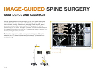
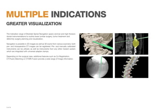
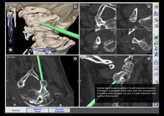
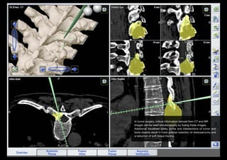
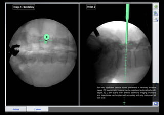
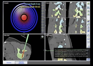
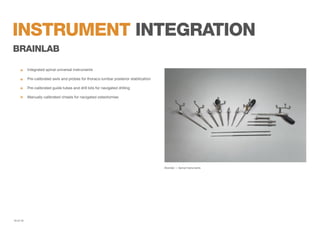
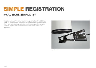
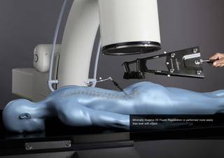
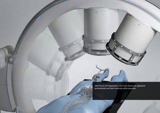
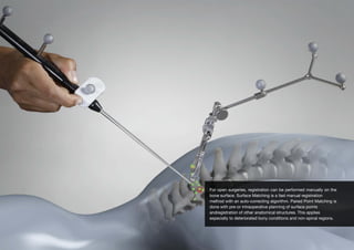
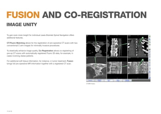
SPINE
NAVIGATION
IMAGE-guided spine surgery
CONFIDENCE AND ACCURACY
Brainlab Spinal Navigation combines state-of-the-art touch screen based image
control with best-in-class registration methods for image-guided surgery. As an
open navigation platform, Brainlab Spinal Navigation enables accurate pedicle
screw placement as well as drastic reduction of X-Ray exposure to both the
surgical team and the patient. Navigation of implants and instruments is possible in
2D images, 3D fluoroscopy scans, MR or CT datasets in all stages of surgery—from
incision planning to implant placement.
The indication range covers pedicle screw placement in any area of the spine,
placement of C1-C2 screws, complex deformity correction, tumor surgery and
surgical planning.

2 of 19
multiple indications
greater visualization
The indication range of Brainlab Spinal Navigation spans cervical and high thoracic
dorsal instrumentations to routine lower lumbar surgery, tumor treatment and
deformity surgery planning and visualization.
Navigation is possible in 2D images as well as 3D scans from various scanners; both
pre- and intraoperative CT images can be registered. Pre- and manually calibrated
instruments can be utilized, as well as instruments from any other implant system
which are integrated with universal adapter clamps.
Depending on the surgical case, additional features such as Co-Registration,
CT-Fluoro Matching or CT/MR-Fusion provide a wide range of image information.

3 of 19
Brainlab Spinal Navigation assists in the safe placement of implants,
especially in anatomically critical areas. Real time visualization of
instruments leads to greater accuracy of implant placement while
sparing X-Ray exposure.
In tumor surgery, critical information derived from CT and MR
images can be used simultaneously by fusing these images.
Additional visualized safety zones and intersections of tumor and
bone objects result in more precise resection of osteosarcoma and
a reduction of soft-tissue trauma.
For easy, confident pedicle screw placement in minimally invasive
cases, 2D fluoroscopic images can be registered automatically with
xSpot; 3D C-arm scans even without additional imaging. Incisions
and trajectories can be planned accurately with any instrument at
skin level.
The combination of pre-planning and quick registration of adjacent
levels allows the support of complex procedures like deformity
corrections. The improved orientation of navigation and enhanced
information from planning is especially valuable with deformed
anatomy, resulting in less soft tissue trauma and greater efficiency.
instrument integration
seAmless workflows
In addition to a range of Brainlab instruments, cooperations with DePuy Synthes
and other selected 3rd party manufacturers also provide navigation-ready integrated
instruments.
Moreover, the open platform principle of Brainlab Spinal Navigation enables any
instrument to be manually calibrated for navigation by attaching adapter clamps.
Pre- or manually calibrated instruments can be integrated into Navigation for smooth
and seamless workflows.


Follow standard clinical workflow

	
Navigate up to eight instruments simultaneously without the need for
	re-selection
	

Visualize pre-calibrated instruments in 3D geometry


Integrate any instrument and implant system with universal adapter clamps

8 of 19
instrument integration
DEPUY SYNTHES
	

Integrated Viper2 // Expedium system instruments


Thoraco-lumbar posterior stabilization
	

Pre-calibrated cannulated and non-cannulated awls, probes, taps


Manually calibrated cannulated and non-cannulated screwdrivers

DePuy Synthes - Viper2 // Expedium

9 of 19
instrument integration
brainlab
	

Integrated spinal universal instruments


Pre-calibrated awls and probes for thoraco-lumbar posterior stabilization
	

Pre-calibrated guide tubes and drill bits for navigated drilling


Manually calibrated chisels for navigated osteotomies

Brainlab — Spinal Instruments

10 of 19
instrument integration
ulrich medical
	

Integrated tangoRS // neon system instruments


Cervical and thoraco-lumbar posterior stabilization

	
tangoRS: pre-calibrated instrumentation for open thoraco-lumbar posterior
	stabilization
	

neon: pre-calibrated instruments for cervical posterior stabilization

ulrich medical - tangoRS // neon

11 of 19
instrument integration
aesCulap
	

Integrated S4 Cervical system instruments


Cervical-thoracic posterior stabilization
	

Pre-calibrated guide sleeves, drill guide & tap


Manually calibrated screwdriver

Aesculap — S4 Cervical

12 of 19
simple registration
practical simplicity
Navigation can be performed in any common image format from 2D and 3D images
to MR, CT and Angio scans, regardless of pre- or intraoperative image acquisition.
After a quick automatic image registration or point based registration, navigation
can begin. Attached to the pointer, SmartClip enables remote control of the
application.

xSpot cArc

13 of 19
Minimally invasive 2D Fluoro Registration is performed more easily
than ever with xSpot.
With Fluoro 3D Registration, 3D C-arm scans are registered
automatically and less invasively within seconds.
For open surgeries, registration can be performed manually on the
bone surface. Surface Matching is a fast manual registration
method with an auto-correcting algorithm. Paired Point Matching is
done with pre-or intraoperative planning of surface points
andregistration of other anatomical structures. This applies
especially to deteriorated bony conditions and non-spinal regions.
fusion and co-registration
image unity
To gain even more insight for individual cases Brainlab Spinal Navigation offers
additional features.
CT-Fluoro Matching allows for the registration of pre-operative CT scans with two
conventional C-arm images for minimally invasive procedures.
To drastically enhance image quality, Co-Registration allows co-registering of
pre-op CT scans with automatically registered Fluoro 3D data, for example, in
cases involving obese patients.
For additional soft tissue information, for instance, in tumor treatment, Fusion
brings full pre-operative MR information together with a registered CT scan.

CT/MR-Fusion

17 of 19
intelligent platforms
touch-based command and control

18 of 19
brainlab.com
© 2013 Brainlab AG. ST_BR_EN_Spine_Image Guided Surgery_Oct13

Image-Guided Spine Navigation Brochure

  • 1.
  • 2.
    IMAGE-guided spine surgery CONFIDENCEAND ACCURACY Brainlab Spinal Navigation combines state-of-the-art touch screen based image control with best-in-class registration methods for image-guided surgery. As an open navigation platform, Brainlab Spinal Navigation enables accurate pedicle screw placement as well as drastic reduction of X-Ray exposure to both the surgical team and the patient. Navigation of implants and instruments is possible in 2D images, 3D fluoroscopy scans, MR or CT datasets in all stages of surgery—from incision planning to implant placement. The indication range covers pedicle screw placement in any area of the spine, placement of C1-C2 screws, complex deformity correction, tumor surgery and surgical planning. 2 of 19
  • 3.
    multiple indications greater visualization Theindication range of Brainlab Spinal Navigation spans cervical and high thoracic dorsal instrumentations to routine lower lumbar surgery, tumor treatment and deformity surgery planning and visualization. Navigation is possible in 2D images as well as 3D scans from various scanners; both pre- and intraoperative CT images can be registered. Pre- and manually calibrated instruments can be utilized, as well as instruments from any other implant system which are integrated with universal adapter clamps. Depending on the surgical case, additional features such as Co-Registration, CT-Fluoro Matching or CT/MR-Fusion provide a wide range of image information. 3 of 19
  • 4.
    Brainlab Spinal Navigationassists in the safe placement of implants, especially in anatomically critical areas. Real time visualization of instruments leads to greater accuracy of implant placement while sparing X-Ray exposure.
  • 5.
    In tumor surgery,critical information derived from CT and MR images can be used simultaneously by fusing these images. Additional visualized safety zones and intersections of tumor and bone objects result in more precise resection of osteosarcoma and a reduction of soft-tissue trauma.
  • 6.
    For easy, confidentpedicle screw placement in minimally invasive cases, 2D fluoroscopic images can be registered automatically with xSpot; 3D C-arm scans even without additional imaging. Incisions and trajectories can be planned accurately with any instrument at skin level.
  • 7.
    The combination ofpre-planning and quick registration of adjacent levels allows the support of complex procedures like deformity corrections. The improved orientation of navigation and enhanced information from planning is especially valuable with deformed anatomy, resulting in less soft tissue trauma and greater efficiency.
  • 8.
    instrument integration seAmless workflows Inaddition to a range of Brainlab instruments, cooperations with DePuy Synthes and other selected 3rd party manufacturers also provide navigation-ready integrated instruments. Moreover, the open platform principle of Brainlab Spinal Navigation enables any instrument to be manually calibrated for navigation by attaching adapter clamps. Pre- or manually calibrated instruments can be integrated into Navigation for smooth and seamless workflows. Follow standard clinical workflow Navigate up to eight instruments simultaneously without the need for re-selection Visualize pre-calibrated instruments in 3D geometry Integrate any instrument and implant system with universal adapter clamps 8 of 19
  • 9.
    instrument integration DEPUY SYNTHES IntegratedViper2 // Expedium system instruments Thoraco-lumbar posterior stabilization Pre-calibrated cannulated and non-cannulated awls, probes, taps Manually calibrated cannulated and non-cannulated screwdrivers DePuy Synthes - Viper2 // Expedium 9 of 19
  • 10.
    instrument integration brainlab Integrated spinaluniversal instruments Pre-calibrated awls and probes for thoraco-lumbar posterior stabilization Pre-calibrated guide tubes and drill bits for navigated drilling Manually calibrated chisels for navigated osteotomies Brainlab — Spinal Instruments 10 of 19
  • 11.
    instrument integration ulrich medical IntegratedtangoRS // neon system instruments Cervical and thoraco-lumbar posterior stabilization tangoRS: pre-calibrated instrumentation for open thoraco-lumbar posterior stabilization neon: pre-calibrated instruments for cervical posterior stabilization ulrich medical - tangoRS // neon 11 of 19
  • 12.
    instrument integration aesCulap Integrated S4Cervical system instruments Cervical-thoracic posterior stabilization Pre-calibrated guide sleeves, drill guide & tap Manually calibrated screwdriver Aesculap — S4 Cervical 12 of 19
  • 13.
    simple registration practical simplicity Navigationcan be performed in any common image format from 2D and 3D images to MR, CT and Angio scans, regardless of pre- or intraoperative image acquisition. After a quick automatic image registration or point based registration, navigation can begin. Attached to the pointer, SmartClip enables remote control of the application. xSpot cArc 13 of 19
  • 14.
    Minimally invasive 2DFluoro Registration is performed more easily than ever with xSpot.
  • 15.
    With Fluoro 3DRegistration, 3D C-arm scans are registered automatically and less invasively within seconds.
  • 16.
    For open surgeries,registration can be performed manually on the bone surface. Surface Matching is a fast manual registration method with an auto-correcting algorithm. Paired Point Matching is done with pre-or intraoperative planning of surface points andregistration of other anatomical structures. This applies especially to deteriorated bony conditions and non-spinal regions.
  • 17.
    fusion and co-registration imageunity To gain even more insight for individual cases Brainlab Spinal Navigation offers additional features. CT-Fluoro Matching allows for the registration of pre-operative CT scans with two conventional C-arm images for minimally invasive procedures. To drastically enhance image quality, Co-Registration allows co-registering of pre-op CT scans with automatically registered Fluoro 3D data, for example, in cases involving obese patients. For additional soft tissue information, for instance, in tumor treatment, Fusion brings full pre-operative MR information together with a registered CT scan. CT/MR-Fusion 17 of 19
  • 18.
  • 19.
    brainlab.com © 2013 BrainlabAG. ST_BR_EN_Spine_Image Guided Surgery_Oct13