Download as PDF, PPTX


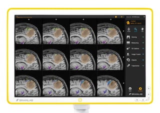


Curve™ Image Guided Surgery is an advanced command and control center that enhances surgical navigation through superior ergonomics and multi-directional touch technology. It integrates various imaging and surgical tools to improve collaboration and patient outcomes while offering flexible setups for diverse surgical environments. With features like high-definition displays, streamlined functionality, and comprehensive connectivity, it empowers surgeons with critical intra-operative information.