Download to read offline

![In general, patients are immobilized with a frame and a single
mean dose of 15 to 19 Gy is prescribed to the 80% isodose
line. The dose is typically delivered by four to eight arcs—for
spherical lesions, conical collimators can be preferred over
high-resolution multi-leaf collimators.
The overall incidence of radiation-induced complications
ranges from 3 to 6% and the reported neurological deficits
10-17
.
are often of transient nature
It is therefore concluded that SRS is a safe and effective
intracranial AVM treatment option, as long as careful
neuroimaging follow-up is guaranteed to monitor the nidus
response.
Intracranial AVMs are often classified according to a grading
20
score developed by Spetzler and Martin and the prescribed
dose is related to the initial AVM grade, since complete nidus
obliteration rates were found to depend mainly on the AVM
11,13,15
.
volume and the SRS dose
Overview of the recent clinical literature on SRS for arteriovenous malformations
Institution
Pedroso
Buis
12
Scarbrough
14
Zabel-du Bois
Huang
13
16
MorenoJiménez
Raza
10
17
% Prior
Treatment
Mean Vol
(cm³)
Mean
Dose (Gy)
#
Fractions
% IDL
covering
PTV
% Complete
Obliteration
1999
50
36
23
16
1
80
45 at 20 months
2004
44
30
18
15
1
80
52 at 37 months
VU University Medical
Center, Amsterdam
15
#
Lesions
David Geffen School of
Medicine, Los Angeles
11
Mobin
Year
University of California,
Davis
Author
2005
31
32
3
19
1
80
77 at 33 months
The Melbourne Cancer
Center, Melbourne
2005
39
8
7
17
1
80
87 at 24 months
University of Heidelberg
2006
22
36
4
18
1
80
65 at 48 months
Ghang Gung Memorial
Hospital, Taiwan
2006
34
14
2
16
1
80
NA
Nat´l Institute of Neurol &
Neurosurg, Mexico
2007
40
40
8
15.4
1
80
63 at 29 months
The Johns Hopkins
Hospital, Baltimore
2007
14
47
25
36
3
NA
36 at 31 months
Complete obliteration implies that the nidus is no longer visible angiographically and that the circulation time and the afferent and efferent vessels that had supplied
the malformation have returned to normal. For angiographically occult lesions like low-flow cavernous malformations, studied by Huang et al., there is currently no
gold standard for demonstrating the obliteration.
References
[1]
[2]
[3]
[4]
[5]
[6]
[7]
[8]
[9]
[10]
Pollock B.E. et al., Stroke 27, 1, 1996
Karhunen P.J. et al., Forensic Sci Int 48, 9, 1990
Fleetwood I.G. et al., Lancet 359, 863, 2002
Thompson R.C. et al., Neurosurgery 43, 202, 1998
Nussbaum E.S. et al., Neurosurgery 43, 347, 1998
Sasaki T. et al., J Neurosurg 88, 285, 1998
Ellis T.L. et al., J Neurosurg 89, 104, 1998
Lawton M.T., Neurosurg 52, 740, 2003
Yu S.C. et al., AJNR Am J Neuroradiol 25, 1139, 2004
Moreno-Jiménez S. et al., Surg Neurol 67, 487, 2007
Europe | +49 89 99 1568 0 | de_sales@brainlab.com
North America | +1 800 784 7700 | us_sales@brainlab.com
Latin America | +55 11 3355 3370 | br_sales@brainlab.com
RT_WP_E_AVM_APR11
[11]
[12]
[13]
[14]
[15]
[16]
[17]
[18]
[19]
[20]
Mobin F. et al., Stereotact Funct Neurosurg 73, 50, 1999
Buis D.R. et al., Int J Radiat Oncol Biol Phys 62(1), 246, 2005
Zabel-du Bois A. et al., Int J Radiat Oncol Biol Phys 65(4), 1206, 2006
Scarbrough T.J. et al., Stereotact Funct Neurosurg 83, 91, 2005
Pedroso A.G. et al., J Neurosurg 101, 425, 2004
Huang Y.C. et al., Clin Neurol Neurosur 108, 750, 2006
Raza S.M. et al., Surg Neurol 68, 24, 2007
Pollock B.E. et al., Neurosurg 38, 652, 1996
Maruyama K. et al., N Engl J Med 352, 146, 2005
Spetzler et al., J Neurosurg 65, 476, 1986
Asia Pacific | +852 2417 1881 | hk_sales@brainlab.com
Japan | +81 3 3769 6900 | jp_sales@brainlab.com](https://image.slidesharecdn.com/whitepaperarteriovenousmalformations-140115050412-phpapp02/85/Stereotactic-Radiosurgery-of-Arteriovenous-Malformations-Clinical-White-Paper-2-320.jpg)

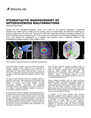
Intracranial arteriovenous malformations (AVMs) are rare but a leading cause of nontraumatic intracerebral hemorrhage in young individuals, requiring aggressive treatment to prevent hemorrhaging. Stereotactic radiosurgery (SRS) has emerged as an effective treatment option, offering a safe method for obliterating AVM nidus primarily through radiation, although careful follow-up is essential due to potential risks during the latency period. Current strategies include microsurgery, embolization, and SRS, with treatment modalities tailored based on lesion characteristics and patient risk.


![PERI-PROSTHETIC FRACTURE NAIL-PLATE CONSTRUCT [NPC].pptx](https://cdn.slidesharecdn.com/ss_thumbnails/drarunkumardrmohamedashrafperiprostheticfrasturenail-plateconstructnpc-260209164459-7e9d15a1-thumbnail.jpg?width=640&height=640&fit=bounds)


