Download to read offline
![The mean treatment dose ranges from 15 to 21 Gy for SRS
and from 45 to 52 Gy for SRT, typically split up in fractions of
1.8 Gy. Depending on the collimator, the dose is delivered by
13,16,18
multiple, non-coplanar arcs or beams, either conformal
15
or intensity-modulated .
The achieved mean local control for both SRS and SRT
treatments is 96% with an average hormonal response rate of
80%. Newly initiated hormonal replacement therapy, as a
result of radiation-induced pituitary deficiency, was required in
5 to 40% of all cases.
This wide range of values reflects the various positions of the
lesions with respect to the organs at risk. Patients with
lesions close the pituitary gland or the hypothalamus are
more likely to suffer radiation-induced toxicity.
From the summarized results it can be concluded that SRS
and SRT, either as a primary treatment or in combination
with surgery, represent an effective, safe and minimally
invasive option for controlling the growth of PAs and reducing
hormone production.
Overview of the recent clinical literature on SRS and SRT for pituitary adenomas
Institution
Year
#
Lesions
% Prior
Surgery
Mean
Volume
(cm³)
Mean
Dose
(Gy)
#
Fractions
% Local
Control
%
Tumor
Response
%
Hormonal
Response
% Pituitary
Deficiency
12
Brigham and Women´s
Hospital, Boston
1998
18
88
1.90
15
1
100 at 47
months
22
50
30
12
Brigham and Women´s
Hospital, Boston
1998
30
88
5.70
45
25
85 at 34
months
20
62
21
Kangnam St. Mary´s
Hospital, Seoul
1998
24
96
NA
21
1
96 at 49
months
63
84
29
University of
Heidelberg
2001
62
11
30.2
50
28
93 at 39
months
30
60
5
University of
Heidelberg
2004
5
100
3.50
15
1
100 at 38
months
60
100
8
13
University of
Heidelberg
2004
20
95
26.2
52
29
100 at 61
months
25
80
8
16
Polyclinique Courlancy,
Reims
2005
110
81
4.20
50
28
99 at 82
months
89
100
37
17
National University,
Seoul
2005
68
96
6.20
50
28
98 at 30
months
38
NA
6
Author
Mitsumori
Mitsumori
14
Yoon
Milker-
18
Zabel
Milker-
13
Zabel
MilkerZabel
Colin
Paek
Because local control doesn´t necessarily imply a reduction of tumor volume, tumor response indicates the percentage of adenomas that present a measurable
reduction of the tumor volume after irradiation. Similarly, hormonal response is defined as a quantitative reduction of the initial hormonal level. The wide range of
values for the pituitary deficiency reflects the various positions of the lesions with respect to the organs at risk. Patients with lesions close the pituitary gland or the
hypothalamus are more likely to suffer radiation-induced toxicity.
References
[1]
[2]
[3]
[4]
[5]
[6]
[7]
[8]
[9]
Scheithauer B.W. et al., Neurosurg 59, 341, 2006
Ezzat S. et al., Cancer 101, 613, 2004
Dworakowska D. et al., Best Pract Res Cl En 23, 525, 2009
Milker-Zabel S. et al., Int J Radiat Oncol Biol Phys 50(5), 1279, 2001
Daly A.F. et al., Best Pract Res Cl En 23, 543, 2009
Wilson C.B., Cancer medicine. Lea & Febiger, Philadelphia 1993, pp. 1131–1137
Lindholm J. et al., J Clin Endocr Metab 86, 117, 2001
Sauer R., Therapy of malignant brain tumors. Springer-Verlag 1987, pp. 195–276
Davis D.H. et al., J Neurosurg 79, 70, 1993
Europe | +49 89 99 1568 0 | de_sales@brainlab.com
North America | +1 800 784 7700 | us_sales@brainlab.com
Latin America | +55 11 3355 3370 | br_sales@brainlab.com
RT_WP_E_PITUITARY_APR11
[10]
[11]
[12]
[13]
[14]
[15]
[16]
[17]
[18]
[19]
Beclere A., Arch Roentgen Radiol 14, 147, 1909
Rush S. et al., Int J Radiat Oncol Biol Phys Biol Phys 37, 1031, 1997
Mitsumori M. et al., Int J Radiat Oncol Biol Phys 42(3), 573, 1998
Milker-zabel S. et al., Int J Radiat Oncol Biol Phys 59(4), 1088, 2004
Yoon S.C. et al., Int J Radiat Oncol Biol Phys 41(4), 849, 1998
Mackley H.B. et al., Int J Radiat Oncol Biol Phys 67(1), 232, 2007
Colin P. et al., Int J Radiat Oncol Biol Phys 62(2), 333, 2005
Paek S.H. et al., Int J Radiat Oncol Biol Phys 61(3), 795, 2005
Milker-zabel S. et al., Int J Radiat Oncol Biol Phys 50(5), 1279, 2001
Tome W.A. et al., Technol Cancer Res Treat 1, 153, 2002
Asia Pacific | +852 2417 1881 | hk_sales@brainlab.com
Japan | +81 3 3769 6900 | jp_sales@brainlab.com](https://image.slidesharecdn.com/whitepaperpituaryadenomas-140115050418-phpapp01/85/Stereotactic-Radiosurgery-and-Radiotherapy-of-Pituitary-Adenomas-Clinical-White-Paper-2-320.jpg)

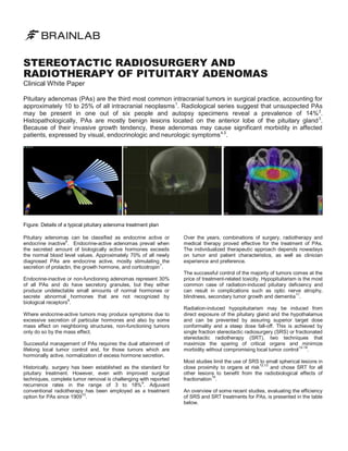
Pituitary adenomas (PAs) are the third most common intracranial tumors, leading to various visual, endocrinologic, and neurologic symptoms due to their invasive nature. Management involves a combination of surgery, stereotactic radiosurgery (SRS), and fractionated stereotactic radiotherapy (SRT), with SRS and SRT showing high local control rates and hormonal response. Treatment-related toxicity, particularly radiation-induced hypopituitarism, is a concern, especially for lesions close to critical organs.


![Hypothalamus short ppt by Dr. Neha [PT].pptx](https://cdn.slidesharecdn.com/ss_thumbnails/hypothalamusbydr-260124145759-b9f94a93-thumbnail.jpg?width=640&height=640&fit=bounds)


