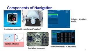
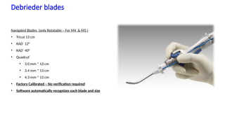
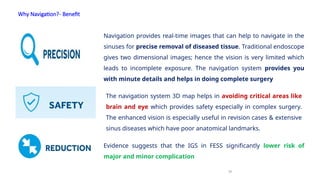
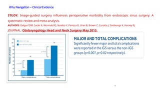
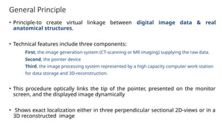
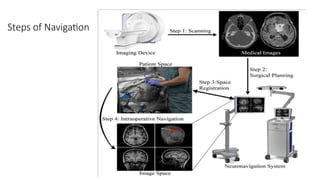
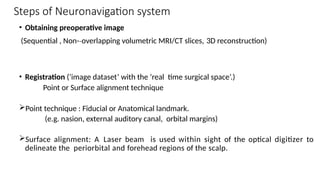
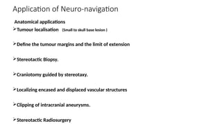
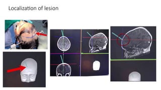
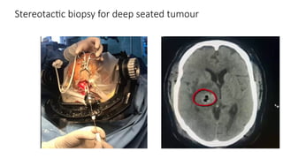
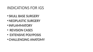
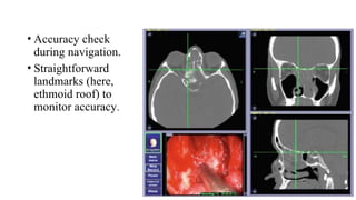
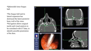
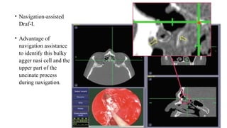
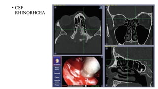
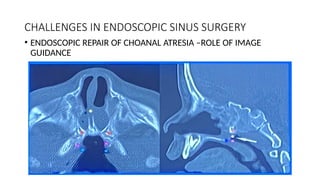
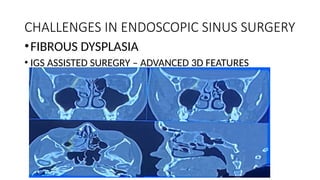
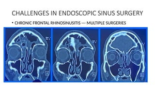
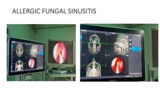
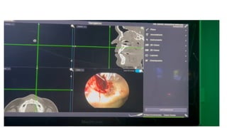
Introduction to Navigation---
ENT & Skull Base
Advanced ENT & Multispeciality Hospital
pvt.ltd.
2
You know driving but what will you do when you find?????
Route, you know is
closed down ?
There is a heavy traffic,
and you want to reach
early & safely. Now
What?
3
Navigation – Is Here to Help you!
Optical System works exactly same as GPS
• How GPS Works?
Image source: http://mason.gmu.edu/~ttruong6/how.html
Trilateration
5
Electromagnetic localization
 The navigation systems using electromagnetic technique
consists of emitter which emits low intensity
electromagnetic field within a limited field of
approximately 50 cm .

 This field has unique field strengths at each point.
 Whenever the navigable instrument is brought into this
field the coils of the instruments or the trackers disrupt
the field and in turn generate EMF i.e. this disruption can
be corelated into position
6
Components of Navigation
A navigation system with a monitor and “localizer”
A patient reference
Software – procedure
specific
Specialized Instruments
Recent imaging data of the patient
7
Scan protocol
8
Scan protocol
CT SCAN SPECIFIC
9
Scan protocol
SCAN REGION
10
ENT Instruments with Instrument Tracker
Registration Probe Straight Suction Curved Suction, 70°
Curved Suction 90° Straight Probe Ostium Seeker
• Malleable Suctions
• Debrider Blades
• Pediatric Reference Frame
• Skull mounted Tracker
11
Debrieder blades
Navigated Blades (only Rotatable – For M4 & M5 )
• Tricut 13 cm
• RAD®
12°
• RAD®
40°
• Quadcut®
• 3.0 mm * 13 cm
• 3.4 mm * 13 cm
• 4.3 mm * 13 cm
• Factory Calibrated – No verification required
• Software automatically recognizes each blade and size
Stealth ENT - See More Do More | December 3, 2020 |
Confidential, for Internal Use Only
Why Navigation?- Benefit
12
Navigation provides real-time images that can help to navigate in the
sinuses for precise removal of diseased tissue. Traditional endoscope
gives two dimensional images; hence the vision is very limited which
leads to incomplete exposure. The navigation system provides you
with minute details and helps in doing complete surgery
The navigation system 3D map helps in avoiding critical areas like
brain and eye which provides safety especially in complex surgery.
The enhanced vision is especially useful in revision cases & extensive
sinus diseases which have poor anatomical landmarks.
Evidence suggests that the IGS in FESS significantly lower risk of
major and minor complication
Why Navigation – Clinical Evidence
13
STUDY: Image-guided surgery influences perioperative morbidity from endoscopic sinus surgery: A
systematic review and meta-analysis.
AUTHORS: Dalgorf DM, Sacks R, Wormald PJ, Naidoo Y, Panizza B, Uren B, Brown C, Curotta J, Snidvongs K, Harvey RJ.
JOURNAL: Otolaryngology Head and Neck Surgery May 2013.
Navigation – help reduce complications
14
• Intracranial
• Orbital
• Hemorrhage
• Lacrimal
Major
Complications
• Orbital
• Epistaxis
• Bronchospasm
• Adhesions
• Other
Minor
Complications
70%
reduction
50%
reduction
 Navigational risks associated with sinus surgery and the clinical effects of implementing a navigational system
for sinus surgery. Reardon EJ. Harvard Medical School, Massachusetts Eye and Ear Infirmary, and Carney Hospital, Boston, MA Laryngoscope. 2002 Jul;112(7 Pt 2):1-19.
 Comparison of endoscopic sinus surgery with and without image guidance. Fried MP, Moharir VM, Shin J, Taylor-Becker M, Morrison P.
Department of Otolaryngology, Montefiore Medical Center, Albert Einstein College of Medicine, Bronx, New York Am J Rhinol 2002 Jul-Aug;16(4):193-7
General Principle
• Principle-to create virtual linkage between digital image data & real
anatomical structures.
• Technical features include three components:
First, the image generation system (CT-scanning or MR imaging) supplying the raw data.
Second, the pointer device
Third, the image processing system represented by a high capacity computer work station
for data storage and 3D-reconstruction.
• This procedure optically links the tip of the pointer, presented on the monitor
screen, and the displayed image dynamically
• Shows exact localization either in three perpendicular sectional 2D-views or in a
3D reconstructed image
Steps of Navigation
Steps of Neuronavigation system
• Obtaining preoperative image
(Sequential , Non-­overlapping volumetric MRI/CT slices,
‐ 3D reconstruction)
• Registration (‘image dataset’ with the ‘real time surgical space’.)
Point or Surface alignment technique
Point technique : Fiducial or Anatomical landmark.
(e.g. nasion, external auditory canal, orbital margins)
Surface alignment: A Laser beam is used within sight of the optical digitizer to
delineate the periorbital and forehead regions of the scalp.
Application of Neuro-navigation
Anatomical applications
Tumour localisation (Small to skull base lesion )
Define the tumour margins and the limit of extension
Stereotactic Biopsy.
Craniotomy guided by stereotaxy.
Localizing encased and displaced vascular structures
Clipping of intracranial aneurysms.
Stereotactic Radiosurgery
Indications:
• American Academy of Otolaryngology Head and Neck Surgery
1. Revision sinus surgery
2. Distorted sinus anatomy of development, postoperative, or traumatic
origin
3. Extensive sino-nasal polyposis
4. Pathology involving the frontal, posterior ethmoid, and sphenoid sinuses
5. Disease abutting the skull base, orbit, optic nerve, or carotid artery
6. Cerebrospinal fluid rhinorrhea or conditions where there is a skull-base
defect
7. Benign and malignant sino-nasal neoplasms
8. Choanal atresia
Localization of lesion
Stereotactic biopsy for deep seated tumour
INDICATIONS FOR IGS
•SKULL BASE SURGERY
•NEOPLASTIC SURGERY
•INFLAMMATORY
• REVISION CASES
• EXTENSIVE POLYPOSIS
•CHALLENGING ANATOMY
• Accuracy check
during navigation.
• Straightforward
landmarks (here,
ethmoid roof) to
monitor accuracy.
•Sphenoidal sinus fungus
ball.
•The fungus ball and its
lateral expansion has
destroyed the latero-posterior
bony wall of the sinus.
•Navigation allows surgical
tactile and visual analysis to
be correlated with CT data, to
identify possible penetration
of the dura.
• Navigation-assisted
Draf-I.
• Advantage of
navigation assistance
to identify this bulky
agger nasi cell and the
upper part of the
uncinate process
during navigation.
• CSF
RHINORHOEA
Surgical challenges & Navigation
•3D PATHOLOGY UNDERSTANDING
•SURGICAL READINESS
•GREATER ANATOMICAL AWARENESS
•INTEGRATED SOLUTIONS (VERSATILE)
CHALLENGES IN ENDOSCOPIC SINUS SURGERY
• ENDOSCOPIC REPAIR OF CHOANAL ATRESIA –ROLE OF IMAGE
GUIDANCE
CHALLENGES IN ENDOSCOPIC SINUS SURGERY
•FIBROUS DYSPLASIA
• IGS ASSISTED SUREGRY – ADVANCED 3D FEATURES
CHALLENGES IN ENDOSCOPIC SINUS SURGERY
• CHRONIC FRONTAL RHINOSINUSITIS --- MULTIPLE SURGERIES
Limitations
• Increased Cost,
• Increased Operative time,
• Accurate surgical navigation depends on robust registration. Bone-
anchored fiducial markers provide the best accuracy. Impractical.
• IGS technology does not change the nature of the surgical procedure.
• All IGS platforms rely on preoperative imaging data sets.
ALLERGIC FUNGAL SINUSITIS
• IN REALITY
• ALL SURGERIES ARE IMAGE
GUIDED……..
• BY IMAGES ON SURGEON’S
RETINA
Future Prospects
• Combining CT and MRI – ENT and Neurosurgery for Anterior skull
base approach to brain tumors.
THANK YOU
“Technology is no substitute to Technique”

INTRODUCTION TO NAVIGATION.pptx DR.RITESH BHAGYAWANT

  • 1.
    Introduction to Navigation--- ENT& Skull Base Advanced ENT & Multispeciality Hospital pvt.ltd.
  • 2.
    2 You know drivingbut what will you do when you find????? Route, you know is closed down ? There is a heavy traffic, and you want to reach early & safely. Now What?
  • 3.
    3 Navigation – IsHere to Help you!
  • 4.
    Optical System worksexactly same as GPS • How GPS Works? Image source: http://mason.gmu.edu/~ttruong6/how.html Trilateration
  • 5.
    5 Electromagnetic localization  Thenavigation systems using electromagnetic technique consists of emitter which emits low intensity electromagnetic field within a limited field of approximately 50 cm .   This field has unique field strengths at each point.  Whenever the navigable instrument is brought into this field the coils of the instruments or the trackers disrupt the field and in turn generate EMF i.e. this disruption can be corelated into position
  • 6.
    6 Components of Navigation Anavigation system with a monitor and “localizer” A patient reference Software – procedure specific Specialized Instruments Recent imaging data of the patient
  • 7.
  • 8.
  • 9.
  • 10.
    10 ENT Instruments withInstrument Tracker Registration Probe Straight Suction Curved Suction, 70° Curved Suction 90° Straight Probe Ostium Seeker • Malleable Suctions • Debrider Blades • Pediatric Reference Frame • Skull mounted Tracker
  • 11.
    11 Debrieder blades Navigated Blades(only Rotatable – For M4 & M5 ) • Tricut 13 cm • RAD® 12° • RAD® 40° • Quadcut® • 3.0 mm * 13 cm • 3.4 mm * 13 cm • 4.3 mm * 13 cm • Factory Calibrated – No verification required • Software automatically recognizes each blade and size Stealth ENT - See More Do More | December 3, 2020 | Confidential, for Internal Use Only
  • 12.
    Why Navigation?- Benefit 12 Navigationprovides real-time images that can help to navigate in the sinuses for precise removal of diseased tissue. Traditional endoscope gives two dimensional images; hence the vision is very limited which leads to incomplete exposure. The navigation system provides you with minute details and helps in doing complete surgery The navigation system 3D map helps in avoiding critical areas like brain and eye which provides safety especially in complex surgery. The enhanced vision is especially useful in revision cases & extensive sinus diseases which have poor anatomical landmarks. Evidence suggests that the IGS in FESS significantly lower risk of major and minor complication
  • 13.
    Why Navigation –Clinical Evidence 13 STUDY: Image-guided surgery influences perioperative morbidity from endoscopic sinus surgery: A systematic review and meta-analysis. AUTHORS: Dalgorf DM, Sacks R, Wormald PJ, Naidoo Y, Panizza B, Uren B, Brown C, Curotta J, Snidvongs K, Harvey RJ. JOURNAL: Otolaryngology Head and Neck Surgery May 2013.
  • 14.
    Navigation – helpreduce complications 14 • Intracranial • Orbital • Hemorrhage • Lacrimal Major Complications • Orbital • Epistaxis • Bronchospasm • Adhesions • Other Minor Complications 70% reduction 50% reduction  Navigational risks associated with sinus surgery and the clinical effects of implementing a navigational system for sinus surgery. Reardon EJ. Harvard Medical School, Massachusetts Eye and Ear Infirmary, and Carney Hospital, Boston, MA Laryngoscope. 2002 Jul;112(7 Pt 2):1-19.  Comparison of endoscopic sinus surgery with and without image guidance. Fried MP, Moharir VM, Shin J, Taylor-Becker M, Morrison P. Department of Otolaryngology, Montefiore Medical Center, Albert Einstein College of Medicine, Bronx, New York Am J Rhinol 2002 Jul-Aug;16(4):193-7
  • 15.
    General Principle • Principle-tocreate virtual linkage between digital image data & real anatomical structures. • Technical features include three components: First, the image generation system (CT-scanning or MR imaging) supplying the raw data. Second, the pointer device Third, the image processing system represented by a high capacity computer work station for data storage and 3D-reconstruction. • This procedure optically links the tip of the pointer, presented on the monitor screen, and the displayed image dynamically • Shows exact localization either in three perpendicular sectional 2D-views or in a 3D reconstructed image
  • 16.
  • 17.
    Steps of Neuronavigationsystem • Obtaining preoperative image (Sequential , Non-­overlapping volumetric MRI/CT slices, ‐ 3D reconstruction) • Registration (‘image dataset’ with the ‘real time surgical space’.) Point or Surface alignment technique Point technique : Fiducial or Anatomical landmark. (e.g. nasion, external auditory canal, orbital margins) Surface alignment: A Laser beam is used within sight of the optical digitizer to delineate the periorbital and forehead regions of the scalp.
  • 18.
    Application of Neuro-navigation Anatomicalapplications Tumour localisation (Small to skull base lesion ) Define the tumour margins and the limit of extension Stereotactic Biopsy. Craniotomy guided by stereotaxy. Localizing encased and displaced vascular structures Clipping of intracranial aneurysms. Stereotactic Radiosurgery
  • 19.
    Indications: • American Academyof Otolaryngology Head and Neck Surgery 1. Revision sinus surgery 2. Distorted sinus anatomy of development, postoperative, or traumatic origin 3. Extensive sino-nasal polyposis 4. Pathology involving the frontal, posterior ethmoid, and sphenoid sinuses 5. Disease abutting the skull base, orbit, optic nerve, or carotid artery 6. Cerebrospinal fluid rhinorrhea or conditions where there is a skull-base defect 7. Benign and malignant sino-nasal neoplasms 8. Choanal atresia
  • 20.
  • 21.
    Stereotactic biopsy fordeep seated tumour
  • 22.
    INDICATIONS FOR IGS •SKULLBASE SURGERY •NEOPLASTIC SURGERY •INFLAMMATORY • REVISION CASES • EXTENSIVE POLYPOSIS •CHALLENGING ANATOMY
  • 23.
    • Accuracy check duringnavigation. • Straightforward landmarks (here, ethmoid roof) to monitor accuracy.
  • 24.
    •Sphenoidal sinus fungus ball. •Thefungus ball and its lateral expansion has destroyed the latero-posterior bony wall of the sinus. •Navigation allows surgical tactile and visual analysis to be correlated with CT data, to identify possible penetration of the dura.
  • 25.
    • Navigation-assisted Draf-I. • Advantageof navigation assistance to identify this bulky agger nasi cell and the upper part of the uncinate process during navigation.
  • 26.
  • 27.
    Surgical challenges &Navigation •3D PATHOLOGY UNDERSTANDING •SURGICAL READINESS •GREATER ANATOMICAL AWARENESS •INTEGRATED SOLUTIONS (VERSATILE)
  • 28.
    CHALLENGES IN ENDOSCOPICSINUS SURGERY • ENDOSCOPIC REPAIR OF CHOANAL ATRESIA –ROLE OF IMAGE GUIDANCE
  • 29.
    CHALLENGES IN ENDOSCOPICSINUS SURGERY •FIBROUS DYSPLASIA • IGS ASSISTED SUREGRY – ADVANCED 3D FEATURES
  • 30.
    CHALLENGES IN ENDOSCOPICSINUS SURGERY • CHRONIC FRONTAL RHINOSINUSITIS --- MULTIPLE SURGERIES
  • 31.
    Limitations • Increased Cost, •Increased Operative time, • Accurate surgical navigation depends on robust registration. Bone- anchored fiducial markers provide the best accuracy. Impractical. • IGS technology does not change the nature of the surgical procedure. • All IGS platforms rely on preoperative imaging data sets.
  • 32.
  • 34.
    • IN REALITY •ALL SURGERIES ARE IMAGE GUIDED…….. • BY IMAGES ON SURGEON’S RETINA
  • 35.
    Future Prospects • CombiningCT and MRI – ENT and Neurosurgery for Anterior skull base approach to brain tumors.
  • 36.
    THANK YOU “Technology isno substitute to Technique”