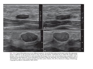
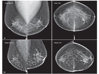
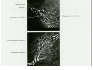
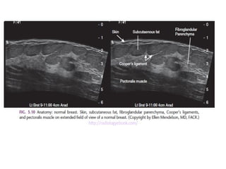
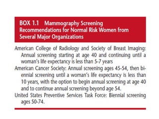
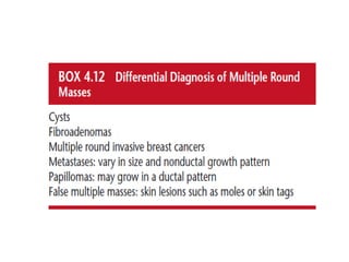
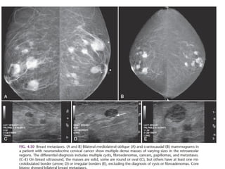
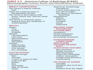
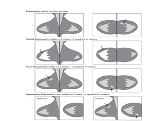
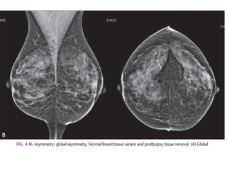
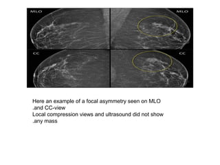
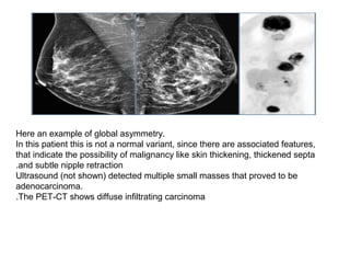
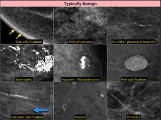
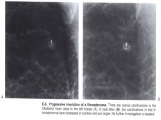
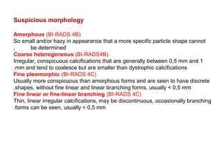
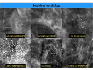
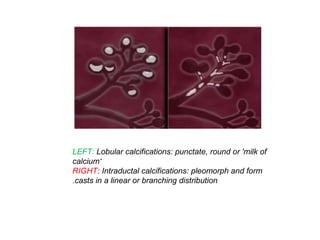
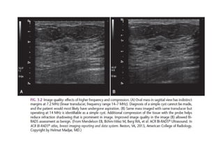
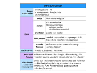
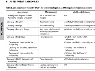
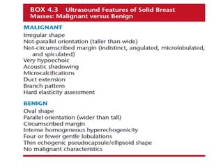
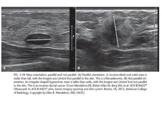
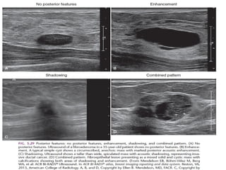
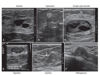
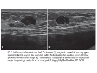
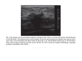
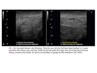
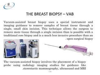
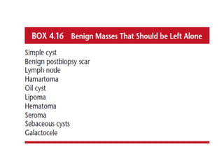
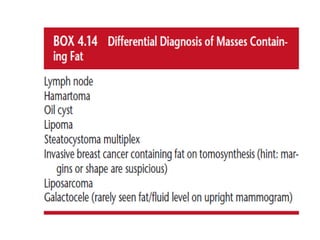
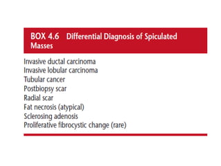
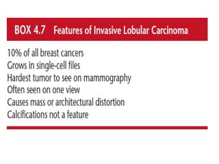
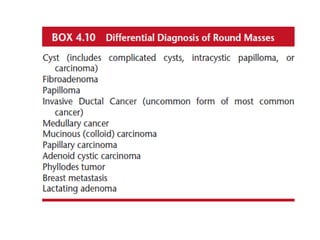
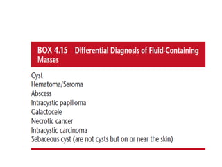
Breast imaging based on
ACR2013
MD .Dr.k.sharifi.radiologist
The shape of a mass is either round, oval or irregular.
Always make sure that a mass that is found on physical examination is
the same as the mass that is found with mammography or ultrasound.
Location and size should be applied in any lesion, that must undergo
biopsy.
The margin of a lesion can be:
Circumscribed (historically well-defined This is a benign finding.
Obscured or partially obscured, when the margin is hidden by superimposed
fibroglandular tissue. Ultrasound can be helpful to define the margin better.
Microlobulated. This implies a suspicious finding.
Indistinct (historically ill-defined).
This is also a suspicious finding.
Spiculated with radiating lines from the mass is a very suspicious finding.
The density of a mass is related to the expected
attenuation of an equal volume of fibroglandular tissue.
High density is associated with malignancy.
It is extremely rare for breast cancer to be low density.
Here multiple round circumscribed low density masses in the right
breast.
These were the result of lipofilling, which is transplantation of
body fat to the breast.
Here a hyperdense mass with an irregular shape and a spiculated margin.
Notice the focal skin retraction.
This was reported as BI-RADS 5 and proved to be an invasive ductal
carcinoma.
The term architectural distortion is used, when the normal architecture is distorted
with no definite mass visible.
This includes thin straight lines or spiculations radiating from a point, and focal
retraction, distortion or straightening at the edges of the parenchyma.
The differential diagnosis is scar tissue or carcinoma.
Architectural distortion can also be seen as an associated feature.
For instance if there is a mass that causes architectural distortion, the likelihood of
malignancy is greater than in the case of a mass without distortion.
Notice the distortion of the normal breast architecture on oblique view (yellow circle)
and magnification view.
A resection was performed and only scar tissue was found in the specimen.
Architectural distortion
Asymmetries
Findings that represent unilateral deposits of fibroglandular tissue not
conforming to the definition of a mass.
Asymmetry as an area of fibroglandulair tissue visible on only one
mammographic projection, mostly caused by superimposition of normal breast
tissue.
Focal asymmetry visible on two projections, hence a real finding rather than
superposition.
This has to be differentiated from a mass.
Global asymmetry consisting of an asymmetry over at least one quarter of the
breast and is usually a normal variant.
Developing asymmetry new, larger and more conspicuous than on a previous
examination.
Here an example of a focal asymmetry seen on MLO
and CC-view.
Local compression views and ultrasound did not show
any mass.
Here an example of global asymmetry.
In this patient this is not a normal variant, since there are associated features,
that indicate the possibility of malignancy like skin thickening, thickened septa
and subtle nipple retraction.
Ultrasound (not shown) detected multiple small masses that proved to be
adenocarcinoma.
The PET-CT shows diffuse infiltrating carcinoma.
Asymmetry versus Mass
All types of asymmmetry have different border contours than true masses and also
lack the conspicuity of masses.
Asymmetries appear similar to other discrete areas of fibroglandulair tissue except that
they are unilateral, with no mirror-image correlate in the opposite breast.
An asymmetry demonstrates concave outward borders and usually is interspersed with
fat, whereas a mass demonstrates convex outward borders and appears denser in the
center than at the periphery.
The use of the term "density" is confusing, as the term "density" should only be used to
describe the x-ray attenuation of a mass compared to an equal volume of
fibroglandular tissue.
Typically benign
Skin, vascular, coarse, large rod like, round or punctate
(< 1mm), rim, dystrophic, milk of calcium and suture
calcifications are typically benign.
There is one exception of the rule: an isolated group of
punctuate calcifications that is new, increasing, linear,
or segmental in distribution, or adjacent to a known
cancer can be assigned as probably benign or
suspicious.
Suspicious morphology
Amorphous (BI-RADS 4B)
So small and/or hazy in appearance that a more specific particle shape cannot
be determined.
Coarse heterogeneous (BI-RADS4B)
Irregular, conspicuous calcifications that are generally between 0,5 mm and 1
mm and tend to coalesce but are smaller than dystrophic calcifications.
Fine pleomorphic (BI-RADS 4C)
Usually more conspicuous than amorphous forms and are seen to have discrete
shapes, without fine linear and linear branching forms, usually < 0,5 mm.
Fine linear or fine-linear branching (BI-RADS 4C)
Thin, linear irregular calcifications, may be discontinuous, occasionally branching
forms can be seen, usually < 0,5 mm.
Distribution of calcifications
The arrangement of calcifications, the distribution, is at least as important as
morphology.
These descriptors are arranged according to the risk of malignancy:
Diffuse: distributed randomly throughout the breast.
Regional: occupying a large portion of breast tissue > 2 cm greatest dimension
Grouped (historically cluster): few calcifications occupying a small portion of breast
tissue: lower limit 5 calcifications within 1 cm and upper limit a larger number of
calcifications within 2 cm.
Linear: arranged in a line, which suggests deposits in a duct.
Segmental: suggests deposits in a duct or ducts and their branches.
The 2013 edition refines the upper limit in size for grouped distribution as 2 cm
(historically 1 cm) while retaining > 2 cm as the lower limit for regional distribution.
LEFT: Lobular calcifications: punctate, round or 'milk of
calcium‘
RIGHT: Intraductal calcifications: pleomorph and form
casts in a linear or branching distribution.
Case 1: BIRADS II lesion
BI-RADS 3
BI-RADS 4A in a 55-year-old woman who presented with a
palpable mass and swelling in her left breast
BI-RADS 4B
BI-RADS 5
Enhancement pattern of a mass
Mass enhancement occurs in 6 main patterns:
Homogeneous enhancement is uniform and confluent enhancement throughout the
mass.
Heterogeneous enhancement is nonuniform enhancement, which varies within the
mass.
Rim enhancement is enhancement mainly concentrated at the periphery of the
mass. This type of enhancement is frequently a feature of high-grade invasive
ductal cancer, fat necrosis, and inflammatory cysts. A lesion with rim enhancement
that is not a typical cyst has a 40% chance of malignancy.
Dark internal septations refers to non-enhancing septations in an enhancing mass.
These are typical for fibroadenomas, especially when the lesion has smooth or
lobulated margins.
Enhancing internal septations are usually a feature of malignancy.
Central enhancement is pronounced enhancement of a nidus within an enhancing
mass. Central enhancement has been associated with high-grade ductal cancer..
LEFT: Fibroadenoma with non-enhancing septations.
RIGHT: Invasive carcinoma with enhancing septations
Homogeneous enhancement
The image on the left shows a homogeneously
enhancing lesion.
This proved to be an invasive ductal
carcinoma.
Invasive lobular carcinoma with
heterogenous enhancemen
Invasive ductal carcinoma with rim enhancement
LEFT: Heterogeneous enhancement in invasive ductal carcinoma
RIGHT: Punctate enhancement in a hamartoma with
Clumped enhancement
Clumped enhancement is the most important non-mass enhancing
pattern to recognize.
It has a 60% chance of cancer (typically DCIS).
On the left two examples of clumped enhancement in DCIS
Clumped enhancement in DCIS
in two patient
Focal DCIS
Enhancing large lymph node (arrow)in a
patient with breast cancer
DCIS bilaterally
The image shows a mass as well as areas of linear
non-mass enhancement.
This proved to be linear DCIS with an invasive ductal
carcinoma.
DCIS bilaterally
Two cases of intraductal carcinomas
Terminal duct carcinoma
Type 1 curve with slow rise and a continued rise with time
Type 3 curve with rapid initial rise, followed
by washout in the delayed phase
•
Type 2
Then there is the type 2 curve, which is in the middle: a slow or
rapid initial rise followed by a plateau in the delayed phase, which
is allowed a variance of 10% up or down.
The chance of a lesion with a type 2 curve being malignant lies
somewhere between the 6% of the type 1 curve and the 29-77%
of the type 3 curve.
Many physicians will biopsy lesions with type 2 curves.
.
•
CAD with a large area of type 3 enhancement
CAD
Computer Aided Detection is a purely kinetic evaluation.
It does not evaluate the anatomy or pathology of the images.
CAD looks at the curves and peak enhancements for the contrast (automated kinetics).
It can do multiplanar reconstruction and subtraction very well and very quickly – it also has a good measurement
package.
The CAD shows a large area of red superimposed on the breast lesion
in the image on the left.
It also has some very nice features, including motion registration during subtraction, which can correct for a
patient's movement during the exam - something not all MRI scanners can do.
In CAD, red is bad: it means type 3 washout, and probably cancer.
Made by DR SHARIFI MD
128
THE BREAST BIOPSY – FINE NEEDLE
The doctor repeats this procedure several times. If the mass is a
cyst, the withdrawn samples will consist mainly of fluid and the cyst
may collapse, relieving any pain the patient feels. If the mass is
solid, the samples will consist primarily of tissue cells.
By analyzing the samples immediately after their withdrawal, a
doctor may be able to determine that they came from a cyst and
simply discard them, diagnosing the growth as benign. In all other
cases, fluid and tissue samples are placed on slides and then
analyzed in a laboratory by a pathologist.
Made by DR SHARIFI MD
129
THE BREAST BIOPSY – FINE NEEDLE
To guide the needle to the site, the doctor simply feels (palpates)
the suspicious area. When a growth is too small or deep to palpate
(feel), the doctor must locate it with one of several imaging
techniques like stereotactic mammography, ultrasound and MRI.
Then the doctor uses a small hollow needle with a syringe to
withdraw (aspirate) fluid and cells from the growth for testing.
When the needle reaches the mass, the doctor suctions out a sample
with the syringe.
Made by DR SHARIFI MD
130
THE BREAST BIOPSY – CORE NEEDLE
SEMIAUTOMATIC SINGLE USE BIOPSY SYSTEM
FULLY AUTOMATIC REUSABLE BIOPSY SYSTEM
To attain a tissue cylinder for fine tissue examination two
instrument systems are available:
Made by DR SHARIFI MD
131
THE BREAST BIOPSY – CORE NEEDLE
PROCEDURE WITH SEMIAUTOMATIC SINGLE USE BIOPSY SYSTEM
1)The piston of the biopsy system is cocked all the
way to the first or second engagement notch
depending on the desired size of biopsy
2)PUNCTURE: the inside cannula is advanced
forward manually so that it will be opened for
aspiration
3)TRIGGER: depressing the cocking piston will
automatically slip the outer cannula over the
lateral opening of the inside cannula
4)EXTRACTION: The tissue sample can be removed
in the protected condition
i
132
THE BREAST BIOPSY – CORE NEEDLE
Made by DR SHRIFI MD
133
THE BREAST BIOPSY – VAB
The surgeon or radiologist then turns a control knob on the biopsy
probe that moves the sampling chamber to a new position. This
procedure is repeated until all desired areas have been sampled. In
this way, samples can be taken all around a suspicious area through
a single insertion of the biopsy probe. With a traditional core biopsy,
sampling of multiple areas would involve repeated insertions of the
biopsy instrument.
The vacuum-assisted biopsy technique is performed under local
anesthesia and leaves a small incision that does not require stitches
for closure. It takes less than an hour to perform, and patients can
usually return to normal activities soon after the procedure.
134
THE BREAST BIOPSY – VAB
Once the biopsy probe
has been positioned, a
vacuum pulls the breast
tissue through an
opening in the probe into
the sampling chamber of
the device. Then a
rotating cutting device in
the instrument removes
the tissue sample, which
is carried through the
biopsy probe to a tissue
collection receptacle.
135
THE BREAST BIOPSY – VAB
Vacuum-assisted breast biopsy uses a special instrument and
imaging guidance to remove samples of breast tissue through a
single, small skin incision. This technique allows the surgeon to
remove more tissue through a single incision than is possible with a
traditional core biopsy and is a much less invasive procedure than an
open surgical biopsy.
The vacuum-assisted biopsy involves the placement of a biopsy
probe using radiology imaging studies for guidance like
stereotactic mammography, ultrasound and MRI.
Breast imaging power point

Breast imaging power point

  • 1.
    Breast imaging basedon ACR2013 MD .Dr.k.sharifi.radiologist
  • 46.
    The shape ofa mass is either round, oval or irregular. Always make sure that a mass that is found on physical examination is the same as the mass that is found with mammography or ultrasound. Location and size should be applied in any lesion, that must undergo biopsy.
  • 47.
    The margin ofa lesion can be: Circumscribed (historically well-defined This is a benign finding. Obscured or partially obscured, when the margin is hidden by superimposed fibroglandular tissue. Ultrasound can be helpful to define the margin better. Microlobulated. This implies a suspicious finding. Indistinct (historically ill-defined). This is also a suspicious finding. Spiculated with radiating lines from the mass is a very suspicious finding.
  • 48.
    The density ofa mass is related to the expected attenuation of an equal volume of fibroglandular tissue. High density is associated with malignancy. It is extremely rare for breast cancer to be low density.
  • 49.
    Here multiple roundcircumscribed low density masses in the right breast. These were the result of lipofilling, which is transplantation of body fat to the breast.
  • 50.
    Here a hyperdensemass with an irregular shape and a spiculated margin. Notice the focal skin retraction. This was reported as BI-RADS 5 and proved to be an invasive ductal carcinoma.
  • 51.
    The term architecturaldistortion is used, when the normal architecture is distorted with no definite mass visible. This includes thin straight lines or spiculations radiating from a point, and focal retraction, distortion or straightening at the edges of the parenchyma. The differential diagnosis is scar tissue or carcinoma. Architectural distortion can also be seen as an associated feature. For instance if there is a mass that causes architectural distortion, the likelihood of malignancy is greater than in the case of a mass without distortion. Notice the distortion of the normal breast architecture on oblique view (yellow circle) and magnification view. A resection was performed and only scar tissue was found in the specimen. Architectural distortion
  • 53.
    Asymmetries Findings that representunilateral deposits of fibroglandular tissue not conforming to the definition of a mass. Asymmetry as an area of fibroglandulair tissue visible on only one mammographic projection, mostly caused by superimposition of normal breast tissue. Focal asymmetry visible on two projections, hence a real finding rather than superposition. This has to be differentiated from a mass. Global asymmetry consisting of an asymmetry over at least one quarter of the breast and is usually a normal variant. Developing asymmetry new, larger and more conspicuous than on a previous examination.
  • 56.
    Here an exampleof a focal asymmetry seen on MLO and CC-view. Local compression views and ultrasound did not show any mass.
  • 57.
    Here an exampleof global asymmetry. In this patient this is not a normal variant, since there are associated features, that indicate the possibility of malignancy like skin thickening, thickened septa and subtle nipple retraction. Ultrasound (not shown) detected multiple small masses that proved to be adenocarcinoma. The PET-CT shows diffuse infiltrating carcinoma.
  • 58.
    Asymmetry versus Mass Alltypes of asymmmetry have different border contours than true masses and also lack the conspicuity of masses. Asymmetries appear similar to other discrete areas of fibroglandulair tissue except that they are unilateral, with no mirror-image correlate in the opposite breast. An asymmetry demonstrates concave outward borders and usually is interspersed with fat, whereas a mass demonstrates convex outward borders and appears denser in the center than at the periphery. The use of the term "density" is confusing, as the term "density" should only be used to describe the x-ray attenuation of a mass compared to an equal volume of fibroglandular tissue.
  • 60.
    Typically benign Skin, vascular,coarse, large rod like, round or punctate (< 1mm), rim, dystrophic, milk of calcium and suture calcifications are typically benign. There is one exception of the rule: an isolated group of punctuate calcifications that is new, increasing, linear, or segmental in distribution, or adjacent to a known cancer can be assigned as probably benign or suspicious.
  • 65.
    Suspicious morphology Amorphous (BI-RADS4B) So small and/or hazy in appearance that a more specific particle shape cannot be determined. Coarse heterogeneous (BI-RADS4B) Irregular, conspicuous calcifications that are generally between 0,5 mm and 1 mm and tend to coalesce but are smaller than dystrophic calcifications. Fine pleomorphic (BI-RADS 4C) Usually more conspicuous than amorphous forms and are seen to have discrete shapes, without fine linear and linear branching forms, usually < 0,5 mm. Fine linear or fine-linear branching (BI-RADS 4C) Thin, linear irregular calcifications, may be discontinuous, occasionally branching forms can be seen, usually < 0,5 mm.
  • 67.
    Distribution of calcifications Thearrangement of calcifications, the distribution, is at least as important as morphology. These descriptors are arranged according to the risk of malignancy: Diffuse: distributed randomly throughout the breast. Regional: occupying a large portion of breast tissue > 2 cm greatest dimension Grouped (historically cluster): few calcifications occupying a small portion of breast tissue: lower limit 5 calcifications within 1 cm and upper limit a larger number of calcifications within 2 cm. Linear: arranged in a line, which suggests deposits in a duct. Segmental: suggests deposits in a duct or ducts and their branches. The 2013 edition refines the upper limit in size for grouped distribution as 2 cm (historically 1 cm) while retaining > 2 cm as the lower limit for regional distribution.
  • 71.
    LEFT: Lobular calcifications:punctate, round or 'milk of calcium‘ RIGHT: Intraductal calcifications: pleomorph and form casts in a linear or branching distribution.
  • 87.
    Case 1: BIRADSII lesion
  • 90.
  • 92.
    BI-RADS 4A ina 55-year-old woman who presented with a palpable mass and swelling in her left breast
  • 94.
  • 96.
  • 102.
    Enhancement pattern ofa mass Mass enhancement occurs in 6 main patterns: Homogeneous enhancement is uniform and confluent enhancement throughout the mass. Heterogeneous enhancement is nonuniform enhancement, which varies within the mass. Rim enhancement is enhancement mainly concentrated at the periphery of the mass. This type of enhancement is frequently a feature of high-grade invasive ductal cancer, fat necrosis, and inflammatory cysts. A lesion with rim enhancement that is not a typical cyst has a 40% chance of malignancy. Dark internal septations refers to non-enhancing septations in an enhancing mass. These are typical for fibroadenomas, especially when the lesion has smooth or lobulated margins. Enhancing internal septations are usually a feature of malignancy. Central enhancement is pronounced enhancement of a nidus within an enhancing mass. Central enhancement has been associated with high-grade ductal cancer..
  • 103.
    LEFT: Fibroadenoma withnon-enhancing septations. RIGHT: Invasive carcinoma with enhancing septations
  • 104.
    Homogeneous enhancement The imageon the left shows a homogeneously enhancing lesion. This proved to be an invasive ductal carcinoma.
  • 105.
    Invasive lobular carcinomawith heterogenous enhancemen
  • 106.
    Invasive ductal carcinomawith rim enhancement
  • 107.
    LEFT: Heterogeneous enhancementin invasive ductal carcinoma RIGHT: Punctate enhancement in a hamartoma with
  • 108.
    Clumped enhancement Clumped enhancementis the most important non-mass enhancing pattern to recognize. It has a 60% chance of cancer (typically DCIS). On the left two examples of clumped enhancement in DCIS
  • 109.
    Clumped enhancement inDCIS in two patient
  • 110.
  • 111.
    Enhancing large lymphnode (arrow)in a patient with breast cancer
  • 112.
  • 113.
    The image showsa mass as well as areas of linear non-mass enhancement. This proved to be linear DCIS with an invasive ductal carcinoma.
  • 114.
  • 115.
    Two cases ofintraductal carcinomas
  • 116.
  • 118.
    Type 1 curvewith slow rise and a continued rise with time
  • 119.
    Type 3 curvewith rapid initial rise, followed by washout in the delayed phase
  • 120.
    • Type 2 Then thereis the type 2 curve, which is in the middle: a slow or rapid initial rise followed by a plateau in the delayed phase, which is allowed a variance of 10% up or down. The chance of a lesion with a type 2 curve being malignant lies somewhere between the 6% of the type 1 curve and the 29-77% of the type 3 curve. Many physicians will biopsy lesions with type 2 curves.
  • 121.
    . • CAD with alarge area of type 3 enhancement CAD Computer Aided Detection is a purely kinetic evaluation. It does not evaluate the anatomy or pathology of the images. CAD looks at the curves and peak enhancements for the contrast (automated kinetics). It can do multiplanar reconstruction and subtraction very well and very quickly – it also has a good measurement package. The CAD shows a large area of red superimposed on the breast lesion in the image on the left. It also has some very nice features, including motion registration during subtraction, which can correct for a patient's movement during the exam - something not all MRI scanners can do. In CAD, red is bad: it means type 3 washout, and probably cancer.
  • 128.
    Made by DRSHARIFI MD 128 THE BREAST BIOPSY – FINE NEEDLE The doctor repeats this procedure several times. If the mass is a cyst, the withdrawn samples will consist mainly of fluid and the cyst may collapse, relieving any pain the patient feels. If the mass is solid, the samples will consist primarily of tissue cells. By analyzing the samples immediately after their withdrawal, a doctor may be able to determine that they came from a cyst and simply discard them, diagnosing the growth as benign. In all other cases, fluid and tissue samples are placed on slides and then analyzed in a laboratory by a pathologist.
  • 129.
    Made by DRSHARIFI MD 129 THE BREAST BIOPSY – FINE NEEDLE To guide the needle to the site, the doctor simply feels (palpates) the suspicious area. When a growth is too small or deep to palpate (feel), the doctor must locate it with one of several imaging techniques like stereotactic mammography, ultrasound and MRI. Then the doctor uses a small hollow needle with a syringe to withdraw (aspirate) fluid and cells from the growth for testing. When the needle reaches the mass, the doctor suctions out a sample with the syringe.
  • 130.
    Made by DRSHARIFI MD 130 THE BREAST BIOPSY – CORE NEEDLE SEMIAUTOMATIC SINGLE USE BIOPSY SYSTEM FULLY AUTOMATIC REUSABLE BIOPSY SYSTEM To attain a tissue cylinder for fine tissue examination two instrument systems are available:
  • 131.
    Made by DRSHARIFI MD 131 THE BREAST BIOPSY – CORE NEEDLE PROCEDURE WITH SEMIAUTOMATIC SINGLE USE BIOPSY SYSTEM 1)The piston of the biopsy system is cocked all the way to the first or second engagement notch depending on the desired size of biopsy 2)PUNCTURE: the inside cannula is advanced forward manually so that it will be opened for aspiration 3)TRIGGER: depressing the cocking piston will automatically slip the outer cannula over the lateral opening of the inside cannula 4)EXTRACTION: The tissue sample can be removed in the protected condition i
  • 132.
    132 THE BREAST BIOPSY– CORE NEEDLE
  • 133.
    Made by DRSHRIFI MD 133 THE BREAST BIOPSY – VAB The surgeon or radiologist then turns a control knob on the biopsy probe that moves the sampling chamber to a new position. This procedure is repeated until all desired areas have been sampled. In this way, samples can be taken all around a suspicious area through a single insertion of the biopsy probe. With a traditional core biopsy, sampling of multiple areas would involve repeated insertions of the biopsy instrument. The vacuum-assisted biopsy technique is performed under local anesthesia and leaves a small incision that does not require stitches for closure. It takes less than an hour to perform, and patients can usually return to normal activities soon after the procedure.
  • 134.
    134 THE BREAST BIOPSY– VAB Once the biopsy probe has been positioned, a vacuum pulls the breast tissue through an opening in the probe into the sampling chamber of the device. Then a rotating cutting device in the instrument removes the tissue sample, which is carried through the biopsy probe to a tissue collection receptacle.
  • 135.
    135 THE BREAST BIOPSY– VAB Vacuum-assisted breast biopsy uses a special instrument and imaging guidance to remove samples of breast tissue through a single, small skin incision. This technique allows the surgeon to remove more tissue through a single incision than is possible with a traditional core biopsy and is a much less invasive procedure than an open surgical biopsy. The vacuum-assisted biopsy involves the placement of a biopsy probe using radiology imaging studies for guidance like stereotactic mammography, ultrasound and MRI.