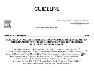
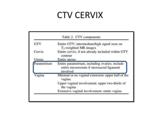
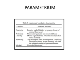
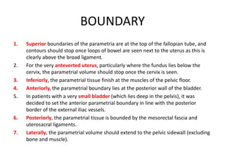
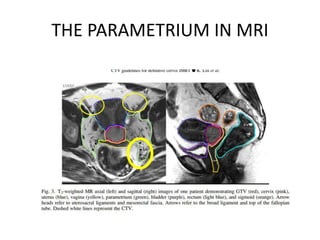
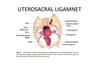
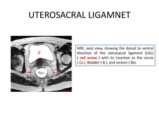
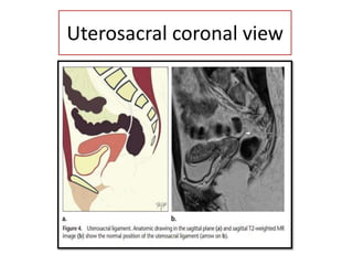
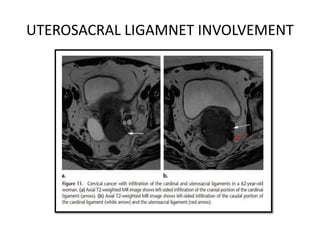
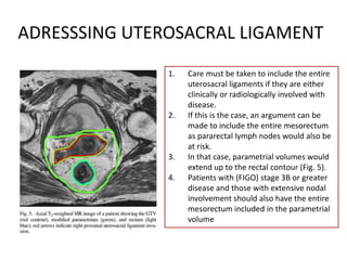
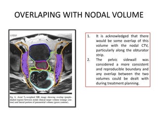
MANAGEMENT OF DIFFUSE GLIOMAS
5/12/2023 1
DR KANHU CHARAN PATRO
MD,DNB (Radiation Oncology),MBA,FICRO,FAROI(USA),PDCR,CEPC
HOD(Radiation Oncology)
Mahatma Gandhi Cancer Hospital And Research Institute, Visakhapatnam
drkcpatro@gmail.com M-9160470564
TARGET DELINEATION OF PARAMETRIUM
What is PARAMETRIUM?
1. The fat and connective tissue that
surrounds the uterus.
2. The parametrium helps connect the uterus
to other tissues in the pelvis.
PARAMETRIUM content?
1. The parametrium includes the superficial uterine pedicle (uterine artery and superficial
uterine vein) and related connective tissue and lymphatic channels.
2. The connective tissue crossing below the ureter is considered paracervix.
GUIDELINE
CTV CERVIX
PARAMETRIUM
BOUNDARY
BOUNDARY
1. Superior boundaries of the parametria are at the top of the fallopian tube, and
contours should stop once loops of bowel are seen next to the uterus as this is
clearly above the broad ligament.
2. For the very anteverted uterus, particularly where the fundus lies below the
cervix, the parametrial volume should stop once the cervix is seen.
3. Inferiorly, the parametrial tissue finish at the muscles of the pelvic floor.
4. Anteriorly, the parametrial boundary lies at the posterior wall of the bladder.
5. In patients with a very small bladder (which lies deep in the pelvis), it was
decided to set the anterior parametrial boundary in line with the posterior
border of the external iliac vessels.
6. Posteriorly, the parametrial tissue is bounded by the mesorectal fascia and
uterosacral ligaments.
7. Laterally, the parametrial volume should extend to the pelvic sidewall (excluding
bone and muscle).
THE PARAMETRIUM IN MRI
UTEROSACRAL LIGAMNET
MRI, axial view, showing the dorsal to ventral
direction of the uterosacral ligament (USL)
( red arrow ) with its insertion to the cervix
( Cx ), Bladder ( B ), and rectum ( Rec
Uterosacral coronal view
UTEROSACRAL LIGAMNET INVOLVEMENT
ADRESSSING UTEROSACRAL LIGAMENT
1. Care must be taken to include the entire
uterosacral ligaments if they are either
clinically or radiologically involved with
disease.
2. If this is the case, an argument can be
made to include the entire mesorectum
as pararectal lymph nodes would also be
at risk.
3. In that case, parametrial volumes would
extend up to the rectal contour (Fig. 5).
4. Patients with (FIGO) stage 3B or greater
disease and those with extensive nodal
involvement should also have the entire
mesorectum included in the parametrial
volume
OVERLAPING WITH NODAL VOLUME
1. It is acknowledged that there
would be some overlap of this
volume with the nodal CTV,
particularly along the obturator
strip.
2. The pelvic sidewall was
considered a more consistent
and reproducible boundary and
any overlap between the two
volumes could be dealt with
during treatment planning.
THANK YOU
TARGET DELINEATION OF PARAMETRIUM

TARGET DELINEATION OF PARAMETRIUM

  • 1.
    MANAGEMENT OF DIFFUSEGLIOMAS 5/12/2023 1 DR KANHU CHARAN PATRO MD,DNB (Radiation Oncology),MBA,FICRO,FAROI(USA),PDCR,CEPC HOD(Radiation Oncology) Mahatma Gandhi Cancer Hospital And Research Institute, Visakhapatnam drkcpatro@gmail.com M-9160470564 TARGET DELINEATION OF PARAMETRIUM
  • 2.
    What is PARAMETRIUM? 1.The fat and connective tissue that surrounds the uterus. 2. The parametrium helps connect the uterus to other tissues in the pelvis.
  • 3.
    PARAMETRIUM content? 1. Theparametrium includes the superficial uterine pedicle (uterine artery and superficial uterine vein) and related connective tissue and lymphatic channels. 2. The connective tissue crossing below the ureter is considered paracervix.
  • 4.
  • 5.
  • 6.
  • 7.
  • 8.
    BOUNDARY 1. Superior boundariesof the parametria are at the top of the fallopian tube, and contours should stop once loops of bowel are seen next to the uterus as this is clearly above the broad ligament. 2. For the very anteverted uterus, particularly where the fundus lies below the cervix, the parametrial volume should stop once the cervix is seen. 3. Inferiorly, the parametrial tissue finish at the muscles of the pelvic floor. 4. Anteriorly, the parametrial boundary lies at the posterior wall of the bladder. 5. In patients with a very small bladder (which lies deep in the pelvis), it was decided to set the anterior parametrial boundary in line with the posterior border of the external iliac vessels. 6. Posteriorly, the parametrial tissue is bounded by the mesorectal fascia and uterosacral ligaments. 7. Laterally, the parametrial volume should extend to the pelvic sidewall (excluding bone and muscle).
  • 9.
  • 10.
  • 11.
    UTEROSACRAL LIGAMNET MRI, axialview, showing the dorsal to ventral direction of the uterosacral ligament (USL) ( red arrow ) with its insertion to the cervix ( Cx ), Bladder ( B ), and rectum ( Rec
  • 12.
  • 13.
  • 14.
    ADRESSSING UTEROSACRAL LIGAMENT 1.Care must be taken to include the entire uterosacral ligaments if they are either clinically or radiologically involved with disease. 2. If this is the case, an argument can be made to include the entire mesorectum as pararectal lymph nodes would also be at risk. 3. In that case, parametrial volumes would extend up to the rectal contour (Fig. 5). 4. Patients with (FIGO) stage 3B or greater disease and those with extensive nodal involvement should also have the entire mesorectum included in the parametrial volume
  • 15.
    OVERLAPING WITH NODALVOLUME 1. It is acknowledged that there would be some overlap of this volume with the nodal CTV, particularly along the obturator strip. 2. The pelvic sidewall was considered a more consistent and reproducible boundary and any overlap between the two volumes could be dealt with during treatment planning.
  • 16.